. 2007 Nov;27(21):7631-40.
doi: 10.1128/MCB.00543-07. Epub 2007 Sep 4.

The Tom1L1-clathrin heavy chain complex regulates membrane partitioning of the tyrosine kinase Src required for mitogenic and transforming activities

Affiliations

The Tom1L1-clathrin heavy chain complex regulates membrane partitioning of the tyrosine kinase Src required for mitogenic and transforming activities

Guillaume Collin et al. Mol Cell Biol. 2007 Nov.

Abstract

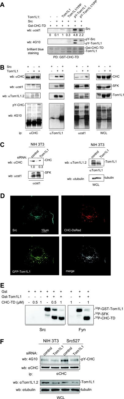
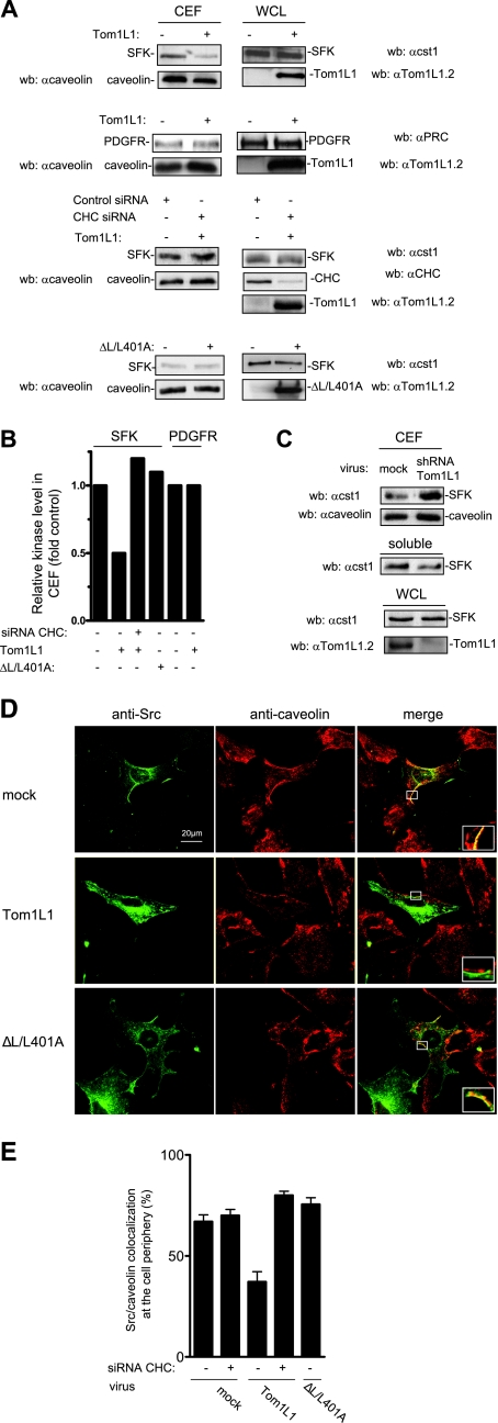
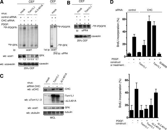
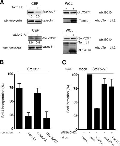
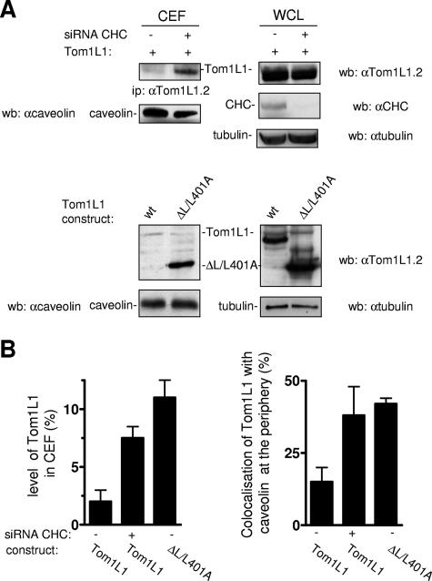
Compartmentalization of Src tyrosine kinases (SFK) plays an important role in signal transduction induced by a number of extracellular stimuli. For example, Src mitogenic signaling induced by platelet-derived growth factor (PDGF) is initiated in cholesterol-enriched microdomain caveolae. How this Src subcellular localization is regulated is largely unknown. Here we show that the Tom1L1-clathrin heavy chain (CHC) complex negatively regulates the level of SFK in caveolae needed for the induction of DNA synthesis. Tom1L1 is both an interactor and a substrate of SFK. Intriguingly, it stimulates Src activity without promoting mitogenic signaling. We found that, upon association with CHC, Tom1L1 reduced the level of SFK in caveolae, thereby preventing its association with the PDGF receptor, which is required for the induction of mitogenesis. Similarly, the Tom1L1-CHC complex reduced also the level of oncogenic Src in cholesterol-enriched microdomains, thus affecting both its capacity to induce DNA synthesis and cell transformation. Conversely, Tom1L1, when not associated with CHC, accumulated in caveolae and promoted Src-driven DNA synthesis. We concluded that the Tom1L1-CHC complex defines a novel mechanism involved in negative regulation of mitogenic and transforming signals, by modulating SFK partitioning at the plasma membrane.


Figures

FIG. 1.
FIG. 1.
Association of CHC with Tom1L1. (A) Modular structure of Tom1L1 wild type and of the mutants used in this study. The VHS and GAT homology domains, the linker region, and the C terminus (C) are indicated. Src binding sites (i.e., PP421LP and Tyr457), the mutations of these Src binding sites (i.e., A420ALA and Phe457 in Tom1L1YPP), and the mutation of the CHC binding site A401AQPSVA (in ΔL/L401A) are indicated. (B) Association of CHC with Tom1L1 in vitro. HeLa cell lysates were incubated with the indicated GST fusion proteins or control GST beads, and the presence of CHC in the pull-down assays was revealed by Western blotting (wb) with a specific antibody. Input (15% of the cell lysate) was loaded as a positive control. αCHC, anti-CHC. (C) Association of CHC with Tom1L1 in NIH 3T3 cells. The CHC level associated with Tom1L1 in NIH 3T3 cells was assessed by Western blotting with anti-CHC antibody after immunoprecipitation of Tom1L1 with control (immunoglobulin G [IgG]) or anti-Tom1L1.3 (αTom1L1.3) antibody as shown. The level of immunoprecipitated Tom1L1 is also shown.
FIG. 2.
FIG. 2.
CHC-Tom1L1 complex formation. (A) Association of CHC with Tom1L1 involves both the linker and the C-terminus sequences. HEK 293 cells were transfected with CHC-DsRed and the indicated Tom1L1 constructs. Tom1L1 proteins were immunoprecipitated (ip) with the anti-Tom1L1.3 (αTom1L1.3) antibodies, and the presence of associated CHC-DsRed was revealed by Western blotting (wb) with anti-CHC (αCHC) antibodies. The levels of associated CHC-Ds-Red, immunoprecipitated Tom1L1, and expressed CHC-DsRed are shown. wt, wild type; WCL, whole-cell lysate. (B) In vitro association of Tom1L1 with Gst-CHC-TD fusion protein. The indicated fusion protein bound to glutathione beads was incubated with the purified Tom1L1. The presence of Tom1L1 was revealed by Western blotting with the indicated antibody. (C) Association of Tom1L1 C terminus with CHC involves a Leu-rich motif at the C terminus. HeLa cell lysates were incubated with indicated GST fusion proteins or control GST beads, and the interaction with CHC was revealed by Western blotting with a specific antibody. Input (5% of the cell lysates) was included as a positive control. (D) Regulation of CHC-Tom1L1 complex formation by the linker and the Leu-rich motif L401LQPSVL. Tom1L1 was immunoprecipitated from lysates of HEK 293 cells transiently expressing the indicated constructs with the anti-Tom1L1.3 antibodies or a control immunoglobulin G (IgG), and the presence of associated CHC was revealed by Western blotting using anti-CHC antibodies. The levels of expressed and associated CHC and immunoprecipitated Tom1L1 are shown. (E) Colocalization of CHC with Tom1L1. Shown is the representative fluorescence of CHC-DsRed, GFP-Tom1L1 (top panels), or a GFP-Tom1L1 mutant that does not associate with CHC (ΔL/L401A) (bottom panel). Also shown is the merged image of a cotranfected NIH 3T3 cell as obtained after deconvolution (Huygens software).
FIG. 3.
FIG. 3.
CHC-Tom1L1-Src ternary complex formation. (A) Src-CHC-Tom1L1 ternary complex formation in vitro. Association of indicated purified proteins with Gst-CHC-TD bound to beads. The presence of Src, phosphorylated Src, and phosphorylated Tom1L1 was shown by Western blotting (wb) with the indicated antibodies (αcst1). Association of Gst-CHC-TD with Tom1L1 was revealed by Coomassie brilliant blue staining of the complex separated on a sodium dodecyl sulfate-polyacrylamide gel electrophoresis gel. (B) Src-CHC-Tom1L1 complex formation in HEK 293 cells that coexpress Src and Tom1L1. Each member of the complex was immunoprecipitated with the indicated antibodies (anti-CHC [α-CHC], anti-Tom1L1 [αTom1L1], and [αcst1]), and the presence of coassociated protein was detected by Western blotting with the antibodies shown. The tyrosine phosphorylation content of each immunoprecipitate is shown. Levels of tyrosine-phosphorylated proteins, CHC, SFK, and Tom1L1 from a whole-cell lysate (WCL) are also shown. (C) Endogenous Tom1L1 bridges SFK to CHC in NIH 3T3 cells. The CHC level associated with SFK was assessed by Western blotting with the indicated antibody after immunoprecipitation of SFK from NIH 3T3 cells that were transfected with the indicated siRNA. The levels of SFK, Tom1L1 and tubulin are also shown. Quantification of associated CHC is indicated. αtubulin, antitubulin antibody. (D) Colocalization of Src, Tom1L1, and CHC. Representative fluorescences of CHC-DsRed, GFP-Tom1L1, and immunostained avian Src are shown with the merged image of an NIH 3T3 cell coexpressing all three components after deconvolution, as described in Materials and Methods. (E) SFK phosphorylates CHC-TD in the presence of Tom1L1. Shown are the results of an in vitro kinase assay using purified Src or Fyn and in the presence of the indicated concentrations of CHC-TD and 1 μM of GST or Gst-Tom1L1, as indicated. Labeled SFK, Gst-Tom1L1, and CHC-TD are shown. (F) Tom1L1 regulates Src-induced CHC phosphorylation in Src-transformed cells. CHC was immunoprecipitated from NIH 3T3 cells or NIH3 3T3 cells stably expressing SrcY527F (Src 527) as shown and that were transfected with control or siRNA Tom1L1 as indicated. The levels of CHC and tyrosine-phosphorylated CHC (pY-CHC) are shown and were assessed by Western blotting with the indicated antibodies. The levels of Tom1L1 and tubulin from indicated cell lysates (WCL) are also shown.
FIG. 4.
FIG. 4.
Tom1L1 reduces SFK localization in caveolae through CHC association. (A) Tom1L1 reduces SFK but not PDGFR level in CEF. Shown are the levels of SFK, PDGFR, and caveolin in CEF purified from HEK 293 cells that were transfected with the indicated constructs and control or CHC siRNAs when indicated. The levels of expressed SFK, PDGFR, and Tom1L1 from the whole-cell lysate (WCL) are also shown. wb, Western blotting. αcaveolin, anticaveolin; αcst1, anti-cst1; αTom1L1.2, anti-Tom1L1.2; αPRC, anti-PRC; αCHC, anti-CHC. (B) Quantitative analysis of SFK and PDGFR levels in CEF obtained from two independent experiments. (C) Depletion of Tom1L1 enhances SFK accumulation in CEF. SFK and caveolin levels in CEF and soluble fractions (fractions 7 to 9 [“soluble”]) purified from HEK 293 cells that were mock infected (luciferase shRNA) or infected with Tom1L1 shRNA as indicated. The level of tubulin and Tom1L1 is also shown. A representative example (D) and its statistical analysis (E) (mean ± standard deviation [n = 3]) of Src-caveolin colocalization at the periphery of NIH 3T3 cells are shown. Cells infected with control (mock) or indicated Tom1L1 retroviruses were seeded on coverslips and transfected with avian Src together with siRNA (when indicated) for 48 h. Cells were then fixed and processed for immunofluorescence as described in Materials and Methods. Shown is a representative example of avian Src immunostaining, caveolin immunostaining, and the merged image from indicated infected NIH 3T3 cells obtained by confocal analysis as described in Materials and Methods. A threefold magnification of the merged image at the cell periphery is also included. The percentage of transfected cells that exhibited Src-caveolin colocalization at the cell periphery was calculated as described in Materials and Methods. Results are expressed as the mean ± standard deviation of three independent experiments. The levels of Tom1L1 and CHC are shown in panel A.
FIG. 5.
FIG. 5.
Tom1L1-CHC inhibits SFK-PDGFR coupling in caveolae and PDGF-induced DNA synthesis. (A) The Tom1L1-CHC complex inhibits SFK-PDGFR complex formation in caveolae. In vitro kinase assays of immunoprecipitated (ip) SFK (left panel) are shown from CEF purified from PDGF-stimulated NIH 3T3 cells that were infected with control (mock) or indicated viruses and transfected with control or CHC siRNA as indicated. The level of caveolin and quantified level of SFK in CEF are shown. (Right panel) Reimmunoprecipitation of the labeled 180-kDa protein observed in SFK immunoprecipitate with the PDGFRβ-specific PR4 antibody (αPR4). Labeled PDGFR and SFK are shown. wb, Western blotting; αcst1, anti-cst1; αcaveolin, anticaveolin. (B) The Tom1L1-CHC does not affect PDGFR activity in caveolae. The results of in vitro kinase assays of immunoprecipitated PDGFRβ with the indicated antibody are shown from CEF purified from PDGF-stimulated NIH 3T3 cells that were infected with control (mock) or indicated viruses and transfected with control or CHC siRNA when indicated. The level of caveolin in CEF is shown. (C) Shown are the levels of CHC, Tom1L1, SFK, and tubulin from whole-cell lysates (WCL) of NIH 3T3 cells infected with control (mock) or indicated viruses and transfected with control or CHC siRNAs when indicated. αtubulin, antitubulin. (D) PDGF mitogenic inhibition induced by Tom1L1 requires association with CHC. NIH 3T3 cells seeded onto coverslips and transfected or not with indicated constructs or siRNA were made quiescent by serum starvation for 30 h, treated or not with the SFK inhibitor SU6656 (2 μM), and stimulated or not with PDGF (5 ng/ml), as indicated, in the presence of BrdU for 18 h. Cells were then fixed and processed for immunofluorescence. The percentage of transfected cells that incorporated BrdU was calculated as described in Materials and Methods. Results are expressed as the mean ± standard deviation of three to five independent experiments.
FIG. 6.
FIG. 6.
Tom1L1-CHC affects SrcY527F membrane partitioning, SrcY527F-induced DNA synthesis, and focus formation. (A) Tom1L1-CHC reduces SrcY527F level in CEF. Shown are the levels of avian Src and caveolin from CEF purified from HEK 293 cells that were transfected with SrcY527F together or not with the indicated Tom1L1 constructs. The levels of expressed Src, Tom1L1, and tubulin expressed from the whole-cell lysate (WCL) are shown. wb, Western blotting; αcaveolin, anticaveolin; αTom1L1.2, anti-Tom1L1.2. (B) Tom1L1-CHC inhibits Src-driven DNA synthesis. NIH 3T3 cells stably transformed by SrcY527F (Src 527), seeded onto coverslips, and transfected or not with the indicated constructs were incubated in 0.5% serum for 30 h and further incubated in the presence of BrdU for 18 h. Cells were then fixed and processed for immunofluorescence. The percentage of transfected cells that incorporated BrdU was calculated as described in Materials and Methods. Results are expressed as the mean ± standard deviation of three independent experiments. (C) Tom1L1-CHC inhibits SrcY527F transforming activity. The statistical analysis of inhibition of SrcY527F-induced foci by Tom1L1-CHC is shown. NIH 3T3 cells were transfected or not with siRNA CHC as shown and then infected with control (mock) or indicated retroviruses. After 12 days of growth, foci were stained and scored as described in Materials and Methods. Focus formation (percentage of foci obtained relative to foci induced by SrcY527F) is represented as the mean ± standard deviation of three independent experiments.
FIG. 7.
FIG. 7.
Tom1L1 that does not associate with CHC accumulates in caveolae. (A and B) Tom1L1 that does not associate with CHC accumulates in caveolae. (A) Tom1L1 and caveolin levels in CEF of HEK 293 cells transfected with Tom1L1 constructs and control or CHC siRNAs, when indicated. The levels of CHC, Tom1L1, and tubulin from the whole-cell lysate (WCL) are also shown. wb, Western blotting; ip, immunoprecipitation; wt, wild type; αcaveolin, anticaveolin; αTom1L1.2, anti-Tom1L1.2; αCHC, anti-CHC; αtubulin, antitubulin. (B, left panel) Statistical analysis (mean ± standard deviation [n = 3]) of the percentage of the Tom1L1 level in CEF shown in panel A. (B, right panel) Statistical analysis of Tom1L1-caveolin colocalization at the periphery of NIH 3T3 cells. Cells seeded on coverslips were transfected with the indicated Tom1L1 construct together or not with the indicated siRNA for 48 h. Cells were then fixed and processed for immunofluorescence as described in Materials and Methods. The percentage of transfected cells that exhibited Tom1L1-caveolin colocalization at the cell periphery was calculated as described in Materials and Methods. The results are expressed as the mean ± standard deviation of three independent experiments.
FIG. 8.
FIG. 8.
Tom1L1 that does not associate with CHC increases Src-driven DNA synthesis. (A) Tom1L1 that does not associate with CHC increases wild-type Src-driven DNA synthesis. NIH 3T3 cells seeded onto coverslips and transfected or not with the indicated Tom1L1 constructs and CHC siRNA, as indicated, were incubated in 0.5% serum for 30 h and further incubated in the presence of BrdU for 18 h. Cells were then fixed and processed for immunofluorescence. (B) CHC does not regulate the capacity of mT antigen to enhance Src-driven DNA synthesis. Shown is BrdU incorporation (mean ± standard deviation [n = 4]) of serum-starved NIH 3T3 cells that were transfected with indicated constructs and the indicated siRNAs as shown. (C) mT antigen is preferentially localized in CEF. Shown are the level of mT antigen in CEF and soluble fractions (fractions 7 to 9 [“soluble”]) from HEK 293 cells expressing mT antigen. The level of caveolin is also shown. wb, Western blotting; αcaveolin, anticaveolin. (D) Tom1L1 enhances DNA synthesis in CHC-depleted NIH 3T3 cells in a Src-dependent manner. NIH 3T3 cells seeded onto coverslips and transfected or not with the indicated constructs and CHC siRNA, as indicated, were incubated in 0.5% serum for 30 h, treated or not with SU6656 (2 μM) as shown, and further incubated in the presence of BrdU for 18 h. Cells were then fixed and processed for immunofluorescence. The percentage of transfected cells that incorporated BrdU was calculated as described in Materials and Methods. The percentage of transfected cells that incorporated BrdU was calculated as described in Materials and Methods. Results are expressed as the mean ± standard deviation of three to four independent experiments. *, P < 0.05, and **, P < 0.01, by Student's t test.

References

    1. Blake, R. A., M. A. Broome, X. Liu, J. Wu, M. Gishizky, L. Sun, and S. A. Courtneidge. 2000. SU6656, a selective Src family kinase inhibitor, used to probe growth factor signaling. Mol. Cell. Biol. 20: 9018-9027. - PMC - PubMed
    1. Boggon, T. J., and M. J. Eck. 2004. Structure and regulation of Src family kinases. Oncogene 23: 7918-7927. - PubMed
    1. Bonifacino, J. S. 2004. The GGA proteins: adaptors on the move. Nat. Rev. Mol. Cell Biol. 5: 23-32. - PubMed
    1. Boureux, A., O. Furstoss, V. Simon, and S. Roche. 2005. c-Abl tyrosine kinase regulates a Rac/JNK and a Rac/Nox pathway for DNA synthesis and c-myc expression induced by growth factors. J. Cell Sci. 118: 3717-3726. - PubMed
    1. Bromann, P. A., H. Korkaya, and S. A. Courtneidge. 2004. The interplay between Src family kinases and receptor tyrosine kinases. Oncogene 23: 7957-7968. - PubMed

Publication types