. 2007 Nov;27(21):7551-9.
doi: 10.1128/MCB.01034-07. Epub 2007 Sep 4.

Wnt/beta-catenin is essential for intestinal homeostasis and maintenance of intestinal stem cells

Affiliations

Wnt/beta-catenin is essential for intestinal homeostasis and maintenance of intestinal stem cells

Tea Fevr et al. Mol Cell Biol. 2007 Nov.

Abstract

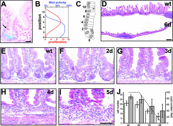
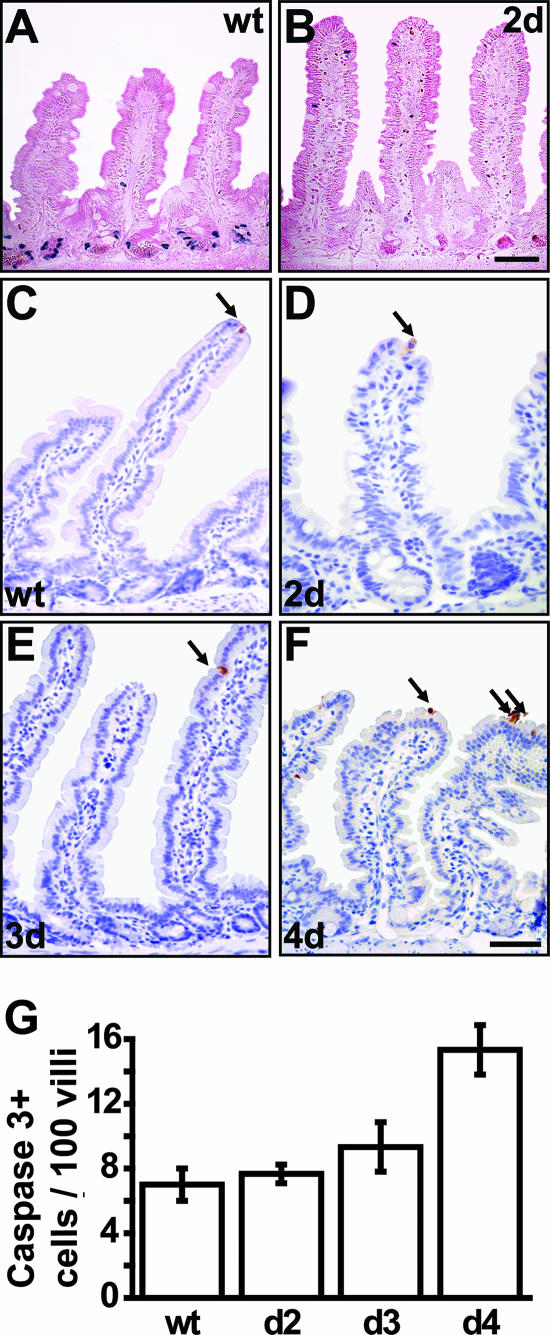
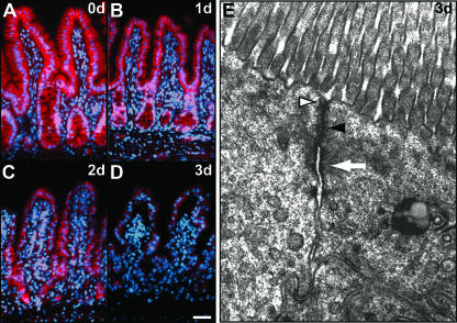
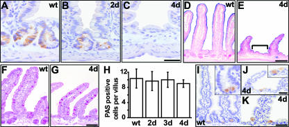
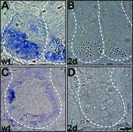
The Wnt signaling pathway is deregulated in over 90% of human colorectal cancers. beta-Catenin, the central signal transducer of the Wnt pathway, can directly modulate gene expression by interacting with transcription factors of the TCF/LEF family. In the present study we investigate the role of Wnt signaling in the homeostasis of intestinal epithelium by using tissue-specific, inducible beta-catenin gene ablation in adult mice. Block of Wnt/beta-catenin signaling resulted in rapid loss of transient-amplifying cells and crypt structures. Importantly, intestinal stem cells were induced to terminally differentiate upon deletion of beta-catenin, resulting in a complete block of intestinal homeostasis and fatal loss of intestinal function. Transcriptional profiling of mutant crypt mRNA isolated by laser capture microdissection confirmed those observations and allowed us to identify genes potentially responsible for the functional preservation of intestinal stem cells. Our data demonstrate an essential requirement of Wnt/beta-catenin signaling for the maintenance of the intestinal epithelium in the adult organism. This challenges attempts to target aberrant Wnt signaling as a new therapeutic strategy to treat colorectal cancer.


Figures

FIG. 1.
FIG. 1.
Active Wnt signaling is required for crypt maintenance. (A to C) Wnt activity versus cell proliferation in the crypts. (A) Localization of Wnt activity using conductin-lacZ reporter mice. The arrow shows the mitotic figure. Bar, 20 μm. (B) Schematic representation of the localization of Wnt activity (blue; arbitrary units) compared to cell proliferation (red; cells in metaphase/anaphase) along the crypt-villus axis, from Paneth cells up to position 10 above Paneth cells in the crypt. (C) Schematic representation of an intestinal crypt. Numbers represent cell positions from Paneth cells at the bottom of the crypt corresponding to position 0 up to position 10 above them. A cell in metaphase/anaphase is represented at position 9. (D) H&E staining of intestinal sections at day 6 after β-catenin ablation. Note that the intestinal epithelium is largely lost in the mutants. Bar, 200 μm. (E to I) H&E staining of the crypt area. Mutants show progressive crypt loss starting from day 2 (F) after induction and leading to the establishment of an extended intervillus region by day 4 (H). Bar, 100 μm. (J) Quantification of morphological changes was performed from 200 crypt/villus units per sample and three mice per group. Left axis: white bars show numbers of epithelial cells per crypt and gray bars show total numbers of epithelial cells which are not part of a villus (intervillus region). Error bars depict standard deviations. Right axis: dashed line represents crypt number per field as a percentage. wt, wild type.
FIG. 2.
FIG. 2.
Cell proliferation and death as a result of β-catenin ablation in the intestinal epithelium. (A and B) Complete loss of cell proliferation as assessed by short-time BrdU incorporation 2 days after β-catenin ablation. (C to F) Immunostaining for active caspase 3 (arrows) shows no increase in apoptotic features in mutant crypts up to day 3 (E). However, a moderate increase is observed at day 4 in mutant villus tips (F). Bars, 50 μm. (G) Quantification of apoptosis expressed as number of caspase 3-positive cells per 100 villi. For quantification, 200 villi were counted per mouse and three mice per group. wt, wild type.
FIG. 3.
FIG. 3.
Loss of intestinal epithelial cells is due to canonical Wnt signaling. (A to D) Immunofluorescence time course for β-catenin protein expression reveals its presence in adherens junctions up to day 2 (C) after induction. Bar, 50 μm. (E) Electron micrograph of a mouse intestinal crypt region. Junctional complexes such as tight junctions (white arrowhead), adherens junctions (black arrowhead), and desmosomes (white arrow) remain unaffected 3 days after β-catenin deletion. Magnification, ×23,000.
FIG. 4.
FIG. 4.
Impact on differentiation upon β-catenin ablation in intestinal epithelium. (A to C) Immunostaining for CD44 reveals progressive loss of the crypt phenotype in mutants (B and C) between day 2 and day 4. Bar, 10 μm. (D and E) The enterocytic differentiation marker alkaline phosphatase (as detected by blue nitroblue tetrazolium-5-bromo-4-chloro-3-indolylphosphate staining) is absent from wild-type (D) crypts but is present in mutant (E) intervillus regions (bracket). (F to H) PAS staining for goblet cells reveals unchanged numbers. The graph shows quantification of goblet cell numbers expressed as numbers of PAS stain-positive cells per villus (H). Two hundred villi were counted per mouse and three mice per group. (I to K) Immunostaining for lysozyme shows Paneth cell localization in wild-type crypts (I) and in mutants (J and K). Insets show higher magnifications for panels I and J, respectively. Bar, 20 μm. wt, wild type.
FIG. 5.
FIG. 5.
LRCs undergo differentiation in mutant mice. (A to C) Autoradiography of mouse intestine injected with tritiated TdR 20 days after labeling. In wild-type epithelium (A), LRCs (arrow) are found in the crypts above Paneth cells. In 4-day mutant epithelium (B), LRCs (arrow) are found in similar numbers but at aberrant positions. Bar, 25 μm. (C) Scheme of the localization pattern of LRCs in control (left) and mutants for which the frequency of LRCs in a certain position is depicted as black lines of different widths. Initially, LRCs in uninduced β-catenin−/lox mice (0d) are localized as in the control. Two days after β-catenin deletion, mutant LRCs are dispersed throughout the crypt area (data not shown), followed by an even distribution along the crypt-villus axis at day 4 (4d). We quantified 300 crypt-villus units for two mice per group. In both groups, about 20% of crypts (control) or crypt-villus units (mutant) were found to contain LRCs, which were scored as positive when retaining more than five grains per nucleus. (D and E) Counterstaining for the enterocytic differentiation marker FabpL demonstrates that LRCs (arrows) are differentiated in the mutant 4 days after β-catenin deletion (E), whereas they remain undifferentiated in the control (D). The picture gives a representative example; all LRCs in the mutants express FabpL at this time point. Bar, 25 μm. (F to I) In vivo intestinal epithelial cell migration assay. Proliferating cells were BrdU pulse-labeled immediately prior to tamoxifen injection, and subsequently migrating cells were localized by immunohistochemistry at day 2 (F and G) and day 3 (H to I) after tamoxifen injection in control (F and H) and mutant (G and I) epithelium. Bar, 50 μm. wt, wild type.
FIG. 6.
FIG. 6.
Rapid downregulation of stem cell markers upon β-catenin ablation. (A and B) Sox4 is located in the bottom part of intestinal wild-type crypts (A) and is strongly repressed in β-catenin mutants at day 2 (B). (C and D) Diap3 is expressed at the stem cell location in wild-type crypts (C) and is strongly reduced in β-catenin mutants (D). Dashed lines mark crypt limits. Bar, 10 μm. wt, wild type.

Similar articles

Cited by

References

    1. Batlle, E., J. T. Henderson, H. Beghtel, M. M. van den Born, E. Sancho, G. Huls, J. Meeldijk, J. Robertson, M. van de Wetering, T. Pawson, and H. Clevers. 2002. Beta-catenin and TCF mediate cell positioning in the intestinal epithelium by controlling the expression of EphB/ephrinB. Cell 111: 251-263. - PubMed
    1. Behrens, J., and B. Lustig. 2004. The Wnt connection to tumorigenesis. Int. J. Dev. Biol. 48: 477-487. - PubMed
    1. Bettess, M. D., N. Dubois, M. J. Murphy, C. Dubey, C. Roger, S. Robine, and A. Trumpp. 2005. c-Myc is required for the formation of intestinal crypts but dispensable for homeostasis of the adult intestinal epithelium. Mol. Cell. Biol. 25: 7868-7878. - PMC - PubMed
    1. Bianchini, M., E. Levy, C. Zucchini, V. Pinski, C. Macagno, P. De Sanctis, L. Valvassori, P. Carinci, and J. Mordoh. 2006. Comparative study of gene expression by cDNA microarray in human colorectal cancer tissues and normal mucosa. Int. J. Oncol. 29: 83-94. - PubMed
    1. Bjerknes, M., and H. Cheng. 1999. Clonal analysis of mouse intestinal epithelial progenitors. Gastroenterology 116: 7-14. - PubMed

Publication types

MeSH terms