. 2007 Nov;27(21):7615-22.
doi: 10.1128/MCB.01239-07. Epub 2007 Sep 4.

Skp2B stimulates mammary gland development by inhibiting REA, the repressor of the estrogen receptor

Affiliations

Skp2B stimulates mammary gland development by inhibiting REA, the repressor of the estrogen receptor

Karina Umanskaya et al. Mol Cell Biol. 2007 Nov.

Abstract

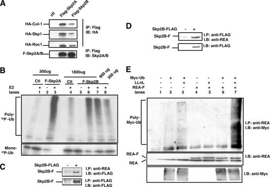
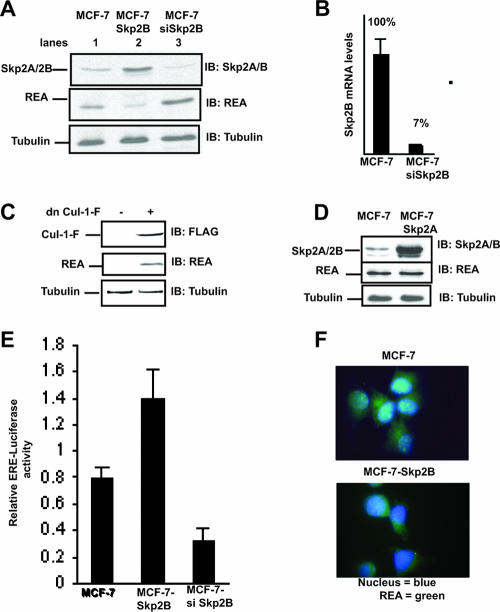
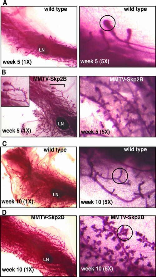
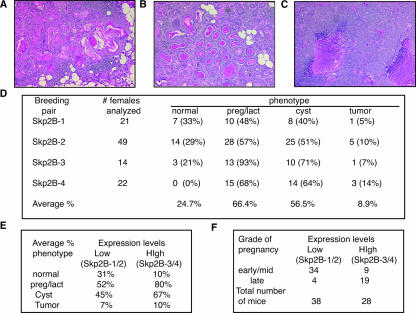
Skp2B, an F-box protein of unknown function, is frequently overexpressed in breast cancer. In order to determine the function of Skp2B and whether it has a role in breast cancer, we performed a two-hybrid screen and established transgenic mice expressing Skp2B in the mammary glands. We found that Skp2B interacts with the repressor of estrogen receptor activity (REA) and that overexpression of Skp2B leads to a reduction in REA levels. In the mammary glands of MMTV-Skp2B mice, REA levels are also low. Our results show that in virgin transgenic females, Skp2B induces lobuloalveolar development and differentiation of the mammary glands normally observed during pregnancy. As this phenotype is identical to what was observed for REA heterozygote mice, our observations suggest that the Skp2B-REA interaction is physiologically relevant. However, in contrast to REA(+/-) mice, MMTV-Skp2B mice develop mammary tumors, suggesting that Skp2B affects additional proteins. These results indicate that the observed expression of Skp2B in breast cancer does contribute to tumorigenesis at least in part by modulating the activity of the estrogen receptor.


Figures

FIG. 1.
FIG. 1.
Skp2B interacts with REA. (A) HEK293T cells were transfected with the indicated plasmids, FLAG-Skp2A and FLAG-Skp2B were immunoprecipitated using FLAG antibody, and their association with either HA-Cul-1, HA-Skp1, or HA-Roc1 was analyzed using anti-HA antibody. ctl, control. (B) HEK293T cells were transfected with either FLAG-Skp2A or FLAG-Skp2B along with Cul-1 and Roc1 followed by anti-FLAG immunoprecipitation. The resulting immunoprecipitations were then used in an in vitro ubiquitination assay using 32P-labeled ubiquitin (32P-Ub). Top, polyubiquitin chain formation; bottom, level of monoubiquitin. Extracts from HEK293T cells transfected with Cul-1 and Roc1 alone were used as controls. (C) Coimmunoprecipitation of endogenous REA with FLAG-Skp2B, where REA was immunoprecipitated (IP) and levels of FLAG-Skp2B were determined by immunoblotting (IB). (D) Coimmunoprecipitation of endogenous REA with FLAG-Skp2B, where FLAG-Skp2B was immunoprecipitated and levels of REA were determined by immunoblotting. (E) HEK293T cells were transfected with the indicated plasmids, and REA was immunoprecipitated followed by immunoblotting using anti-Myc antibody to detect ubiquitinated proteins.
FIG. 2.
FIG. 2.
Skp2B but not Skp2A overexpression leads to a reduction in REA and activation of the ER. (A) Western analysis of the levels of REA and Skp2B in MCF-7 cells, MCF-7-Skp2B cells, and MCF-7 cells transfected with siRNA against Skp2B (siSkp2B). (B) The reduction of Skp2B mRNA following the transfection of siRNA against SKP2B was determined by quantitative RT-PCR. (C) MCF-Skp2B cells were transfected with FLAG-tagged dominant negative cul-1 (dn Cul-1-F), and the levels of dn-Cul-1-F and REA were determined by immunoblotting (IB). (D) Immunoblot analysis of the levels of REA and Skp2A in MCF-7 and MCF7-Skp2A cells. (E) ER activity was monitored using an ERE luciferase reporter transfected in MCF-7, MCF-7-Skp2B, and MCF-7 cells cotransfected with siRNA Skp2B. (F) Immunofluorescence of REA in MCF-7 and MCF-7-Skp2B cells.
FIG. 3.
FIG. 3.
Skp2B expression in the mammary glands reduces REA levels. (A) Four separate MMTV-Skp2B founders were used for breeding, and the mammary gland of one female of the second generation from each line was tested for Skp2B expression by quantitative RT-PCR using Skp2B-specific primers. WT, wild type. (B) The expression of Skp2B was tested by immunohistochemistry on mammary gland sections of a wild-type female (left) and a female from the Skp2B-4 transgenic line (right) by use of anti-Skp2 antibody. (C) Western analysis of the levels of REA in the mammary glands of a wild-type mouse and of three MMTV-Skp2B transgenic mice.
FIG. 4.
FIG. 4.
Expression of Skp2B accelerates ductal invasion of the fat pad and promotes lobuloalveolar development. (A) Mammary gland from a wild-type female at week 5 was stained by whole-mount staining and visualized at magnifications of ×1 and ×5. LN indicates the position of the lymph node, while a TEB is indicated by the circle. (B) Mammary gland from an MMTV-Skp2B female at week 5 was stained by whole-mount staining and visualized at magnifications of ×1 and ×5. (C) Mammary gland from a wild-type female at week 10 was stained by whole-mount staining and visualized at magnifications of ×1 and ×5. A site of side branching is indicated by the circle. (D) Mammary gland from an MMTV-Skp2B female at week 10 was stained by whole-mount staining and visualized at magnifications of ×1 and ×5. Lobules are indicated by the circle. In each case, one representative gland is shown.
FIG. 5.
FIG. 5.
Expression of Skp2B in virgin females promotes a pregnancy phenotype for the mammary glands. (A) Whole mount and H&E stained-section of the mammary glands from a 12-month-old MMTV-Skp2B transgenic female. (B) Whole mount and H&E-stained section of the mammary glands from a 2-day-pregnant wild-type female. (C) Whole mount and H&E-stained section of the mammary glands from a 14-month-old MMTV-Skp2B transgenic female. (D) H&E-stained sections of the mammary glands from a wild-type female that had been nursing her pups for 2 days after delivery and from a 16-day-pregnant female. Pictures were taken at magnifications of ×1, ×4, and ×10 as indicated.
FIG. 6.
FIG. 6.
Cyst formation and deregulation of STAT5 and IGFBPs in virgin MMTV-Skp2B females. (A) H&E staining of mammary gland from 5-month-old and 9-month-old virgin MMTV-Skp2B females. (B) The mammary glands from three transgenic females were selected based on the phenotypes indicated. The levels of STAT5, phospho-STAT5, and IGFBP-2 and -4 were determined by Western blotting (immunoblotting [IB]) and compared to the levels observed for wild-type females. Morphology abbreviations: C, cyst alone; C+P, cyst and pregnancy; P+L, pregnancy and lactation without cyst.
FIG. 7.
FIG. 7.
MMTV-Skp2B mice develop mammary carcinoma. (A) H&E staining of mammary gland from a virgin MMTV-Skp2B female with squamous cell carcinoma. (B) H&E staining of mammary gland from a virgin MMTV-Skp2B female with glandular carcinoma that displays a secretory component. (C) H&E staining of mammary gland from a virgin MMTV-Skp2B female with high-grade adenocarcinoma with a necrotic area. (D) Summary of the phenotypes observed for the mammary glands of MMTV-Skp2B transgenic mice. The ages of the mice analyzed ranged from 3 to 16 months for each transgenic group. (E) The average frequencies of each phenotype in transgenic lines that expressed low levels of Skp2B (Skp2B-1/2) and transgenic lanes that expressed high levels of Skp2B (Skp2B-3/4) are shown. preg/lact, pregnant/lactating. (F) The numbers of mice that expressed low levels of Skp2B (Skp2B-1/2) and high levels of Skp2B (Skp2B-3/4) were subclassified according to the severity of the pregnancy phenotype.

References

    1. Allar, M. A., and T. L. Wood. 2004. Expression of the insulin-like growth factor binding proteins during postnatal development of the murine mammary gland. Endocrinology 145: 2467-2477. - PubMed
    1. Boutinaud, M., J. H. Shand, M. A. Park, K. Phillips, J. Beattie, D. J. Flint, and G. J. Allan. 2004. A quantitative RT-PCR study of the mRNA expression profile of the IGF axis during mammary gland development. J. Mol. Endocrinol. 33: 195-207. - PubMed
    1. Brennan, K. R., and A. M. Brown. 2004. Wnt proteins in mammary development and cancer. J. Mammary Gland Biol. Neoplasia 9: 119-131. - PubMed
    1. Bruzzi, P., L. Dogliotti, C. Naldoni, L. Bucchi, M. Costantini, A. Cicognani, M. Torta, G. F. Buzzi, and A. Angeli. 1997. Cohort study of association of risk of breast cancer with cyst type in women with gross cystic disease of the breast. BMJ 314: 925-928. - PMC - PubMed
    1. Carrano, A. C., E. Eytan, A. Hershko, and M. Pagano. 1999. SKP2 is required for ubiquitin-mediated degradation of the CDK inhibitor p27. Nat. Cell Biol. 1: 193-199. - PubMed

Publication types

MeSH terms