The role of B cells in the development of CD4 effector T cells during a polarized Th2 immune response
- PMID: 17785819
- PMCID: PMC2258088
- DOI: 10.4049/jimmunol.179.6.3821
The role of B cells in the development of CD4 effector T cells during a polarized Th2 immune response
Abstract
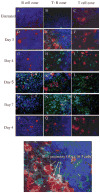
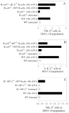
Previous studies have suggested that B cells promote Th2 cell development by inhibiting Th1 cell differentiation. To examine whether B cells are directly required for the development of IL-4-producing T cells in the lymph node during a highly polarized Th2 response, B cell-deficient and wild-type mice were inoculated with the nematode parasite, Nippostrongylus brasiliensis. On day 7, in the absence of increased IFN-gamma, IL-4 protein and gene expression from CD4 T cells in the draining lymph nodes were markedly reduced in B cell-deficient mice and could not be restored by multiple immunizations. Using a DO11.10 T cell adoptive transfer system, OVA-specific T cell IL-4 production and cell cycle progression, but not cell surface expression of early activation markers, were impaired in B cell-deficient recipient mice following immunization with N. brasiliensis plus OVA. Laser capture microdissection and immunofluorescent staining showed that pronounced IL-4 mRNA and protein secretion by donor DO11.10 T cells first occurred in the T cell:B cell zone of the lymph node shortly after inoculation of IL-4-/- recipients, suggesting that this microenvironment is critical for initial Th2 cell development. Reconstitution of B cell-deficient mice with wild-type naive B cells, or IL-4-/- B cells, substantially restored Ag-specific T cell IL-4 production. However, reconstitution with B7-1/B7-2-deficient B cells failed to rescue the IL-4-producing DO11.10 T cells. These results suggest that B cells, expressing B7 costimulatory molecules, are required in the absence of an underlying IFN-gamma-mediated response for the development of a polarized primary Ag-specific Th2 response in vivo.
Conflict of interest statement
Disclosures
The authors have no financial conflict of interest. The opinions or assertions contained within are the private views of the authors and should not be construed as official or necessarily reflecting the views of the University of Medicine and Dentistry of New Jersey or the Department of Agriculture.
Figures


Similar articles
-
Nippostrongylus brasiliensis can induce B7-independent antigen-specific development of IL-4-producing T cells from naive CD4 T cells in vivo.J Immunol. 2002 Dec 15;169(12):6959-68. doi: 10.4049/jimmunol.169.12.6959. J Immunol. 2002. PMID: 12471130
-
IL-2 and autocrine IL-4 drive the in vivo development of antigen-specific Th2 T cells elicited by nematode parasites.J Immunol. 2005 Feb 15;174(4):2242-9. doi: 10.4049/jimmunol.174.4.2242. J Immunol. 2005. PMID: 15699158 Free PMC article.
-
The role of OX40 ligand interactions in the development of the Th2 response to the gastrointestinal nematode parasite Heligmosomoides polygyrus.J Immunol. 2003 Jan 1;170(1):384-93. doi: 10.4049/jimmunol.170.1.384. J Immunol. 2003. PMID: 12496423
-
Dendritic cell-intrinsic expression of NF-kappa B1 is required to promote optimal Th2 cell differentiation.J Immunol. 2005 Jun 1;174(11):7154-9. doi: 10.4049/jimmunol.174.11.7154. J Immunol. 2005. PMID: 15905559
-
Rapid in vivo conversion of effector T cells into Th2 cells during helminth infection.J Immunol. 2012 Jan 15;188(2):615-23. doi: 10.4049/jimmunol.1101164. Epub 2011 Dec 7. J Immunol. 2012. PMID: 22156341
Cited by
-
Cytokine-producing effector B cells regulate type 2 immunity to H. polygyrus.Immunity. 2009 Mar 20;30(3):421-33. doi: 10.1016/j.immuni.2009.01.006. Epub 2009 Feb 26. Immunity. 2009. PMID: 19249230 Free PMC article.
-
Effector and regulatory B cells: modulators of CD4+ T cell immunity.Nat Rev Immunol. 2010 Apr;10(4):236-47. doi: 10.1038/nri2729. Epub 2010 Mar 12. Nat Rev Immunol. 2010. PMID: 20224569 Free PMC article. Review.
-
Dendritic cells and B cells: unexpected partners in Th2 development.J Immunol. 2014 Aug 15;193(4):1531-7. doi: 10.4049/jimmunol.1400149. J Immunol. 2014. PMID: 25086176 Free PMC article. Review.
-
Anti-CD20 treatment attenuates Th2 cell responses: implications for the role of lung follicular mature B cells in the asthmatic mice.Inflamm Res. 2024 Mar;73(3):433-446. doi: 10.1007/s00011-023-01847-4. Epub 2024 Feb 12. Inflamm Res. 2024. PMID: 38345634
-
CD19 expression in B cells regulates atopic dermatitis in a mouse model.Am J Pathol. 2013 Jun;182(6):2214-22. doi: 10.1016/j.ajpath.2013.02.042. Epub 2013 Apr 12. Am J Pathol. 2013. PMID: 23583649 Free PMC article.
References
-
- Steinman RM, Pack M, Inaba K. Dendritic cells in the T-cell areas of lymphoid organs. Immunol Rev. 1997;156:25–37. - PubMed
-
- Jenkins MK, Khoruts A, Ingulli E, Mueller DL, McSorley SJ, Reinhardt RL, Itano A, Pape KA. In vivo activation of antigen-specific CD4 T cells. Annu Rev Immunol. 2001;19:23–45. - PubMed
-
- Garside P, Ingulli E, Merica RR, Johnson JG, Noelle RJ, Jenkins MK. Visualization of specific B and T lymphocyte interactions in the lymph node. Science. 1998;281:96–99. - PubMed
-
- Crawford A, MacLeod M, Schumacher T, Corlett L, Gray D. Primary T cell expansion and differentiation in vivo requires antigen presentation by B cells. J Immunol. 2006;176:3498–3506. - PubMed
-
- O’Neill SK, Shlomchik MJ, Glant TT, Cao Y, Doodes PD, Finnegan A. Antigen-specific B cells are required as APCs and autoantibody-producing cells for induction of severe autoimmune arthritis. J Immunol. 2005;174:3781–3788. - PubMed
Publication types
MeSH terms
Substances
Grants and funding
LinkOut - more resources
Full Text Sources
Molecular Biology Databases
Research Materials

