. 2007 Sep 15;179(6):3831-40.
doi: 10.4049/jimmunol.179.6.3831.

Induction of the CTLA-4 gene in human lymphocytes is dependent on NFAT binding the proximal promoter

Affiliations

Induction of the CTLA-4 gene in human lymphocytes is dependent on NFAT binding the proximal promoter

Heather M Gibson et al. J Immunol. .

Abstract

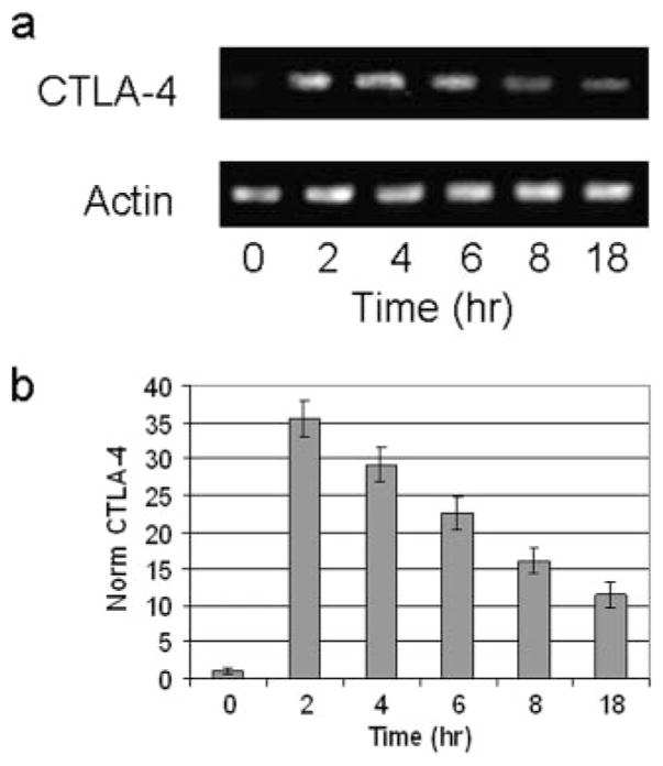
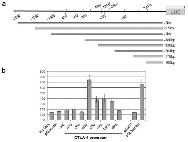
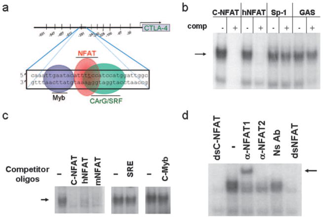
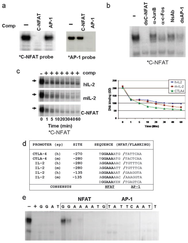
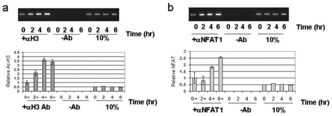
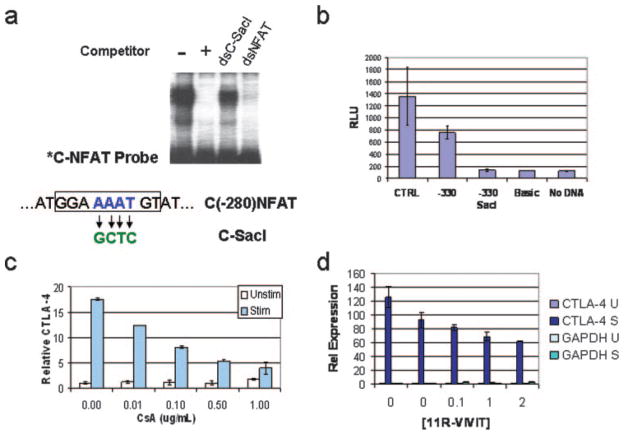
CTLA-4 is a member of the costimulatory family, has homology to CD28, and binds the B7 family of ligands. Unlike CD28, CTLA-4 ligation transmits a negative signal in T cells. CTLA-4 expression, while inducible in most T cells, is expressed constitutively on T cells with a regulatory phenotype. The mechanism controlling CTLA-4 expression in human T cells is poorly characterized, thus we sought to better understand the mechanism of activation of the CTLA-4 gene. By cloning the 5' upstream promoter and creating promoter-deletion reporter constructs, we show that the proximal promoter is critical for activating the CTLA-4 gene. Within this region, we identify a NFAT consensus sequence that binds NFAT with high affinity that differs from other NFAT sequences and does not recruit AP-1. Analysis of the chromatin proteins in the native CTLA-4 gene shows that this promoter region becomes associated with acetylated histones by chromatin immunoprecipitation assays. In addition, NFAT1 binds to the promoter of the CTLA-4 gene after stimulation by chromatin immunoprecipitation. The functional requirement of the NFAT site for CTLA-4 transcription was demonstrated by mutations in the NFAT site that abolished the activity of the promoter. Furthermore, inhibitors of NFAT suppressed CTLA-4 gene expression, indicating that NFAT plays a critical role in regulating the induction of the CTLA-4 gene in lymphocytes. The identification of NFAT as a critical regulator of the CTLA-4 gene suggests that targeting NFAT function may lead to novel approaches to modulate the CTLA-4 gene to control the immune response.


Conflict of interest statement

Disclosures

The authors have no financial conflict of interest.

Figures

FIGURE 1
FIGURE 1
CTLA-4 is rapidly induced in primary human T lymphocytes. a, Surface level CTLA-4 expression as determined in representative normal PBMCs by flow cytometry. PBMCs were stimulated with PMA/A23187 and stained with Abs to CD3 and CTLA-4. The level of CTLA-4 expression is undetectable in unstimulated lymphocytes, but increases in stimulated CD3 lymphocytes in PBMCs from normal volunteers. b, The average level of expression of surface CTLA-4 in T lymphocytes at 18 h after stimulation. Results represent analysis from 12 individuals, with mean level = 15%, SD ± 0.1%. c, Total CTLA-4 protein expression from PBMCs as measured by immunoblots. Actin immunoblot was performed to normalize for protein level. PBMCs were stimulated and lysed at 6 h. Equal protein was separated by SDS-PAGE and immunoblots were performed as described in Materials and Methods. Representative findings are from three independent experiments. d, Densitometric measurement of CTLA-4 immunoblot shown in c as determined using NIH ImageJ software. Levels of CTLA-4 are relative to the expression of the actin level that has been normalized by the OD among the samples.
FIGURE 2
FIGURE 2
CTLA-4 transcription is rapidly increased after stimulation. RNA analysis of CTLA-4 expression in freshly isolated human PBMCs. PBMCs were isolated and stimulated with PMA/A23187 for the indicated times. Total RNA was isolated as described in Materials and Methods. a, CTLA-4 levels as determined by RT-PCR and PCR products visualized on ethidium bromide-stained agarose gel. Results are representative of three independent experiments. b, CTLA-4 expression measured by quantitative PCR. Real-time PCR was performed as described in Materials and Methods and the results represent average induction in PBMCs from 15 normal volunteers.
FIGURE 3
FIGURE 3
Characterization of the 5′ proximal promoter of the human CTLA-4 gene in primary cells. a, A schematic of the human CTLA-4 promoter is shown, along with the 5′ deletions of the promoter that have been constructed into luciferase reporter plasmids. The details of cloning are described in Materials and Methods. b, CTLA-4 promoter activity in normal human PBMCs. Purified human PBMCs were pretreated with PMA/A23187 as described in Materials and Methods and electroporated with individual CTLA-4 promoter reporter plasmids with pEGFP cotransfected to monitor transfection efficiency. Cell lysates were prepared and luciferase assays were performed as described in Materials and Methods. Results are representative of three separate experiments. Relative light unit background is shown in the lane with no DNA. Relative light unit was determined by normalizing to transfection efficiency and equal protein levels.
FIGURE 4
FIGURE 4
Identifying NFAT as the factor that binds to the proximal CTLA-4 promoter. a, Sequence of the proximal CTLA-4 promoter important for transcription and potential transcription factor recognition motifs predicted by Transcription ESS is shown from nucleotides −264 to −297 from the translational start site. This sequence has consensus sites for NFAT, c-Myb, and SRE and is defined as C(−280)NFAT. b, NFAT sequences compete away DNA binding to C(−280)NFAT probe. EMSA results with γ-32P-labeled dsC(−280)NFAT probe, with extracts prepared from stimulated human PBMCs. Competition assays were performed using excess double-stranded unlabeled oligonucleotides for C(−280)NFAT = C-NFAT, human IL-2 NFAT = hNFAT, Sp-1, or GAS sites. c, C(−280)NFAT oligonucleotide interacts with DNA-binding activity for NFAT and is competed by other NFAT sequences (hIL-2 NFAT and mIL-2 NFAT). Competition with excess c-Myb or SRE oligonucleotide does not alter DNA binding to the C(−280)NFAT probe. d, NFAT1 binds to C(−280)NFAT. EMSA performed in the presence of Abs specific to NFAT1 yields a supershifted band (arrow) whereas nonspecific Ab (Ns Ab) or anti-NFAT2 does not.
FIGURE 5
FIGURE 5
C(−280)NFAT-binding activity does not depend on AP-1 cooperativity and has a higher affinity than NFAT sites with AP-1 cooperativity. a, NFAT binding to C(−280)NFAT is independent of AP-1. AP-1 sequence failed to compete for binding by C(−280)NFAT in EMSA using human PBMC extracts and vice versa. C(−280)NFAT failed to compete against AP-1 binding. *C-NFAT probe, C(−280)NFAT; *AP-1 probe, AP-1 oligonucleotide. b, NFAT-binding complex does not contain AP-1 components. Abs to Jun-B or c-Fos when incubated with PBMC extracts in EMSAs do not alter binding to the C(−280)NFAT probe. c, NFAT DNA-binding kinetic analysis. NFAT binding to C(−280)NFAT is more stable than to NFAT:AP-1 sites from the human and murine IL-2 promoter in PBMC extracts. Excess competitor for the indicated sites was added and the duration of binding was measured by EMSA. Right panel, The absorbance (OD) of the binding complex after addition of excess competitor. d, NFAT promoter sequence comparison. C(−280)NFAT sequence has four adenosine bases (shadowed) in common when aligned with NFAT IL-2 promoter sequences. Underlined adenosine bases are critical for DNA binding as determined by EMSA (e). Flanking AP-1 sites, shown in teal, are present in listed IL-2 NFAT sites but not in C(−280)NFAT. IL-2 sequences adapted from Rao et al. (25). Sp = species; h = human; m = mouse; D = A,T; N = A,C,T,G; X = A,T,C; a = noncoding sequence; b = EMSA competitor. e, Mutation analysis of C(−280)NFAT to identify nucleotides important for DNA binding. Single-base pair substitutions (C↔A and G↔T) across the C(−280)NFAT sequence were introduced into the indicated nucleotide positions and the corresponding double-stranded oligonucleotides containing mutations were used as competitor sequence in EMSA.
FIGURE 6
FIGURE 6
The native CTLA-4 promoter region in primary human PBMCs is associated with acetylated histones and NFAT1. Analysis of the chromatin status of the C(−280)NFAT site shows that the endogenous promoter is associated with acetylated histones in vivo and is directly associated with NFAT1. Normal human PBMCs were stimulated at the indicated times with PMA and A23187 followed by ChIP assays as described in Materials and Methods. a, Amplified CTLA-4 promoter fragment coimmunoprecipitated with anti-histone H3 Ab revealed by ethidium-stained agarose gel after stimulation (top panel). Lanes (–Ab) show immunoprecipitation using beads without Ab. The 10% control represents normalization from the same number of cells. Lower panel, Quantitation of the promoter fragment by real-time PCR analysis on Applied Biosystems 7000. Results represent data obtained from three independent studies. b, Amplified CTLA-4 promoter fragment precipitated with anti-NFAT1 separated by ethidium-stained agarose gel after stimulation (top panel). Lanes (–Ab) show precipitation with beads alone. The 10% control represents normalization from the same number of cells. Lower panel, Quantitation of the promoter fragment by real-time PCR analysis. Results represent data obtained from three independent studies.
FIGURE 7
FIGURE 7
C-NFAT is necessary for activation of the CTLA-4 promoter in PBMCs. a, Site-directed mutagenesis of the C(−280)NFAT site by changing the nucleotides to create a SacI site as shown. Double-stranded competitor with SacI oligonucleotide (dsC-SacI) knocks out the ability to compete in EMSA, whereas the dsNFAT site competes effectively. b, Cloning the SacI mutation into the CTLA-4 reporter plasmids with the highest promoter activity abolishes the promoter activity. Luciferase reporter assays were performed as described in Materials and Methods in PBMCs transfected with the indicated plasmids to study the effect of specific mutation to the −280 NFAT site. The −330-mut reporter plasmid with the SacI restriction site is inactive compared with the parent −330Luc which is active in transient electroporation. c, CsA suppresses CTLA-4 expression in normal human lymphocytes. Normal PBMCs were preincubated with CsA in increasing concentrations as shown and stimulated using PMA/A23187 for 2 h. Total RNA was purified with TRIzol as described in Materials and Methods. The level of RNA was determined by quantitative PCR as described in Materials and Methods. d, Soluble peptide inhibitor of NFAT suppresses CTLA-4 expression. Normal PBMCs were preincubated with 11R-VIVIT-peptide for 18 h in increasing concentrations as shown. The cells were stimulated using PMA/A23187 and incubated for 2 h. Total RNA was isolated and quantitative RT-PCR was performed as described in Materials and Methods.

References

    1. Greenfield EA, Nguyen KA, Kuchroo VK. CD28/B7 costimulation: a review. Crit Rev Immunol. 1998;18:389–418. - PubMed
    1. Slavik JM, Hutchcroft JE, Bierer BE. CD28/CTLA-4 and CD80/CD86 families: signaling and function. Immunol Res. 1999;19:1–24. - PubMed
    1. Yu X, Fournier S, Allison JP, Sharpe AH, Hodes RJ. The role of B7 costimulation in CD4/CD8 T cell homeostasis. J Immunol. 2000;164:3543–3553. - PubMed
    1. Chambers CA, Allison JP. Costimulatory regulation of T cell function. Curr Opin Cell Biol. 1999;11:203–210. - PubMed
    1. Teft WA, Kirchhof MG, Madrenas J. A molecular perspective of CTLA-4 function. Annu Rev Immunol. 2006;24:65–97. - PubMed

Publication types

MeSH terms