Comparative Study
. 2007 Aug;9(8):634-42.
doi: 10.1593/neo.07397.

Upregulation of NAD(P)H:quinone oxidoreductase by radiation potentiates the effect of bioreductive beta-lapachone on cancer cells

Affiliations
Comparative Study

Upregulation of NAD(P)H:quinone oxidoreductase by radiation potentiates the effect of bioreductive beta-lapachone on cancer cells

Eun K Choi et al. Neoplasia. 2007 Aug.

Abstract

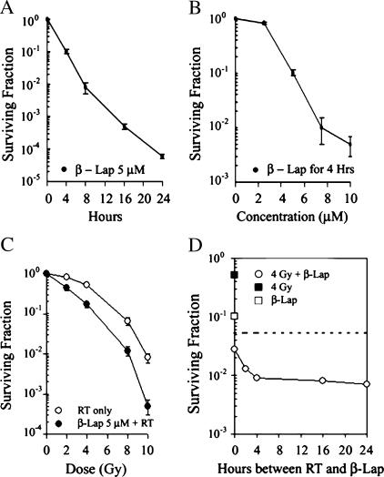
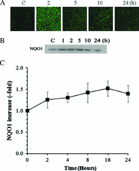
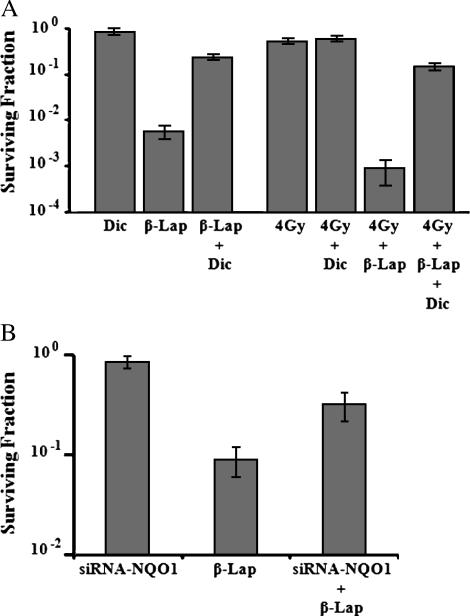
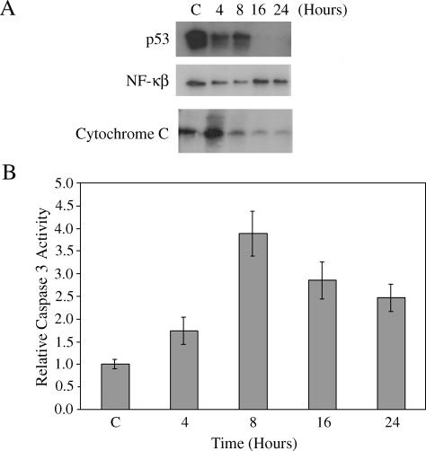
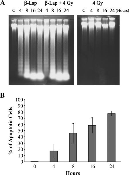
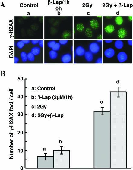
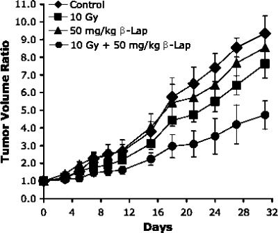
We found that beta-lapachone (beta-lap), a novel bioreductive drug, caused rapid apoptosis and clonogenic cell death in A549 human lung epithelial cancer cells in vitro in a dose-dependent manner. The clonogenic cell death caused by beta-lap could be significantly inhibited by dicoumarol, an inhibitor of NAD(P)H:quinone oxido-reductase (NQO1), and also by siRNA for NQO1, demonstrating that NQO1-induced bioreduction of beta-lap is an essential step in beta-lap-induced cell death. Irradiation of A549 cells with 4 Gy caused a long-lasting upregulation of NQO1, thereby increasing NQO1-mediated beta-lap-induced cell deaths. Although the direct cause of beta-lap-induced apoptosis is not yet clear, beta-lap treatment reduced the expression of p53 and NF-kappaB, whereas it increased cytochrome C release, caspase-3 activity, and gammaH2AX foci formation. Importantly, beta-lap treatment immediately after irradiation enhanced radiation-induced cell death, indicating that beta-lap sensitizes cancer cells to radiation, in addition to directly killing some of the cells. The growth of A549 tumors induced in immunocompromised mice could be markedly suppressed by local radiation therapy when followed by beta-lap treatment. This is the first study to demonstrate that combined radiotherapy and beta-lap treatment can have a significant effect on human tumor xenografts.

Keywords: A549 cells; Bioreductive drug; NQO1; radiation; β-lapachone.


Figures

Figure 1
Figure 1
Effect of β-lap alone or in combination with irradiation on the survival of A549 cells. (A) Survival curve of A549 cells treated with 5 µM β-lap for various lengths of time. A549 cells were treated with 5 µM β-lap for 4 to 24 hours at 37°C and cultured for 7 to 9 days, and surviving fractions were calculated. The averages of seven experiments with 1 SE are shown. (B) Survival curve of A549 cells treated with different concentrations of β-lap for varying lengths of time. Cells were treated with 2 to 10 µM β-lap for 4 to 24 hours at 37°C and cultured for 7 to 9 days, and surviving fractions were calculated. The averages of seven experiments with 1 SE are shown. (C) Survival curves of cells treated with radiation alone or in combination with β-lap. Cells were treated with different doses of radiation alone (RT only) or irradiated and immediately incubated with 5 µM β-lap for 4 hours (β-lap 5 µM + RT). The cells were then cultured for 7 to 9 days, and surviving fractions were calculated. The survival curve for the combined treatment was normalized for cell deaths caused by β-lap alone. The averages of seven experiments with 1 SE are shown. (D) Effects of β-lap treatment applied at different times after the irradiation of cells. Cells were treated with 4-Gy radiation alone (4 Gy) at 4-hour incubation with 5 µM β-lap alone (β-lap), or irradiated with 4 Gy and treated with 5 µM β-lap for 4 hours at different times after irradiation (4 Gy + β-lap). Cells were then incubated for 7 to 9 days, and surviving fractions were calculated. The averages of six experiments with 1 SE are shown. The dotted line corresponds to a hypothetical cell survival when the combined effect of 4-Gy irradiation and 5 µM β-lap treatment is assumed to be additive.
Figure 2
Figure 2
Effects of radiation on the expression and enzymatic activity of NQO1 in A549 cells. (A) NQO1 expression at various times after 4-Gy irradiation in A549 cells. Control and irradiated cells were labeled with anti-NQO1 antibody, followed by labeling with secondary antibody conjugated with FITC and examined with confocal microscopy. (B) Typical example of Western blot analysis for NQO1 at various times after 4-Gy irradiation in A549 cells. The cells were dissolved in solubilizing buffer, and lysates were subjected to Western blot analysis. Blots from control and irradiated cells were labeled with anti-NQO1 antibody and then with horseradish peroxidase-conjugated secondary antibody, and immunoreactive bands were visualized using chemiluminescence. (C) Enzymatic activity of NQO1 at various times after 4-Gy irradiation in A549 cells. Control and irradiated cells were sonicated, and enzyme activity in S9 supernatants was determined. The enzyme activity in irradiated cells relative to that in control cells (9.5 ± 1.3 µmol of cytochrome C reduced per minute per milligram of protein) is shown. The averages of six experiments with 1 SE are shown.
Figure 3
Figure 3
Effects of dicoumarol or siRNA-NQO1 on β-lap-induced clonogenic death in A549 cells. (A) Effects of dicoumarol: Dic—cells were treated with 50 µM dicoumarol for 4 hours; β-lap—cells were treated with 10 µM β-lap for 4 hours; β-lap + Dic—cells were treated with 10 µM β-lap plus 50 µM dicoumarol for 4 hours; 4 Gy—cells were irradiated with 4 Gy; 4 Gy + Dic—cells were irradiated and then treated with 50 µM dicoumarol for 4 hours; 4 Gy + β-lap—cells were irradiated with 4 Gy and immediately incubated with 10 µM β-lap for 4 hours; 4 Gy + β-lap + Dic—cells were irradiated with 4 Gy and immediately treated with 10 µM β-lap plus 50 µM dicoumarol for 4 hours. Cells were washed after the treatments and incubated for 7 to 9 days, and surviving fractions were calculated. The averages of six experiments with 1 SE are shown. (B) Effects of siRNA-NQO1: siRNA-NQO1—cells transfected with siRNA-NQO1 were incubated with regular medium for 4 hours; β-lap—cells were incubated with 5 µM β-lap for 4 hours; siRNA-NQO1 + β-lap—cells transfected with siRNA-NQO1 were incubated with 5 µM β-lap for 4 hours. After the treatments, cells were washed and incubated for 7 to 9 days, and surviving fractions were calculated. The averages of four experiments with duplicate cultures with 1 SE are shown.
Figure 4
Figure 4
Changes in the expression of p53 and NF-κB, release of cytochrome C, caspase-3 activity and DNA laddering by β-lap, or radiation exposure in A549 cells. (A) Cells were treated with 5 µM β-lap for 4 hours, harvested at different times, and washed. The cells were then dissolved in solubilizing buffer, and lysates were subjected to Western blot analysis. The hours shown are those from the start of a 4-hour β-lap incubation. Thus, “4 hours” implies 0 hour after 4 hours of incubation with β-lap. The result shown is representative of five individual experiments. (B) Cells were treated with 5 µM β-lap for 4 hours, harvested, washed, and lysed. Caspase-3 activity was determined using a commercially available assay kit (see text). Relative changes in caspase-3 activity are shown. The hours shown are those from the start of a 4-hour incubation with β-lap. The averages of five measurements with 1 SE are shown.
Figure 5
Figure 5
Apoptosis caused by β-lap alone or in combination with irradiation in A549 cells. (A) β-Lap—cells were treated with 5 µM β-lap for 4 hours; β-lap + 4 Gy—cells were irradiated with 4 Gy and immediately incubated with 5 µM β-lap for 4 hours; 4 Gy—cells were irradiated with 4 Gy. Cells were washed, lysed, and subjected to agarose electrophoresis for detection of DNA fragmentation. The times shown for β-lap treatment groups are hours from the start of a 4-hour incubation with β-lap. (B) Percentage of apoptotic cells after a 4-hour incubation with 5 µM β-lap. Apoptosis was determined with flow cytometry method. The cells in sub-G1 phase were taken as apoptotic cells. The hours indicate the time from the start of a 4-hour incubation with β-lap. The averages of four experiments with 1 SE are shown.
Figure 6
Figure 6
Effects of irradiation and β-lap alone or combined on γH2AX foci formation in A549 cells. (A) Upper panel: γH2AX foci expression in representative cells from each experimental group; lower panel: the nucleus of corresponding cells stained with 4′,6-diamidino-2-phenylindole. (a) Control cells; (b) 1-hour treatment with 2 µM β-lap; (c) 1 hour after 2-Gy irradiation; (d) 2-Gy irradiation followed by 1-hour incubation with 2 µM β-lap. (B) Number of γH2AX foci per cell. (a) Control cells; (b) 1-hour treatment with 2 µM β-lap; (c) 1 hour after 2-Gy irradiation; (d) 2-Gy irradiation followed by 1-hour incubation with 2 µM β-lap. The averages of four experiments with 1 SE are shown.
Figure 7
Figure 7
Effects of various treatments on the growth of A549 human lung tumor xenografts in the hind legs of nude mice. 10 Gy—tumors were locally irradiated with 10 Gy in a single exposure; 50 mg/kg β-lap—the host mice were injected intraperitoneally with β-lap at 50 mg/kg; 10 Gy + 50 mg/kg β-lap—tumors were locally irradiated with 10 Gy in a single exposure and then the host mice were injected intraperitoneally with β-lap at 50 mg/kg. The averages of five to seven tumors per group with 1 SE are shown.

References

    1. Begleiter A, Fourie J. Induction of NQO1 in cancer cells. Methods Enzymol. 2004;382:321–351. - PubMed
    1. Rauth AM, Goldberg Z, Mirsa V. DT-diaphorase: possible roles in cancer chemotherapy and carcinogenesis. Oncol Res. 1997;9:339–349. - PubMed
    1. Ross D, Siegel D. NAD(P)H:quinone oxidoreductase 1 (NQO1, DT-diaphorase), functions and pharmacogenetics. Methods Enzymol. 2004;382:115–144. - PubMed
    1. Siegel D, Beall H, Senekowitach C, Kasai M, Arai H, Gibson NW, Ross D. Bioreductive activation of mitomycin C by DT-diaphorase. Biol Chem. 1992;31:7879–7885. - PubMed
    1. Robertwon N, Stratford IJ, Houlbrook S, Carmichael J, Adams GE. The sensitivity of human tumour cells to quinone bioreductive drugs: what role for DT-diaphorase? Biochem Pharmacol. 1992;44:409–412. - PubMed

Publication types

LinkOut - more resources