. 2007 Sep;117(9):2445-58.
doi: 10.1172/JCI31901.

Beta-arrestin-mediated beta1-adrenergic receptor transactivation of the EGFR confers cardioprotection

Affiliations

Beta-arrestin-mediated beta1-adrenergic receptor transactivation of the EGFR confers cardioprotection

Takahisa Noma et al. J Clin Invest. 2007 Sep.

Abstract

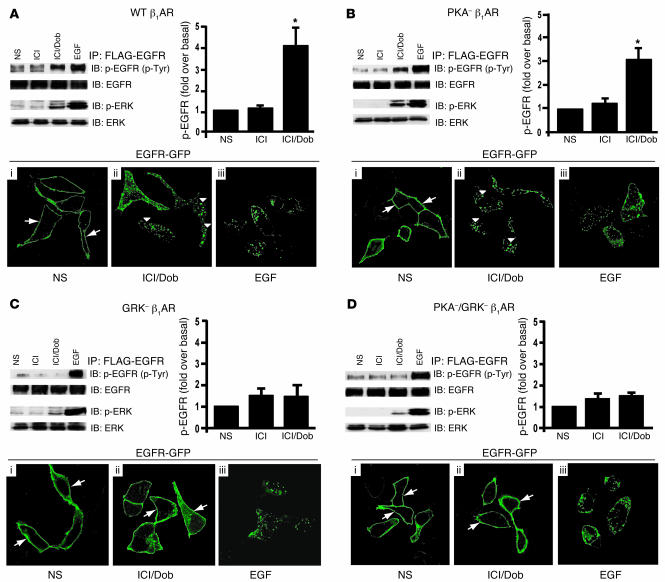
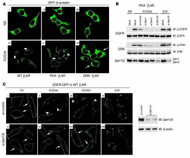
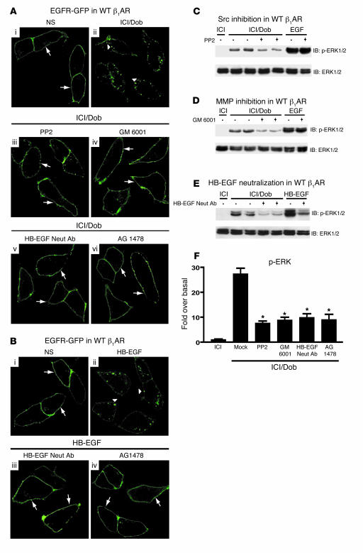
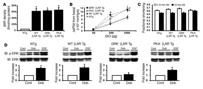
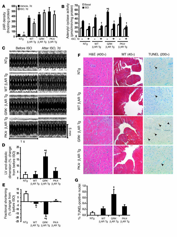
Deleterious effects on the heart from chronic stimulation of beta-adrenergic receptors (betaARs), members of the 7 transmembrane receptor family, have classically been shown to result from Gs-dependent adenylyl cyclase activation. Here, we identify a new signaling mechanism using both in vitro and in vivo systems whereby beta-arrestins mediate beta1AR signaling to the EGFR. This beta-arrestin-dependent transactivation of the EGFR, which is independent of G protein activation, requires the G protein-coupled receptor kinases 5 and 6. In mice undergoing chronic sympathetic stimulation, this novel signaling pathway is shown to promote activation of cardioprotective pathways that counteract the effects of catecholamine toxicity. These findings suggest that drugs that act as classical antagonists for G protein signaling, but also stimulate signaling via beta-arrestin-mediated cytoprotective pathways, would represent a novel class of agents that could be developed for multiple members of the 7 transmembrane receptor family.


Figures

Figure 1
Figure 1. β1AR-mediated transactivation of EGFR requires GRK phosphorylation sites.
HEK293 cells stably expressing WT β1AR (A), PKAβ1AR (B), GRKβ1AR (C), or PKA GRKβ1AR (D) and transfected with FLAG-EGFR were treated with ICI and Dob (as described in Methods) and compared with cells with no stimulation (NS) or EGF stimulation. WT β1AR (A) and PKAβ1AR (B) induced increases in phospho-EGFR and phospho-ERK1/2 after 5 minutes in response to treatment with Dob, while GRK β1AR (C) and PKA/GRK β1AR (D) lacked this effect; *P < 0.05 versus NS. EGFR internalization following Dob or EGF stimulation for 30 minutes was visualized using confocal microscopic analysis of HEK293 cells stably expressing the above β1AR mutants and transfected with EGFR-GFP. In the absence of agonist, EGFR-GFP was visualized on the membrane in each stable cell line (AD, i, arrows), while EGF stimulation resulted in redistribution of EGFR-GFP into cellular aggregates (AD, iii). Treatment of either WT β1AR or PKA β1AR cells with Dob resulted in a similar redistribution of EGFR into cellular aggregates (A and B, ii, arrowheads), an effect that was absent in GRK β1AR and PKA/GRK β1AR cells, where EGFR-GFP remained on the membrane (C and D, ii, arrows). Original magnification, ×100.
Figure 2
Figure 2. β-Arrestin is required for β1AR-mediated EGFR transactivation.
(A) HEK293 cells stably expressing WT β1AR, PKA β1AR, or GRK β1AR were transfected with GFP–β-arrestin. In the absence of agonist, GFP–β-arrestin (green) had a cytosolic distribution (i–iii). Ten minutes of agonist stimulation (Dob) resulted in redistribution of GFP–β-arrestin to the membrane in cells expressing WT β1AR and PKA β1AR (iv and v, arrowheads), whereas no redistribution was observed in cells expressing GRK β1AR (vi, arrows). Original magnification, ×100. (B) HEK293 cells stably expressing PKA β1AR were transfected with FLAG-EGFR alone (Mock) or with siRNAs targeting β-arrestin1 (si-βarr1), β-arrestin2, β-arrestin1/2, or scrambled siRNA (si-control). Reduced Dob-stimulated phospho-EGFR and phospho-ERK1/2 were observed in cells transfected with siRNA targeting β-arrestin. (C) HEK293 cells stably expressing WT β1AR were transfected with EGFR-GFP and si-control or si-βarr1/2 to knock down expression (right panel). In the absence of agonist, EGFR-GFP was located at the membrane (i and v, arrows), while EGF stimulation induced EGFR-GFP redistribution into aggregates (iv and viii). Treatment of si-control–transfected cells with Dob or ISO also resulted in redistribution of EGFR into aggregates (ii and iii, arrowheads), an effect that was diminished in si-βarr1/2–transfected cells, where EGFR-GFP remained at the membrane (vi and vii, arrows). Original magnification, ×150.
Figure 3
Figure 3. GRK5 and -6 are required for β-arrestin–mediated EGFR transactivation.
(A) Cells stably expressing WT β1AR and GRK β1AR transfected with constitutively active β-arrestin, rβarrR169E, both respond to Dob stimulation by increasing association of β-arrestin and AP2, the clathrin adapter protein, which is involved in βAR internalization. (B) GRK β1AR cells were transfected with EGFR-GFP, localized to the plasma membrane (i, arrows), and rβarrR169E-YFP, localized in the cytosol (ii, arrowheads). EGF stimulation induced redistribution of EGFR-GFP from plasma membrane to aggregates (vii, arrows), with no effect on rβarrR169E-YFP (viii, arrowheads). Conversely, Dob stimulation resulted in redistribution of rβarrR169E-YFP from the cytosol to the plasma membrane (v, arrowheads) with no change in EGFR-GFP distribution (iv, arrows). Original magnification, ×100. (C) Transfection of GRK β1AR cells with constitutively active rβarrR169E did not restore EGFR transactivation in response to Dob stimulation. (D) HEK293 cells transiently expressing PKA β1AR were transfected with FLAG-EGFR alone (Mock) or with siRNAs targeting ubiquitous GRKs (si-GRK2, si-GRK3, si-GRK5, and si-GRK6) or a scrambled siRNA sequence (si-control). (E and F) Summary of 6 independent experiments showing significant inhibition of EGFR transactivation (E) and ERK1/2 activation (F) upon Dob stimulation in the cells transfected with siRNA targeting GRK5 or -6; *P < 0.001 versus Dob-stimulated Mock, si-control, si-GRK2, and si-GRK3.
Figure 4
Figure 4. Src, MMP, and HB-EGF are downstream from β-arrestin in β1AR-mediated EGFR transactivation.
(A) HEK293 cells stably expressing WT β1AR were transfected with EGFR-GFP. In the absence of agonist, EGFR-GFP was located at the membrane (i, arrows), while Dob stimulation induced EGFR-GFP redistribution into aggregates (ii, arrowheads). Pretreatment with inhibitors of Src (PP2), MMP (GM 6001), HB-EGF (HB-EGF neutralizing antibody [Neut Ab]), or EGFR (AG 1478) prevented the redistribution of EGFR-GFP into aggregates following Dob stimulation (iii–vi, arrows). (B) Treatment with HB-EGF (1 ng/ml) induced EGFR-GFP internalization (ii, arrowheads) compared with control (i, arrows), while pretreatment with either HB-EGF neutralizing antibody or AG 1478 abrogated this effect (iii and iv, arrows). Original magnification (A and B), ×150. ERK activation was blocked by PP2 (C), GM 6001 (D), and HB-EGF neutralizing antibody (E). (F) Quantification of phospho-ERK response. *P < 0.001 versus Mock (Dob stimulation alone); n ≥ 9 each condition.
Figure 5
Figure 5. In vivo β1AR-mediated EGFR transactivation requires GRK5, GRK6, and β-arrestin2.
β-Arrestin2–knockout mice (βarr2 KO; A), GRK5 KO mice (B), or GRK6 KO mice (C) and their WT littermate controls were injected with Dob following ICI pretreatment or with EGF alone. Myocardial lysates were immunoblotted with anti–phospho- and anti–total ERK1/2 antibodies. Accompanying histograms show summary data of 6 independent experiments depicting the fold increase in ERK1/2 phosphorylation following Dob treatment; *P < 0.05 versus ICI. Dob-mediated ERK1/2 phosphorylation was completely blocked in βarr2 KO, GRK5 KO, and GRK6 KO mice compared with their WT littermate controls.
Figure 6
Figure 6. Cardiac characteristics of Tg mice overexpressing mouse WT β1AR, GRK β1AR, or PKA β1AR.
(A) Myocardial expression levels of βAR were equivalent in WT β1AR Tg (n = 6), GRK β1AR Tg (n = 5), and PKA β1AR Tg (n = 7) mice and approximately 14-fold greater than in their NTg littermates (n = 15); *P < 0.05 versus NTg littermates. (B) In vivo hemodynamics show βAR responsiveness as monitored by the increase in LV contractility (LV dP/dtmax) in WT β1AR (filled circles; n = 11), GRK β1AR (filled squares; n = 5), PKA β1AR (open squares; n = 7) Tg mice and NTg littermates (open circles; n = 18). Both GRK β1AR and PKA β1AR Tg mice showed enhanced contractile response; *P < 0.05 versus NTg littermates. (C) Conscious echocardiography in 5- to 6- and 12-month-old mice indicated no significant differences in fractional shortening among NTg and Tg mice at each age in sedentary conditions. (D) Immunoblotting of LV lysates of NTg and the 3 lines of β1AR Tg mice given i.p. injections of Dob (1 mg/kg, 10 minutes) or EGF (30 μg/kg, 15 minutes) revealed increased ERK1/2 phosphorylation in NTg, WT β1AR Tg, and PKA β1AR Tg mice. Tg overexpression of GRK β1AR prevented Dob-mediated ERK1/2 activation in the heart. Histograms depict the summaries of fold increase in phospho-ERK1/2 in response to Dob stimulation (n = 4–8); *P < 0.05 versus control (Cont).
Figure 7
Figure 7. Deterioration of cardiac function in GRK β1AR Tg mice following chronic ISO treatment.
(A) WT β1AR Tg and NTg mice showed βAR downregulation in the LV membrane fraction following chronic ISO treatment (1 week), whereas GRK β1AR Tg and PKA β1AR Tg mice did not; P < 0.01 versus vehicle in corresponding group. (B) Adenylyl cyclase activity following acute ISO stimulation was enhanced in all vehicle-treated mice but desensitized in all chronic ISO-treated NTg and the 3 lines of β1AR Tg mice; *P < 0.05 versus vehicle-treated NTg ISO; P < 0.05 versus vehicle-treated WT β1AR Tg and PKA β1AR Tg ISO; P < 0.01 versus vehicle-treated ISO in each corresponding group. (C) Representative M-mode echocardiography before and after chronic ISO treatment in β1AR-Tg mice and NTg littermates. Percent changes from pre-ISO treatment in LV end-diastolic dimension (D) and fractional shortening (E) indicate significant LV dilatation and decreased fractional shortening in GRK β1AR Tg mice following chronic ISO treatment; P < 0.01 versus all Tg groups; *P < 0.05 versus NTg littermates. (F) Representative H&E, Masson trichrome (MT), and TUNEL staining following chronic ISO treatment reveals increased interstitial fibrosis (blue stain, MT panels) and apoptosis (arrows, TUNEL panels) in GRK β1AR Tg mice. (G) Percentage of TUNEL-positive nuclei following chronic ISO treatment in NTg and the 3 lines of β1AR Tg mice (n = 5 each); P < 0.05 versus all groups.
Figure 8
Figure 8. Pharmacological inhibition of EGFR causes dilated cardiomyopathy following chronic ISO treatment.
(A) NTg mice were pretreated for 1 hour with erlotinib (20 mg/kg; EGFR antagonist) or DMSO (10%; control) i.p., followed by i.p. injection of Dob (1 mg/kg, 10 minutes) or EGF (30 μg/kg, 15 minutes). Immunoblotting of the cardiac lysates revealed increases in Dob- and EGF-stimulated ERK1/2 phosphorylation, which was blocked by erlotinib pretreatment; *P < 0.05 versus control (n = 4–6 each). Serial echocardiographic parameters, LV end-diastolic dimension (B) and fractional shortening (C), following chronic ISO treatment in conjunction with erlotinib (20 mg/kg/d) indicated that erlotinib treatment mimics the cardiac phenotype observed in chronic ISO-treated GRK β1AR Tg mice (Figure 6); *P < 0.05 versus each group at each time point. (D) Representative TUNEL staining following chronic ISO with or without erlotinib shows increased apoptosis (arrowheads) in LV sections (×200) from NTg mice undergoing chronic ISO with erlotinib treatment, as described above. (E) Percent TUNEL-positive nuclei in LV sections from NTg mice undergoing the following chronic treatments: vehicle plus DMSO (n = 6), vehicle plus erlotinib (n = 8), ISO plus DMSO (n = 7), and ISO plus erlotinib (n = 9); *P < 0.05 versus DMSO in same group.
Figure 9
Figure 9. Signal transduction pathway of β1AR–stimulated EGFR transactivation.
Ligand stimulation of the β1AR leads to GRK5/6-mediated receptor phosphorylation and β-arrestin recruitment. β-Arrestin recruits Src, which leads to MMP activation to promote HB-EGF shedding. HB-EGF binds with the EGFR to induce dimerization and autophosphorylation and subsequent downstream signaling.

Comment in

References

    1. Rockman H.A., Koch W.J., Lefkowitz R.J. Seven-transmembrane-spanning receptors and heart function. Nature. 2002;415:206–212. - PubMed
    1. Koch W.J., Lefkowitz R.J., Rockman H.A. Functional consequences of altering myocardial adrenergic receptor signaling. Annu. Rev. Physiol. 2000;62:237–260. - PubMed
    1. Lefkowitz R.J. G protein-coupled receptors. III. New roles for receptor kinases and beta-arrestins in receptor signaling and desensitization. . J. Biol. Chem. 1998;273:18677–18680. - PubMed
    1. Luttrell L.M., et al. Activation and targeting of extracellular signal-regulated kinases by beta-arrestin scaffolds. Proc. Natl. Acad. Sci. U. S. A. 2001;98:2449–2454. - PMC - PubMed
    1. Maudsley S., et al. The beta(2)-adrenergic receptor mediates extracellular signal-regulated kinase activation via assembly of a multi-receptor complex with the epidermal growth factor receptor. J. Biol. Chem. 2000;275:9572–9580. - PubMed

Publication types