. 2007 Sep;117(9):2459-67.
doi: 10.1172/JCI31960.

Histone deacetylase degradation and MEF2 activation promote the formation of slow-twitch myofibers

Affiliations

Histone deacetylase degradation and MEF2 activation promote the formation of slow-twitch myofibers

Matthew J Potthoff et al. J Clin Invest. 2007 Sep.

Abstract

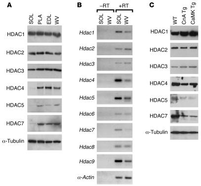
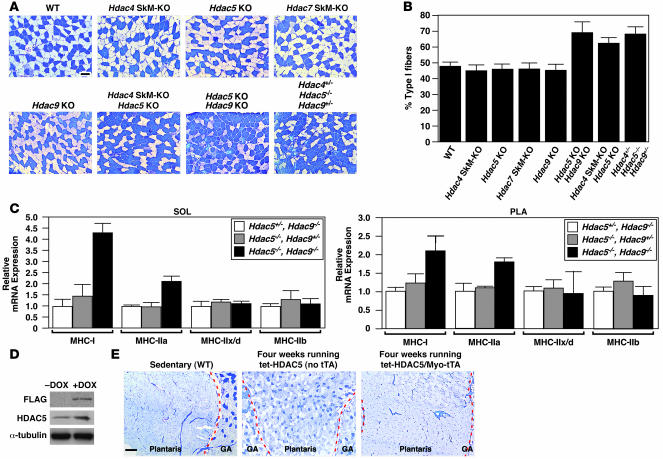
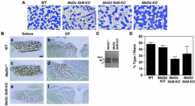
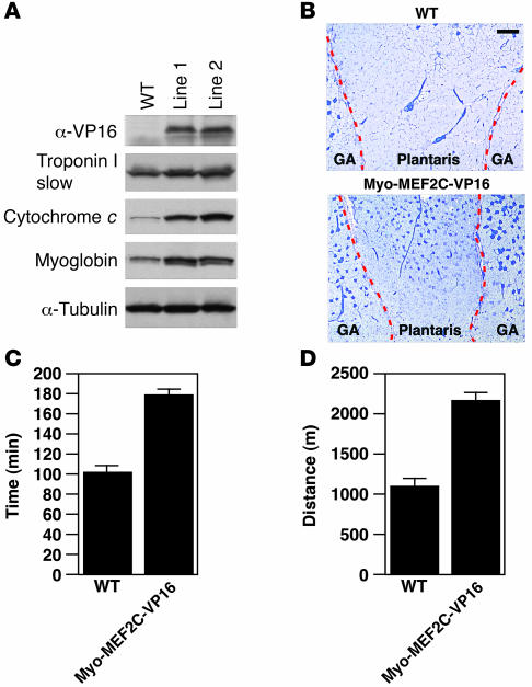
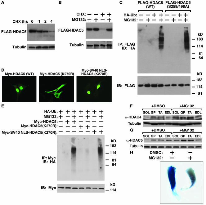
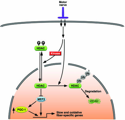
Skeletal muscle is composed of heterogeneous myofibers with distinctive rates of contraction, metabolic properties, and susceptibility to fatigue. We show that class II histone deacetylase (HDAC) proteins, which function as transcriptional repressors of the myocyte enhancer factor 2 (MEF2) transcription factor, fail to accumulate in the soleus, a slow muscle, compared with fast muscles (e.g., white vastus lateralis). Accordingly, pharmacological blockade of proteasome function specifically increases expression of class II HDAC proteins in the soleus in vivo. Using gain- and loss-of-function approaches in mice, we discovered that class II HDAC proteins suppress the formation of slow twitch, oxidative myofibers through the repression of MEF2 activity. Conversely, expression of a hyperactive form of MEF2 in skeletal muscle of transgenic mice promotes the formation of slow fibers and enhances running endurance, enabling mice to run almost twice the distance of WT littermates. Thus, the selective degradation of class II HDACs in slow skeletal muscle provides a mechanism for enhancing physical performance and resistance to fatigue by augmenting the transcriptional activity of MEF2. These findings provide what we believe are new insights into the molecular basis of skeletal muscle function and have important implications for possible therapeutic interventions into muscular diseases.


Figures

Figure 1
Figure 1. Posttranscriptional downregulation of class II HDACs in soleus muscle.
Soleus (SOL), PLA, EDL, and WV muscles were dissected from the hind limbs of adult WT mice (8–10 weeks of age). (A) Protein expression of HDACs was assayed using antibodies specific for individual HDAC proteins. α-Tubulin level indicated equal loading. (B) RNA expression of HDACs in SOL versus WV was analyzed by RT-PCR in the presence (+) or absence (–) of reverse transcriptase (RT). Skeletal α-actin primers were used to show equivalent cDNA input. (C) Immunoblots of HDACs using WV muscle extracts from WT and 2 transgenic mouse models (10 weeks old) overexpressing active calcineurin A (CnA Tg) or CaMKIV (CaMK Tg).
Figure 2
Figure 2. Class II HDACs redundantly regulate slow, oxidative fiber expression.
(A) Soleus muscles from WT, Hdac5–/– (Hdac5 KO), Hdac9–/– (Hdac9 KO), Hdac4fl/–;Myo-Cre (Hdac4 SkM-KO), Hdac7fl/–;Myo-Cre(Hdac7 SkM-KO), and class II HDAC compound mutant mice were analyzed by metachromatic ATPase staining. Type I fibers stain dark blue. Type II fibers stain light blue. Original magnification, ×10. Scale bar: 100 μm. (B) Quantification of fiber-type distribution based on fiber-type analysis in A. (C) Transcripts of MHC isoforms were determined in soleus and PLA muscles from mice of the indicated genotypes by quantitative real-time PCR. (D) Anti-FLAG M2 antibody on a Western blot analysis of proteins isolated from GP muscles of 4-week-old Myo-tTA/tet-HDAC5 mice treated with DOX or 10 days after removal of DOX. Tubulin served as a loading control. (E) Metachromatic ATPase staining of GP muscles harvested from sedentary WT, 4-week-exercised control (tet-HDAC5 [no tTA]), and 4-week-exercised HDAC5 transgenic (Myo-tTA/tet-HDAC5) mice. Original magnification, ×4. Scale bar: 300 μm. Dashed red lines delineate gastrocnemius (GA) muscle from PLA.
Figure 3
Figure 3. Requirement of MEF2 for establishing slow, oxidative myofiber identity.
Muscles from individual MEF2 knockout mice: Mef2a–/–, Mef2c SkM-KO (Mef2cfl/-;Myo-Cre), and Mef2d SkM-KO (Mef2dfl/fl;Myo-Cre) skeletal muscle conditional knockout mice were analyzed for fiber-type composition. (A) Metachromatic ATPase staining of soleus muscle. Type I fibers stain dark blue. Type II fibers stain light blue. Original magnification, ×10. Scale bar: 100 μm. (B) Immunohistochemistry of soleus and GP muscles of Mef2c SkM-KO and WT littermates using an MHC-I specific antibody. Original magnification, ×2.5. Scale bar: 300 μm. (C) Glycerol gradient silver staining of protein extracts from soleus of WT and Mef2c SkM-KO mice. MHC-I, -IIa/x, and -IIb isoforms are indicated. (D) Quantification of fiber-type distribution based on metachromatic ATPase staining of MEF2 knockout mice.
Figure 4
Figure 4. Activated MEF2 is sufficient to increase slow-fiber expression.
(A) Western blot analysis of Myo-MEF2C-VP16 transgene expression using an anti-VP16 antibody. Expression of the slow-fiber–specific troponin I and oxidative markers myoglobin and cytochrome c in protein extract of GP muscles of Myo-MEF2C-VP16 transgenic mice. (B) Metachromatic ATPase staining of gastrocnemius and PLA muscles of WT and Myo-MEF2C-VP16 transgenic mice. Original magnification, ×4. Scale bar: 300 μm. Dashed red lines delineate gastrocnemius muscle from PLA. (C and D) Exercise endurance and muscle performance showing total time running (min; C) and total distance run (m; D) of Myo-MEF2-VP16 transgenic muscles were analyzed by forced treadmill exercise. Eight-week-old Myo-MEF2C-VP16 transgenic and WT male mice with similar body weights were subjected to forced treadmill exercise (n = 5 for each group) on a 10% incline.
Figure 5
Figure 5. Ubiquitination and degradation of class II HDACs in vitro and in vivo.
(A) C2C12 cells stably expressing FLAG-tagged HDAC5 (C2C12-HDAC5) were treated with cycloheximide (CHX, 25 μM) for 0, 1, 2, or 4 hours before cells were lysed and FLAG-HDAC5 expression was measured by Western blot analysis using anti-FLAG M2 antibody. Tubulin immunoblot showed equivalent loading of each lane. (B) C2C12-HDAC5 cells were treated with cycloheximide and MG132, singly or in combination for 4 hours, and FLAG-HDAC5 expression was analyzed. (C) C2C12-HDAC5 and C2C12 cells stably expressing CaMK-resistant HDAC5 (S259/498A) were transfected with or without HA-tagged ubiquitin (HA-Ub) and treated with or without MG-132 (25 μM) for 4 hours; the ubiquitination status of WT and mutant HDAC5 was analyzed. FLAG expression in inputs shows equal loading. (D) Subcellular localization of Myc-HDAC5 (WT), Myc-HDAC5(K270R), or Myc-SV40 NLS-HDAC5(K270R) in C2C12 cells. (E) The ubiquitination status of cytoplasmic HDAC5 [Myc-HDAC5(K270R)] or nuclear HDAC5 [Myc-SV40 NLS-HDAC5(K270R)] was analyzed. (F and G) WT C57BL/6 males (8 weeks old) were IP injected with DMSO or MG132 for 6 hours. Protein was isolated from SOL, GP, tibialis anterior (TA), and EDL muscles and analyzed for expression of (F) HDAC4 or (G) HDAC5. Tubulin shows equal loading. (H) Treatment with MG132 decreases MEF2 activation. Six hours after DesMEF-lacZ mice were injected with DMSO or MG132, mice were run for approximately 3 hours using forced treadmill exercise. Skeletal muscles were then isolated from DMSO- and MG132-treated DesMEF mice and analyzed for lacZ expression. LacZ expression was reduced in MG132-treated muscles (where class II HDAC expression was increased).
Figure 6
Figure 6. A model for the control of slow and oxidative fibers by MEF2 and class II HDACs.
Motor nerve activity regulates MEF2 activity and myofiber identity through ubiquitination (Ub) and degradation of class II HDACs.

Comment in

Similar articles

Cited by

References

    1. Pette D., Staron R.S. Myosin isoforms, muscle fiber types, and transitions. Microsc. Res. Tech. 2000;50:500–509. - PubMed
    1. Schiaffino S., Reggiani C. Molecular diversity of myofibrillar proteins: gene regulation and functional significance. Physiol. Rev. 1996;76:371–423. - PubMed
    1. Baldwin K.M., Haddad F. Effects of different activity and inactivity paradigms on myosin heavy chain gene expression in striated muscle. J. Appl. Physiol. 2001;90:345–357. - PubMed
    1. Sugiura T., Miyata H., Kawai Y., Matoba H., Murakami N. Changes in myosin heavy chain isoform expression of overloaded rat skeletal muscles. Int. J. Biochem. 1993;25:1609–1613. - PubMed
    1. Talmadge R.J. Myosin heavy chain isoform expression following reduced neuromuscular activity: potential regulatory mechanisms. Muscle Nerve. 2000;23:661–679. - PubMed

Publication types

MeSH terms