. 2007 Sep;117(9):2517-25.
doi: 10.1172/JCI32282.

Normal human dermis contains distinct populations of CD11c+BDCA-1+ dendritic cells and CD163+FXIIIA+ macrophages

Affiliations

Normal human dermis contains distinct populations of CD11c+BDCA-1+ dendritic cells and CD163+FXIIIA+ macrophages

Lisa C Zaba et al. J Clin Invest. 2007 Sep.

Abstract

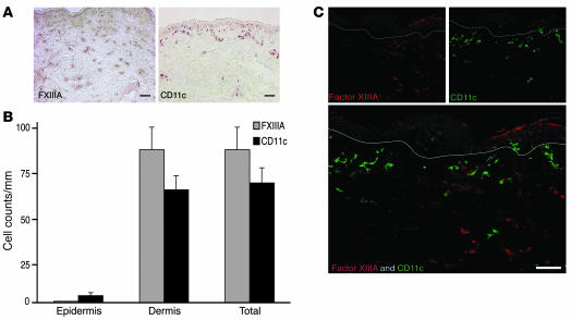
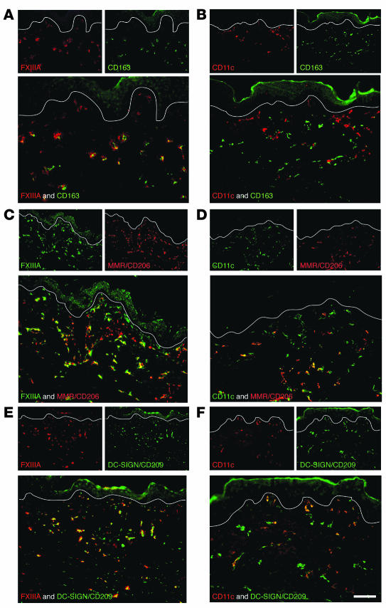
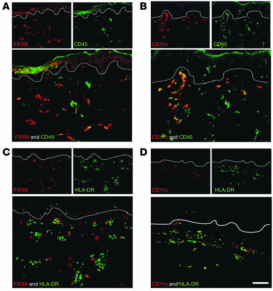
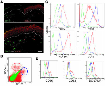
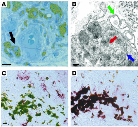
We used a panel of monoclonal antibodies to characterize DCs in the dermis of normal human skin. Staining for the CD11c integrin, which is abundant on many kinds of DCs, revealed cells in the upper dermis. These cells were positive for blood DC antigen-1 (BDCA-1; also known as CD1c), HLA-DR, and CD45, markers that are also expressed by circulating myeloid DCs. A small subset of CD11c+ dermal cells expressed DEC-205/CD205 and DC-lysosomal-associated membrane glycoprotein/CD208 (DC-LAMP/CD208), suggesting some differentiation or maturation. When BDCA-1+ cells were selected from collagenase digests of normal dermis, they proved to be strong stimulators for T cells in a mixed leukocyte reaction. A second major population of cells located throughout the dermis was positive for factor XIIIA (FXIIIA), but lacked CD11c and BDCA-1. They expressed the macrophage scavenger receptor CD163 and stained weakly for HLA-DR and CD45. Isolated CD163+ dermal cells were inactive in stimulating T cell proliferation, but in biopsies of tattoos, these cells were selectively laden with granular pigments. Plasmacytoid DCs were also present in the dermis, marked by CD123 and BDCA-2. In summary, the normal dermis contains typical immunostimulatory myeloid DCs identified by CD11c and BDCA-1, as well as an additional population of poorly stimulatory macrophages marked by CD163 and FXIIIA.


Figures

Figure 1
Figure 1. FXIIIA+ and CD11c+ cells are unique dermal populations.
(A) Immunohistochemistry on normal human skin using FXIIIA (left panel) and CD11c (right panel) antibodies (n = 15). FXIIIA+ cells were spread throughout the dermis, while CD11c+ cells were mainly localized to the superficial dermis. (B) There were similar numbers of CD11c+ and FXIIIA+ cells per mm in normal dermis. Error bars indicate SEM. (C) FXIIIA and CD11c identified 2 discrete populations. White lines denote dermo-epidermal junction. Scale bars: 100 μm.
Figure 2
Figure 2. FXIIIA+ and CD11c+ populations are not LCs or plasmacytoid DCs.
Neither FXIIIA nor CD11c showed coexpression with Langerhans antigen CD1a (A and B) or plasmacytoid antigen CD123 (C and D). Scale bar: 100 mm.
Figure 3
Figure 3. CD11c+ cells are defined by BDCA-1, DC-LAMP/CD208, and DEC-205/CD205.
FXIIIA did not overlap with BDCA-1 (A), DC-LAMP/CD208 (C), or DEC-205/CD205 (E). Most CD11c+ cells coexpressed BDCA-1 (B). Small subsets of CD11c+ cells coexpressed DC-LAMP/CD208 (D) and DEC-205/CD205 (F). Scale bar: 100 μm.
Figure 4
Figure 4. The macrophage marker CD163 defines FXIIIA+ cells.
FXIIIA+ cells expressed macrophage marker CD163 (A), and a subset overlapped with MMR/CD206 (C) and DC-SIGN/CD209 (E). CD11c did not overlap with CD163 (B), but a subset overlapped with MMR/CD206 (D) and DC-SIGN/CD209 (F). Scale bar: 100 μm.
Figure 5
Figure 5. HLA-DR and CD45 mark both CD11c+ and FXIIIA+ cells.
FXIIIA+ cells had a lower expression level of CD45 (A) than did CD11c+ cells (B). HLA-DR was also expressed to a lower extent on FXIIIA+ cells (C) than on CD11c+ cells (D). Scale bar: 100 μm.
Figure 6
Figure 6. Cutaneous DCs compared with blood DCs.
(A) There were 3 nonoverlapping DC populations in peripheral blood, gating on LinCD11c+HLA-DR+ cells: BDCA-1+, CD16hi, and BDCA-3+. (B) Blood BDCA-1+ (left) and BDCA-3+ (right) cells were both HLA-DR+CD14. (C) In dermal single-cell suspensions, BDCA-1+ cells acquired CD16. (D) BDCA-1+ (left) dermal cells increased HLA-DR expression, and BDCA-3+ (right) dermal cells acquired low-level CD14 expression.
Figure 7
Figure 7. BDCA-1 and CD163 are alternative markers for CD11c and FXIIIA, respectively.
(A) BDCA-1 and CD163 identified discrete populations of dermal cells. (B) BDCA-1+ cells (red circle) and CD163+ cells (blue circle) were also discrete populations in dermal single-cell suspensions. (C) FACS histograms gated on BDCA-1+ cells (red line), CD163+ cells (blue line), or isotype (green line). BDCA-1+ cells were CD11chi, FXIIIAlo, HLA-DRhi, and CD45hi. CD163+ cells were CD11cmid, FXIIIAhi, HLA-DRmid, and CD45lo. (D) A subset of BDCA-1+ cells was CD86hi, CD83+, and DC-LAMPhi. Representative graphs from 3 experiments. Scale bar: 100 μm.
Figure 8
Figure 8. BDCA-1+ cells are more immunostimulatory.
(A) Post-sort dot plot of dermal cells from normal skin into BDCA-1+ and CD163+ populations (red, left and right panels, respectively) compared with isotype (blue). (B) Positive control (monocyte-derived mature DC) for MLR on day 8 after sorting at a 1:100 stimulator/responder ratio. Gate contains CD3+ proliferating T cells with left-shifted CFSE. (C) Using BDCA-1+ sorted cells as stimulators (1:10 ratio), 9.1% of the T cells proliferated; using CD163+ cells (1:10 ratio), 2.1% of live T cells proliferated. Background proliferation of T cells alone without stimulation was 1.0%. (D) After cells had been sorted and cultured for 2 days with cytokines to induce maturation, there was a marked increase in the T cell stimulatory capacity of BDCA-1+ cells (25.2%, 1:100 ratio) versus CD163 (2.2%, 1:250 ratio). Representative graphs from 3 experiments.
Figure 9
Figure 9. CD163+ cells phagocytose large particles and have the structural features of macrophages.
(A) Tattoo skin section (0.5 μm) stained with toludine blue. Cells containing green tattoo dye in their cytoplasm (black arrow) surrounded a blood vessel. (B) Electron microscopy of a tattoo showed that dye particles (red arrow) were membrane bound (blue arrow) within the cytoplasm of a cell with multiple microvillus protrusions (green arrow). (C and D) Cells containing green tattoo dye particles stained for CD163 (D) but not BDCA-1 (C). Scale bar: 10 μm (A, C, and D); 200 nm (B).

Comment in

References

    1. Larregina A.T., Falo L.D. Changing paradigms in cutaneous immunology: adapting with dendritic cells. J. Invest. Dermatol. 2005;124:1–12. - PubMed
    1. Valladeau J., Saeland S. Cutaneous dendritic cells. Semin. Immunol. 2005;17:273–283. - PubMed
    1. Hunger R.E., et al. Langerhans cells utilize CD1a and langerin to efficiently present nonpeptide antigens to T cells. J. Clin. Invest. 2004;113:701–708. doi: 10.1172/JCI200419655. - DOI - PMC - PubMed
    1. Cerio R., Griffiths C.E., Cooper K.D., Nickoloff B.J., Headington J.T. Characterization of factor XIIIa positive dermal dendritic cells in normal and inflamed skin. Br. J. Dermatol. 1989;121:421–431. - PubMed
    1. Henriksson P., Becker S., Lynch G., McDonagh J. Identification of intracellular factor XIII in human monocytes and macrophages. J. Clin. Invest. 1985;76:528–534. - PMC - PubMed

Publication types

MeSH terms