. 2007 Sep;117(9):2526-38.
doi: 10.1172/JCI31570.

Aberrant activation of integrin alpha4beta7 suppresses lymphocyte migration to the gut

Affiliations

Aberrant activation of integrin alpha4beta7 suppresses lymphocyte migration to the gut

Eun Jeong Park et al. J Clin Invest. 2007 Sep.

Abstract

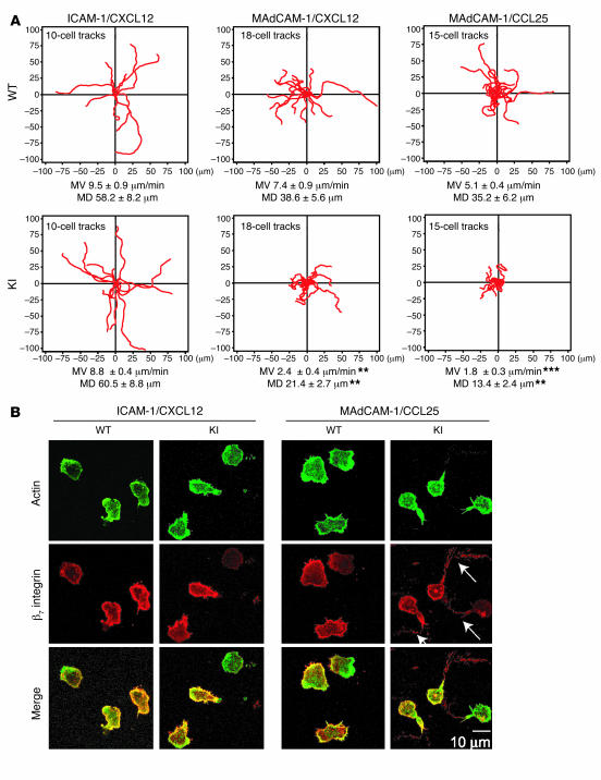
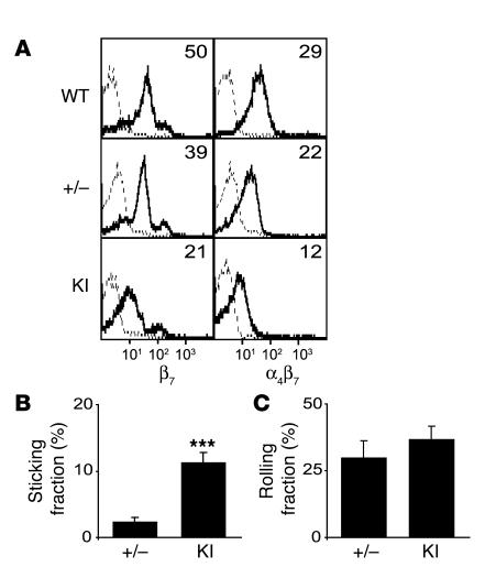
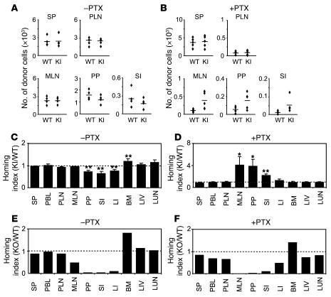
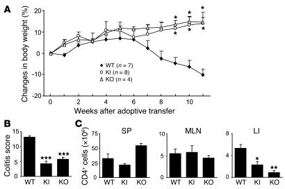
Integrin adhesion molecules mediate lymphocyte migration and homing to normal and inflamed tissues. While the ligand-binding activity of integrins is known to be modulated by conformational changes, little is known about how the appropriate balance of integrin adhesiveness is maintained in order to optimize the migratory capacity of lymphocytes in vivo. In this study we examined the regulation of the gut homing receptor alpha4beta7 integrin by manipulating at the germline level an integrin regulatory domain known as adjacent to metal ion-dependent adhesion site (ADMIDAS). ADMIDAS normally serves to raise the activation threshold of alpha4beta7, thereby stabilizing it in the default nonadhesive state. Lymphocytes from knockin beta7 (D146A) mice, which harbor a disrupted ADMIDAS, not only expressed an alpha4beta7 integrin that persistently adhered to mucosal addressin cell adhesion molecule-1 (MAdCAM-1), but also exhibited perturbed cell migration along MAdCAM-1 substrates resulting from improper de-adhesion of the lymphocyte trailing edge. In vivo, aberrantly activated alpha4beta7 enhanced adhesion to Peyer's patch venules, but suppressed lymphocyte homing to the gut, diminishing the capacity of T cells to induce colitis. Our results underscore the importance of a proper balance in the adhesion and de-adhesion of the alpha4beta7 integrin, both for lymphocyte trafficking to the gut and for colitis progression.


Figures

Figure 1
Figure 1. Generation of β7 (D146A) mice.
(A) Targeted insertion to the Itgb7 locus of the floxed ACN cassette, WT exon 3, and the mutated exon 4 (4*) that contains β7-D146A. The targeting vector, the WT Itgb7 locus, the targeted Itgb7 allele containing floxed ACN cassette, and the mutated Itgb7 (D146A) allele are shown. Exons are shown as filled boxes. Long arm (LA) and short arm (SA) of homology as well as the diphtheria toxin (DT) are also shown. The floxed ACN cassette is deleted in chimeric male mice during spermatogenesis, leaving 1 loxP site. An engineered EcoRI site (E*) was designed to identify the targeted allele by Southern blot analysis. X, XhoI; E, EcoRI; B, BglII; EV, EcoRV. The thick black line indicates the probe used to screen for homologous recombinations. (B) Genotyping and confirmation of deleted ACN cassette by PCR. Genomic DNA isolated from tails was used for PCR analyses. PCR bands are shown for WT (WT/WT, 270 bp), heterozygote (KI/WT, 350 and 270 bp), and homozygote (KI/KI, 350 bp) samples. (C) Sequencing analysis of WT and β7 (D146A) KI mice. DNA sequencing confirmed an aspartate to alanine substitution at position 146 of the mouse β7 integrin gene (boxed regions).
Figure 2
Figure 2. Histology showing reduced T cell numbers in the gut of β7 (D146A) mice.
Representative histology sections (original magnification, ×200) of the SI of WT and β7 (D146A) KI mice were analyzed by (A) hematoxylin and eosin staining and (B) immunofluorescent staining with Cy3-conjugated anti-CD3ε mAb.
Figure 3
Figure 3. Expression of integrins and activation markers in β7 (D146A) mice.
(A) Cell surface expression of integrins on WT and β7 (D146A) KI lymphocytes from the SP and from SI-IEL and SI-LPL compartments. (B) Cell surface expression of activation markers on lymphocytes from SP. (C) mRNA expression of β7 integrins in splenocytes. Real-time quantitative RT-PCR was performed by iCycler (Bio-Rad). The mRNA expression of the β7 integrin was normalized to that of GAPDH. Dots represent 3 independent data sets; bars denote mean values. (D) Total (cell surface plus intracellular) protein expression of β7 integrins in splenocytes. Total expression was examined by immunofluorescent flow cytometry using permeabilized cells. (E) Effects of a proteasome inhibitor on the cell surface expression of integrins. WT and KI splenocytes were treated for 8 hours with epoxomicin (0.5 μM) in the presence of PMA (20 nM) and ionomycin (1 μM). Surface expression of αL and β7 integrins was examined by flow cytometry. (F) Effects of CD3/CD28/RA treatment on the expression of cell surface receptors. Splenocytes were stimulated with mAbs to CD3 and CD28 for 2 days and treated for 3 days with RA in the absence of mAb stimulation. (A, B, D, and F) Numbers denote mean fluorescent intensity (MFI). Binding of isotype control antibodies is shown with dashed lines. (E) Numbers denoting MFI values for vehicle-treated (dashed lines) and epoxomicin-treated (solid lines) samples are shown with and without parentheses, respectively.
Figure 4
Figure 4. Enhanced adhesive interactions with MAdCAM-1 of β7 (D146A) cells.
Splenocytes (A) and RA-treated memory/effector T cells (B) from WT and β7 (D146A) KI mice were infused in 1 mM Mg2+/Ca2+ or 1 mM Mn2+ into a flow chamber and allowed to accumulate on MAdCAM-1 substrates for 30 seconds at 0.3 dyn/cm2. Shear stress was incrementally increased from 1 to 32 dyn/cm2, and adhesive interactions were recorded and analyzed offline. Data are mean ± SEM of 3 independent experiments. *P < 0.05, **P < 0.01, ***P < 0.001 versus WT. In A, cells were pretreated with either DATK32 (20 μg/ml) or isotype control IgG (20 μg/ml) for 30 minutes at room temperature.
Figure 5
Figure 5. Perturbed transmigration of β7 (D146A) splenocytes through MAdCAM-1.
Transmigration of WT and KI splenocytes toward a CCL25 gradient through ICAM-1–coated (A), MAdCAM-1–coated (B), and bEnd.3 endothelial monolayer–seeded (C) permeable inserts was examined using a modified Boyden chamber assay with a Transwell tissue culture system. Data are mean ± SEM of triplicates from 3 independent experiments. *P < 0.05, ***P < 0.001 versus WT.
Figure 6
Figure 6. Perturbed migration of β7 (D146A) T lymphocytes on MAdCAM-1.
(A) Two-dimensional tracking of T lymphocytes migrating on ICAM-1/CXCL12, MAdCAM-1/CXCL12, and MAdCAM-1/CCL25 substrates. Mean velocity (MV) and mean displacement (MD) were determined as described in Methods. Data are mean ± SEM. **P < 0.01, ***P < 0.001 versus WT. (B) Representative confocal images of T lymphocytes migrating on ICAM-1/CXCL12 and MAdCAM-1/CCL25. Samples were fixed as described in Methods and stained with Cy3-conjugated antibody for β7 integrin and with Alexa Fluor 488–conjugated phalloidin for actin. Scale bar: 10 μm.
Figure 7
Figure 7. Increased firm adhesion of β7 (D146A) cells in vivo to PP venules.
(A) Cell surface expression of β7 integrins in WT, β7+/– (+/–), and KI lymphocytes. MFI values are shown. Staining with anti-integrin and isotype control mAbs is shown by solid and dashed lines, respectively. (B and C) Adhesive interactions of β7+/– and KI cells to PP venules studied by intravital microscopy. Fractions of sticking (B) and rolling (C) cells out of total flowing cells are shown. Data are mean ± SEM of 7 different PP venules analyzed per group in 2 independent experiments. ***P < 0.001 versus β7 +/–.
Figure 8
Figure 8. In vivo homing of β7 (D146A) lymphocytes to the gut is suppressed.
Competitive homing assay to compare β7 (D146A) KI and WT (AD) or β7 KO and WT lymphocytes (E and F). Equal numbers (2 × 10⁷) of fluorescently labeled cells, either untreated (–PTX; A, C, and E) or treated with PTX (+PTX; B, D, and F), were mixed and injected into C57BL/6J-CD45.1+ congenic mice. The number of homed donor cells (A and B) and homing indices (CF) were determined 18 hours after injection. PBL, peripheral blood lymphocytes; LIV, liver; LUN, lung. Data are mean ± SEM of at least 3 independent experiments. *P < 0.05; **P < 0.01 versus SP.
Figure 9
Figure 9. Reduced capacity of β7 (D146A) T cells to induce colitis.
Rag1–/– mice were given CD4+CD45RBhigh T cells (1 × 10⁵) isolated from WT, β7 (D146A) KI, and β7 KO splenocytes, and monitored for 11 weeks. (A) Body weight changes. Data are mean ± SEM from 2 independent experiments. (B) Quantitative histopathologic grading of colitis severity (54). Data are mean ± SEM. (C) The numbers of CD4+ T cells in the SP, MLN, and lamina propria of the colon (LI) of recipient mice at week 11 after T cell transfer. Data are mean ± SEM of 4 mice per group. *P < 0.05, **P < 0.01, ***P < 0.001 versus WT cell recipients.

Comment in

References

    1. Hynes R.O. Integrins: bi-directional, allosteric, signalling machines. Cell. 2002;110:673–687. - PubMed
    1. Carman C.V., Springer T.A. Integrin avidity regulation: are changes in affinity and conformation underemphasized? Curr. Opin. Cell Biol. 2003;15:547–556. - PubMed
    1. Kinashi T. Intracellular signalling controlling integrin activation in lymphocytes. Nat. Rev. Immunol. 2005;5:546–559. - PubMed
    1. Sanchez-Madrid F., del Pozo M.A. Leukocyte polarization in cell migration and immune interactions. EMBO J. 1999;18:501–511. - PMC - PubMed
    1. Lu C., Springer T.A. The α subunit cytoplasmic domain regulates the assembly and adhesiveness of integrin lymphocyte function-associated antigen-1 (LFA-1). J. Immunol. 1997;159:268–278. - PubMed

Publication types

MeSH terms