. 2007 Sep;117(9):2553-61.
doi: 10.1172/JCI32959.

Recovery from diabetes in mice by beta cell regeneration

Affiliations

Recovery from diabetes in mice by beta cell regeneration

Tomer Nir et al. J Clin Invest. 2007 Sep.

Abstract

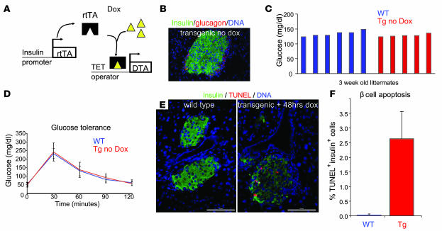
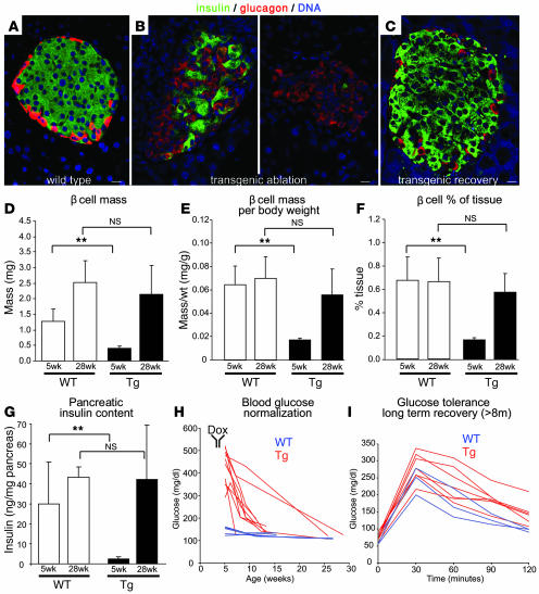
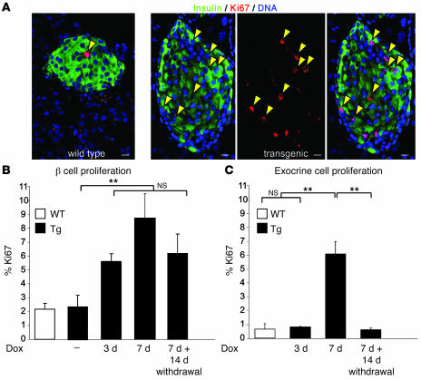
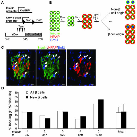
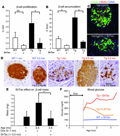
The mechanisms that regulate pancreatic beta cell mass are poorly understood. While autoimmune and pharmacological destruction of insulin-producing beta cells is often irreversible, adult beta cell mass does fluctuate in response to physiological cues including pregnancy and insulin resistance. This plasticity points to the possibility of harnessing the regenerative capacity of the beta cell to treat diabetes. We developed a transgenic mouse model to study the dynamics of beta cell regeneration from a diabetic state. Following doxycycline administration, transgenic mice expressed diphtheria toxin in beta cells, resulting in apoptosis of 70%-80% of beta cells, destruction of islet architecture, and diabetes. Withdrawal of doxycycline resulted in a spontaneous normalization of blood glucose levels and islet architecture and a significant regeneration of beta cell mass with no apparent toxicity of transient hyperglycemia. Lineage tracing analysis indicated that enhanced proliferation of surviving beta cells played the major role in regeneration. Surprisingly, treatment with Sirolimus and Tacrolimus, immunosuppressants used in the Edmonton protocol for human islet transplantation, inhibited beta cell regeneration and prevented the normalization of glucose homeostasis. These results suggest that regenerative therapy for type 1 diabetes may be achieved if autoimmunity is halted using regeneration-compatible drugs.


Figures

Figure 1
Figure 1. A transgenic mouse system for specific and conditional ablation of pancreatic β cells.
(A) Application of doxycycline (Dox) to the drinking water of Insulin-rtTA;TET-DTA mice induces the expression of DTA specifically in β cells, causing apoptotic β cell death. (B) Normal islet morphology in a 4-week-old double-transgenic mouse untreated with doxycycline. (C) Normal blood glucose levels (measured during the day) in 3-week-old mice in the absence of doxycycline. Untreated transgenic mice at 1.5 years had blood glucose levels indistinguishable from those of age-matched wild-type mice (not shown). (D) In the absence of doxycycline, 4-week-old double-transgenic mice had normal fasting blood glucose levels and glucose tolerance. Values are mean ± SD (n = 5–7). Single-transgenic TET-DTA littermates are referred to as wild-type. (E) β Cell specificity of ablation. Four-week-old mice were treated with doxycycline for 48 hours, then sacrificed and assessed for apoptotic cell death. Extensive β cell apoptosis was detected by TUNEL staining in double-transgenic mice. No apoptosis was seen in non-β cells in islets or in the exocrine pancreas. Doxycycline-treated single-transgenic littermates showed no sign of apoptosis. (F) Quantification of β cell apoptosis detected 48 hours after the administration of doxycycline to 4-week-old mice. Values are mean ± SD from 5-week-old mice exposed to doxycycline for 48 hours before sacrifice (n = 3 per group). For each mouse, about 2,000 β cells were counted. Scale bars: 100 μm.
Figure 2
Figure 2. Ablation and regeneration of β cells.
(AC) Morphology of islets. Wild-type (A) and transgenic islets in the absence of doxycycline (Figure 1B) showed the typical organization of central β cells and peripheral α cells. Islets from 5-week-old transgenic mice treated with doxycycline for 7 days (i.e., from 4 weeks of age) showed a decrease in the abundance of β cells and a mixed-islet phenotype with multiple non-β cells at the center (B). Twenty-three weeks after the withdrawal of doxycycline (about 28 weeks of age), transgenic islets had a near-perfect architecture (C). Scale bars: 10 μm. (DF) Morphometric assessment of β cell mass (D), β cell mass normalized to body weight (E), and the fraction of pancreas tissue area covered by β cells (F) in 5- and 28-week-old mice that received doxycycline between weeks 4 and 5 (n = 5–11 per group). Note a significant spontaneous normalization of β cell mass in transgenic mice relative to wild-type littermates undergoing the same treatment. **P < 0.01. (G) Pancreatic insulin content in 5- and 28-week-old mice that received doxycycline between weeks 4 and 5 (n > 3 per group). Values are mean ± SD. (H) Fed blood glucose levels following doxycycline withdrawal in mice treated with doxycycline between 4 and 5 weeks. (I) Glucose tolerance following long-term (>8 months) recovery from diabetes. Some transgenic mice regained not only normal fed and fasting blood glucose levels, but also normal glucose tolerance.
Figure 3
Figure 3. Cell proliferation during regeneration.
(A) Proliferation of differentiated (insulin+) β cells in 5-week-old mice treated with doxycycline between 4 and 5 weeks of age. Arrowheads denote proliferating β cells. (B) Proliferation of β cells following cell ablation and regeneration. Three days after the addition of doxycycline to the drinking water of 4-week-old mice, while fed and fasting glucose levels as well as glucose tolerance were still normal (not shown), the rate of β cell proliferation increased about 2.5-fold. The high rate of β cell proliferation was also seen after 7 days of doxycycline treatment and persisted 2 weeks after doxycycline withdrawal (i.e., 7-week-old mice, treated between weeks 4 and 5). (C) Exocrine cell proliferation during β cell ablation and regeneration. At the peak of β cell ablation (i.e., in 5-week-old mice, treated with doxycycline between weeks 4 and 5), a transient increase in exocrine cell proliferation was observed, possibly the result of an inflammatory process. Values are mean ± SD from 3 mice per time point. More than 1,000 β cells and 5,000 exocrine cells were counted per mouse. **P < 0.01.
Figure 4
Figure 4. New β cells come predominantly from preexisting β cells.
(A) Genetic lineage tracing system, used in conjunction with the ablation system, to determine the cellular origin of new β cells. The experimental protocol for the ablation-lineage tracing experiment is shown below. Tam, tamoxifen. (B) Experiment design and possible interpretations of lineage tracing results. For simplicity, the β cell recombination rate (labeling cells by HPAP expression) is presented as 100% and the rate of new β cell accumulation as 25%. (C) Representative confocal image of an islet from a 2-month-old Insulin-rtTA;TET-DTA;Insulin-CreERTM;Z/AP transgenic mouse exposed to doxycycline to ablate β cells, injected with tamoxifen to label surviving β cells, and treated with BrdU for 2 weeks to label new cells as shown in A. Arrows denote HPAP+BrdU+ β cells, the progeny of surviving and proliferating β cells; arrowheads mark HPAPBrdU+ β cells, derived from either non-β cells or nonlabeled β cells. Because the Insulin-rtTA and Insulin-CreERTM driver strains are distinct transgenes, their efficiency of β cell killing and recombination, respectively, was not expected to be identical. (D) Quantification of the fraction of labeled (HPAP+) β cells compared with the fraction of labeled cells among newly born (BrdU+) β cells in 5 mice (n denotes number of β cells counted per mouse). The similar percentages of labeled cells indicates that new β cells are derived primarily by proliferation of surviving β cells.
Figure 5
Figure 5. SirTac inhibits β cell regeneration.
(A) Treatment with SirTac for 14 days immediately after doxycycline withdrawal caused an approximately 80% decrease in β cell proliferation in both wild-type and diabetic mice. Mice were treated with doxycycline from birth to 1 month of age, and sacrificed 2 weeks later. (B) Accumulation of β cells during SirTac treatment, as assessed by administering BrdU to the drinking water between doxycycline withdrawal and sacrifice, concomitant with SirTac administration as in A. Values are mean ± SD (n = 3–6). More than 500 β cells were counted per mouse. *P < 0.05. (C) Representative images of new β cell accumulation in transgenic mice recovering from diabetes in the absence or presence of SirTac. Arrowheads denote BrdU+Insulin+ cells. (D) Islet morphology at 1 and 3.5 months of age with or without SirTac treatment for months 1–3.5. Note the persistent ablated islet phenotype following SirTac treatment, in contrast to the spontaneous recovery of islet morphology in its absence. Slides were stained for insulin (brown) and hematoxylin (blue). Original magnification, ×350. (E) Abrogation of β cell regeneration in SirTac-treated transgenic mice. Doxycycline was administered from birth to 1 month, after which mice were sacrificed (n = 13) or allowed to recover for 2.5 months in the absence (n = 4) or presence (n = 6) of SirTac. (F) Blood glucose levels of mice treated as in D and E. n = 5 (SirTac-treated wild-type); 4 (untreated transgenic); 9 (SirTac-treated transgenic).

Comment in

References

    1. Rood P.P., et al. Facilitating physiologic self-regeneration: a step beyond islet cell replacement. Pharm. Res. 2006;23:227–242. - PubMed
    1. Parsons J.A., Brelje T.C., Sorenson R.L. Adaptation of islets of Langerhans to pregnancy: increased islet cell proliferation and insulin secretion correlates with the onset of placental lactogen secretion. Endocrinology. 1992;130:1459–1466. - PubMed
    1. Parsons J.A., Bartke A., Sorenson R.L. Number and size of islets of Langerhans in pregnant, human growth hormone-expressing transgenic, and pituitary dwarf mice: effect of lactogenic hormones. Endocrinology. 1995;136:2013–2021. - PubMed
    1. Gupta R.K., et al. Expansion of adult beta-cell mass in response to increased metabolic demand is dependent on HNF-4alpha. Genes Dev. 2007;21:756–769. - PMC - PubMed
    1. Maclean N., Ogilvie R.F. Quantitative estimation of the pancreatic islet tissue in diabetic subjects. Diabetes. 1955;4:367–376. - PubMed

Publication types

Substances