

Evaluation of a computerized measurement technique for joint alignment before and during periacetabular osteotomy
- PMID: 17786597
- PMCID: PMC2716292
- DOI: 10.3109/10929080701541855
Evaluation of a computerized measurement technique for joint alignment before and during periacetabular osteotomy
Abstract
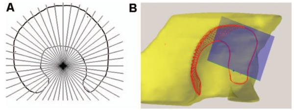
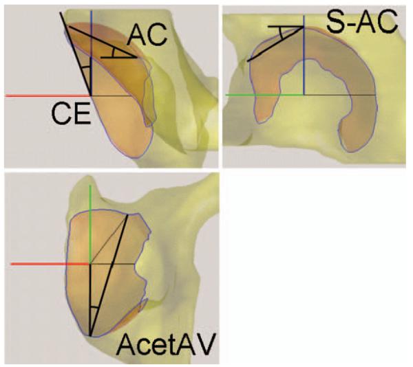
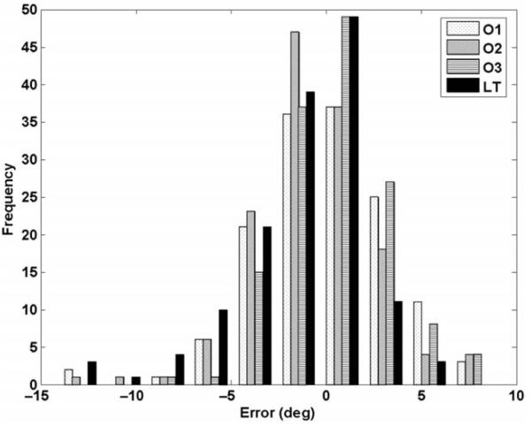
Periacetabular osteotomy (PAO) is intended to treat a painful dysplastic hip. Manual radiological angle measurements are used to diagnose dysplasia and to define regions of insufficient femoral head coverage for planning PAO. No method has yet been described that recalculates radiological angles as the acetabular bone fragment is reoriented. In this study, we propose a technique for computationally measuring the radiological angles from a joint contact surface model segmented from CT-scan data. Using oblique image slices, we selected the lateral and medial edge of the acetabulum lunate to form a closed, continuous, 3D curve. The joint surface is generated by interpolating the curve, and the radiological angles are measured directly using the 3D surface. This technique was evaluated using CT data for both normal and dysplastic hips. Manual measurements made by three independent observers showed minor discrepancies between the manual observations and the computerized technique. Inter-observer error (mean difference +/- standard deviation) was 0.04 +/- 3.53 degrees for Observer 1; -0.46 +/- 3.13 degrees for Observer 2; and 0.42 +/- 2.73 degrees for Observer 3. The measurement error for the proposed computer method was -1.30 +/- 3.30 degrees . The computerized technique demonstrates sufficient accuracy compared to manual techniques, making it suitable for planning and intraoperative evaluation of radiological metrics for periacetabular osteotomy.
Figures


References
-
- Ganz R, Klaue K, Vinh T, Mast J. A new periacetabular osteotomy for the treatment of hip dysplasias. Technique and preliminary results. Clin Orthop Relat Resh. 1988;(232):26–36. - PubMed
-
- Trousdale RT, Cabanela ME. Lessons learned after more than 250 periacetabular osteotomies. Acta Orthop Scand. 2003;74(2):119–126. - PubMed
-
- Klaue K, Wallin A, Ganz R. CT evaluation of coverage and congruency of the hip prior to osteotomy. Clin Orthop Relat Res. 1988;(232):15–25. - PubMed
-
- Wiberg G. Studies on dysplastic acetabula and congenital subluxation of the hip joint with special reference to the complication of osteoarthitis. Acta Chir Scand. 1939;83(Suppl 58):1–132.
-
- Sharp IK. Acetabular dysplasia. The acetabular angle. J Bone Joint Surg. 1961;43-B(2):268–272.
Publication types
MeSH terms
Grants and funding
LinkOut - more resources
Full Text Sources