. 2007 Sep 7;130(5):918-31.
doi: 10.1016/j.cell.2007.07.009.

NF-kappaB is a negative regulator of IL-1beta secretion as revealed by genetic and pharmacological inhibition of IKKbeta

Affiliations

NF-kappaB is a negative regulator of IL-1beta secretion as revealed by genetic and pharmacological inhibition of IKKbeta

Florian R Greten et al. Cell. .

Abstract

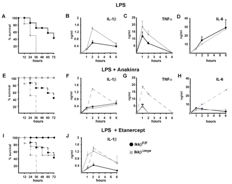
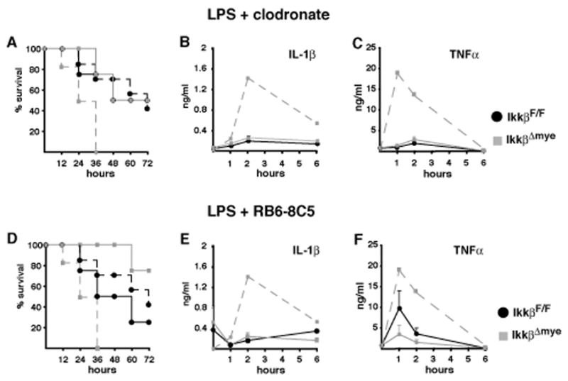
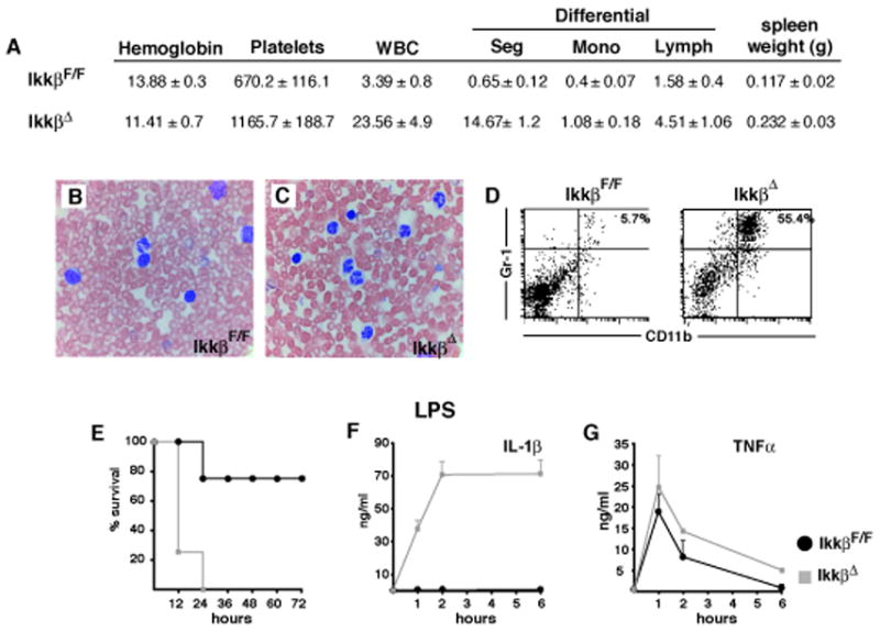
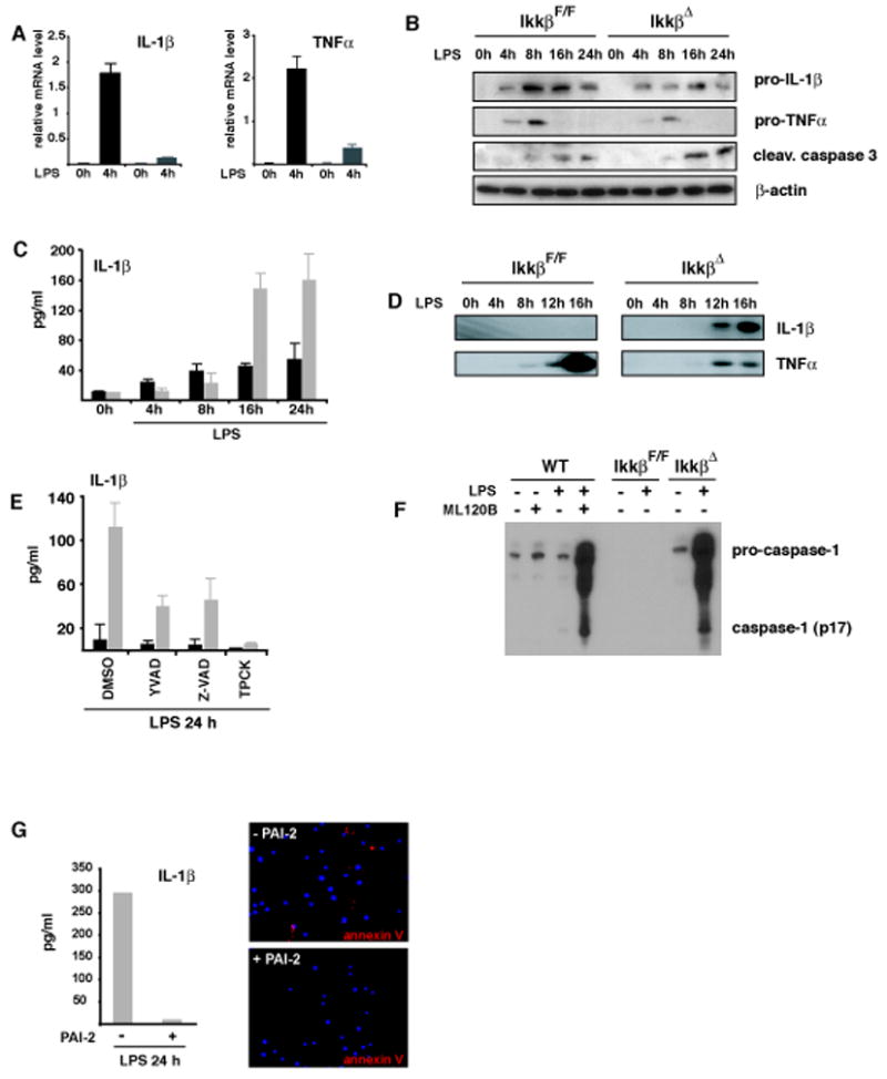
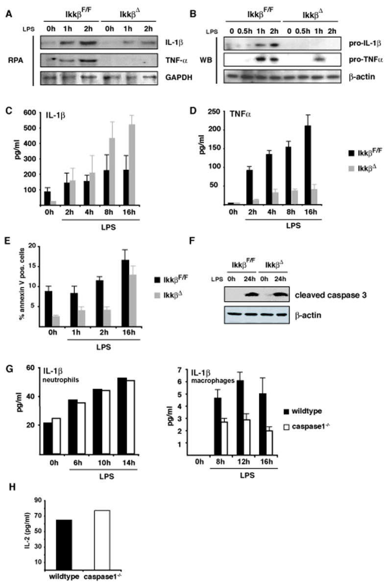
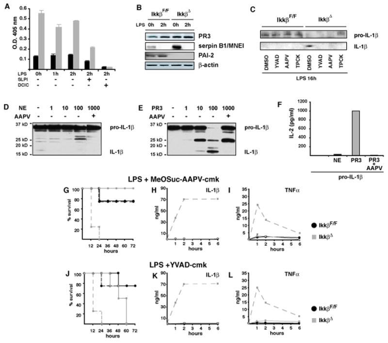
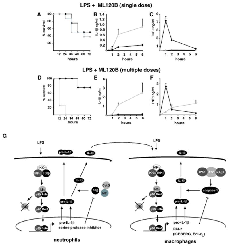
IKKbeta-dependent NF-kappaB activation plays a key role in innate immunity and inflammation, and inhibition of IKKbeta has been considered as a likely anti-inflammatory therapy. Surprisingly, however, mice with a targeted IKKbeta deletion in myeloid cells are more susceptible to endotoxin-induced shock than control mice. Increased endotoxin susceptibility is associated with elevated plasma IL-1beta as a result of increased pro-IL-1beta processing, which was also seen upon bacterial infection. In macrophages enhanced pro-IL-1beta processing depends on caspase-1, whose activation is inhibited by NF-kappaB-dependent gene products. In neutrophils, however, IL-1beta secretion is caspase-1 independent and depends on serine proteases, whose activity is also inhibited by NF-kappaB gene products. Prolonged pharmacologic inhibition of IKKbeta also augments IL-1beta secretion upon endotoxin challenge. These results unravel an unanticipated role for IKKbeta-dependent NF-kappaB signaling in the negative control of IL-1beta production and highlight potential complications of long-term IKKbeta inhibition.


Figures

Figure 1
Figure 1. Increased endotoxin-induced mortaility is associated with elevated circulating IL-1β in IkkβΔmye mice
(A) Survival after endotoxin injection (30 mg/kg, E. coli O111:B4) of IkkβF/F (black) and IkkβΔmye mice (grey), (n = 4–6). (B) IL-1β, (C) TNF-α and (D) IL-6 plasma levels after LPS administration. (E, I) Survival of IkkβF/F (solid black lines) and IkkβΔmye mice (solid grey lines) after LPS administration in the presence of IL-1ra (E) and sTNFRII (I). Dashed lines represent survival without inhibitors. (F, J) IL-1β, (G) TNF-α and (H) IL-6 plasma levels in mice given LPS plus the indicated inhibitors. Dashed line represents respective plasma levels of IkkβΔmye mice without inhibitor. Data are averages of at least 4 animals per time point.
Figure 2
Figure 2. Depletion of either macrophages or neutrophils improves survival in IkkβΔmye mice
(A, D) Survival of IkkβF/F (black lines) and IkkβΔmye mice (grey lines) depleted of macrophages (A) or neutrophils (D). Dashed lines represent survival without depletion. (B, E) IL-1β and (C, F) TNF-α plasma levels after LPS administration to macrophage-depleted (B, C) or neutrophil-depleted (E, F) mice. Dashed line represents respective plasma levels in IkkβΔmye mice without depletion. Data are averages of at least 4 animals per time point.
Figure 3
Figure 3. IkkβΔ mice develop granulocytosis and show massively increased circulating IL-1β after endotoxin exposure
(A) Complete and differential blood counts and spleen weights of IkkβF/F and IkkβΔ mice. Data are average values for 5 mice of each genotype examined two weeks after a single poly-(I:C) injection. (B, C) Blood smears stained with Wright-Giemsa and (D) number of CD11b+/Gr-1+ cells in spleens of of IkkβF/F and IkkβΔ mice analyzed two weeks after poly-(I:C) injection. (E) Survival after endotoxin injection (20 mg/kg, E. coli O111:B4) of IkkβF/F (black) and IkkβΔ mice (grey), (n = 4–6). (F) IL-1β and (G) TNF-α plasma levels 1, 2 and 6 hrs after LPS administration.
Figure 4
Figure 4. Increased IL-1β release correlates with elevated apoptosis of IKKβ-deficient macrophages and is inhibited by PAI-2
(A) Relative levels of IL-1β and TNF-α mRNA after incubation of IkkβF/F (black bars) and IkkβΔ (grey bars) BMDM with LPS (100 ng/ml). (B) Immunoblot analysis of intracellular pro-IL-1β, pro-TNF-α and cleaved caspase-3 in macrophages after LPS stimulation (100 ng/ml). (C) IL-1β levels in supernatants of IkkβF/F (black bars) and IkkβΔ (grey bars) macrophages after LPS stimulation. (D) Immunoblot analysis of processed IL-1β and TNF-α in supernatants of cultured macrophages. (E) IL-1β in supernatants of LPS stimulated IkkβF/F (black) and IkkβΔ (grey) macrophages in the presence of Ac-YVAD-cmk (100 μM), Z-VAD-fmk (10 μM) and TPCK (10 μM). Data are averages of at least three animals. (F) Loss of IKKβ activity enhances caspase-1 activation. WT or IKKβ-deficient macrophages were pretreated with ML120B (30 μM) or DMSO and either left unstimulated or incubated with LPS (100 ng/ml). After 22 hrs, culture supernatants were collected and analyzed by immunoblotting for secretion of activated caspase-1 (p17). The highest mobility represents uncleaved pro-caspase-1. (G) Reconstitution of IKKβ-deficient macrophages with PAI-2 blocks apoptosis and IL-1β release. Bone marrow of IkkβΔ mice was retrovirally transduced with EGFP-PAI-2. EGFP-positive cells were sorted, differentiated into macrophages and stimulated with LPS (100 ng/ml) for 24 hrs. Apoptosis was determined by annexin V staining and IL-1β levels in supernatants were determined by ELISA.
Figure 5
Figure 5. Increased IL-1β release is independent of apoptosis in IKKβ-deficient neutrophils
(A) Levels of IL-1β and TNF-α mRNAs in neutrophils after LPS stimulation were examined by RNase protection. (B) Immunoblot analysis of intracellular pro-IL-1β and pro-TNF-α in neutrophils after LPS stimulation. (C, D) IL-1β and TNF-α secretion by LPS-stimulated neutrophils determined by ELISA. (E) Number of annexin V positive Gr-1+ cells determined by flow cytometry. (F) Immunoblot analysis of cleaved caspase-3 in neutrophils after LPS stimulation (100 ng/ml). (G) IL-1β secretion by LPS-stimulated wt and caspase-1−/− macrophages and neutrophils. (H) Bioassay of supernatants of LPS-stimulated wt and caspase-1−/− neutrophils using EL4 cells.
Figure 6
Figure 6. Elevated serine protease activity in IKKβ-deficient neutrophils accounts for enhanced IL-1β secretion
(A) Hydrolysis of MeOSuc-AAPV-pNA was used to determine serine protease activity in IkkβF/F (black bars) and IkkβΔ neutrophils (grey bars) treated with LPS and the protease inhibitors SLPI (1μg/ml) or 3, 4-DCIC (10 μM). (B) PR3, serpin B1/MNEI and PAI-2 expression in LPS–stimulated neutrophils determined by immunoblotting. (C) Immunoblot analysis of IL-1β expression in supernatants of neutrophils 16 hrs after LPS stimulation in the presence of Ac-YVAD-cmk (100 μM), MeOSuc-AAPV-cmk (500 μM) or TPCK (10 μM). (D, E) Immunoblot analysis of IL-1β produced in HEK293 cells after incubation with increasing amounts of purified (D) NE and (E) PR3 without or in the presence or AAPV. (F) Bioassay of extracts used in (D) and (E) in EL4 cells. (G–L) Survival of IkkβF/F mice (black lines) and IkkβΔ mice (grey lines) after pretreatment with the serine protease inhibitor MeOSuc-AAPV-cmk (1 mg/mouse) 1 hr before LPS application (G) or Ac-YVAD-cmk (J) and corresponding IL-1β (H, K) and TNF-α (I, J) plasma levels. Dashed lines in (G and J) represent survival without inhibitor and in (H, I) and (K, L) they represent plasma IL-1β and plasma TNF-α levels of IkkβΔmye mice without inhibitor, respectively.
Figure 7
Figure 7. Prolonged pharmacological inhibition of IKKβ enhances IL-1β secretion and endotoxic shock
(A–C) Survival and corresponding IL-1β and TNF-α plasma levels of LPS-challenged wt mice given 300 mg/kg of the IKKβ inhibitor ML120B by oral gavage 1 hr prior to LPS administration (30 mg/kg). (D) Survival and corresponding IL-1β (E) and TNF-α (F) plasma levels in wt mice given 300 mg/kg ML120B by oral gavage twice daily for four days before LPS challenge. (G) Schematic representation of the proposed dual role of IKKβ-dependent NF-κB activation in regulation of IL-1β secretion by macrophages and neutrophils: in neutrophils, which appear to be a rapid but minor (albeit critical) source of IL-1β, IKKβ driven NF-κB positively regulates the transcription of pro-IL-1β mRNA and serine protease inhibitor genes whose products inhibit the activity of PR3, which can process pro-IL-1β. Secretion of biologically active IL-1β by neutrophils acts together with LPS to augment IL-1β secretion by macrophages, which represent the major source of this cytokine. In the macrophage, NF-κB controls pro-IL-1β mRNA synthesis as well as the expression of genes such as PAI-1, Bcl-xL and ICEBERG whose products inhibit caspase-1 activation.

Similar articles

Cited by

References

    1. Arkan MC, Hevener AL, Greten FR, Maeda S, Li ZW, Long JM, Wynshaw-Boris A, Poli G, Olefsky J, Karin M. IKK-beta links inflammation to obesity-induced insulin resistance. Nat Med. 2005;11:191–198. - PubMed
    1. Black RA, Kronheim SR, Cantrell M, Deeley MC, March CJ, Prickett KS, Wignall J, Conlon PJ, Cosman D, Hopp TP, et al. Generation of biologically active interleukin-1 beta by proteolytic cleavage of the inactive precursor. J Biol Chem. 1988;263:9437–9442. - PubMed
    1. Bonizzi G, Karin M. The two NF-kappaB activation pathways and their role in innate and adaptive immunity. Trends Immunol. 2004;25:280–288. - PubMed
    1. Bruey JM, Bruey-Sedano N, Luciano F, Zhai D, Balpai R, Xu C, Kress CL, Bailly-Maitre B, Li X, Osterman A, et al. Bcl-2 and Bcl-XL regulate proinflammatory caspase-1 activation by interaction with NALP1. Cell. 2007;129:45–56. - PubMed
    1. Chen LW, Egan L, Li ZW, Greten FR, Kagnoff MF, Karin M. The two faces of IKK and NF-kappaB inhibition: prevention of systemic inflammation but increased local injury following intestinal ischemia-reperfusion. Nat Med. 2003;9:575–581. - PubMed

Publication types

MeSH terms