. 2007 Sep 7;27(5):731-44.
doi: 10.1016/j.molcel.2007.06.033.

Docking of the proteasomal ATPases' carboxyl termini in the 20S proteasome's alpha ring opens the gate for substrate entry

Affiliations

Docking of the proteasomal ATPases' carboxyl termini in the 20S proteasome's alpha ring opens the gate for substrate entry

David M Smith et al. Mol Cell. .

Abstract

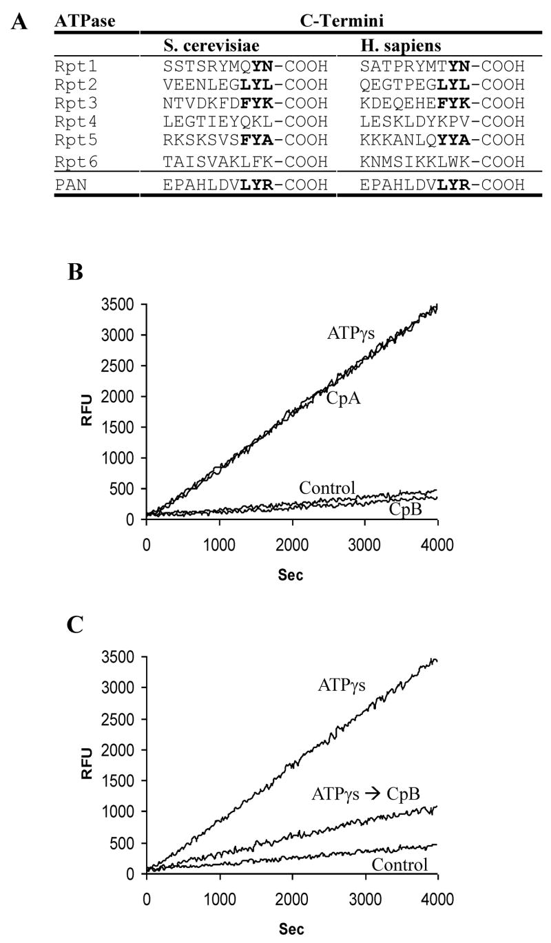
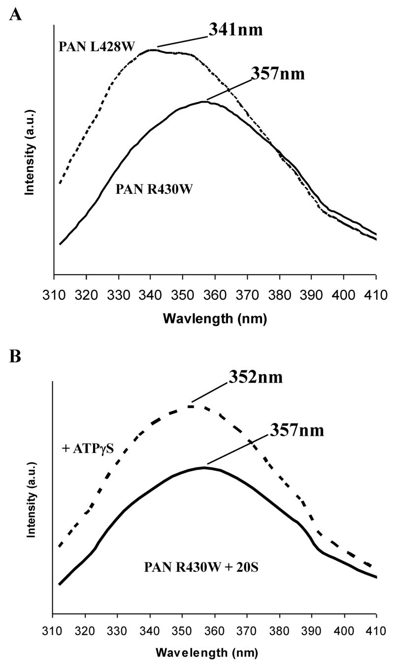
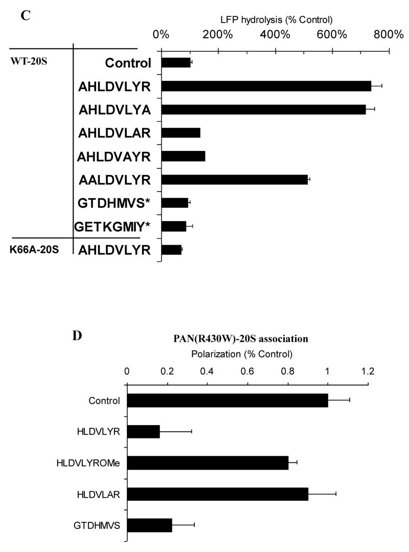
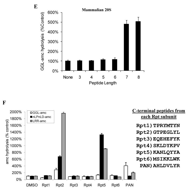
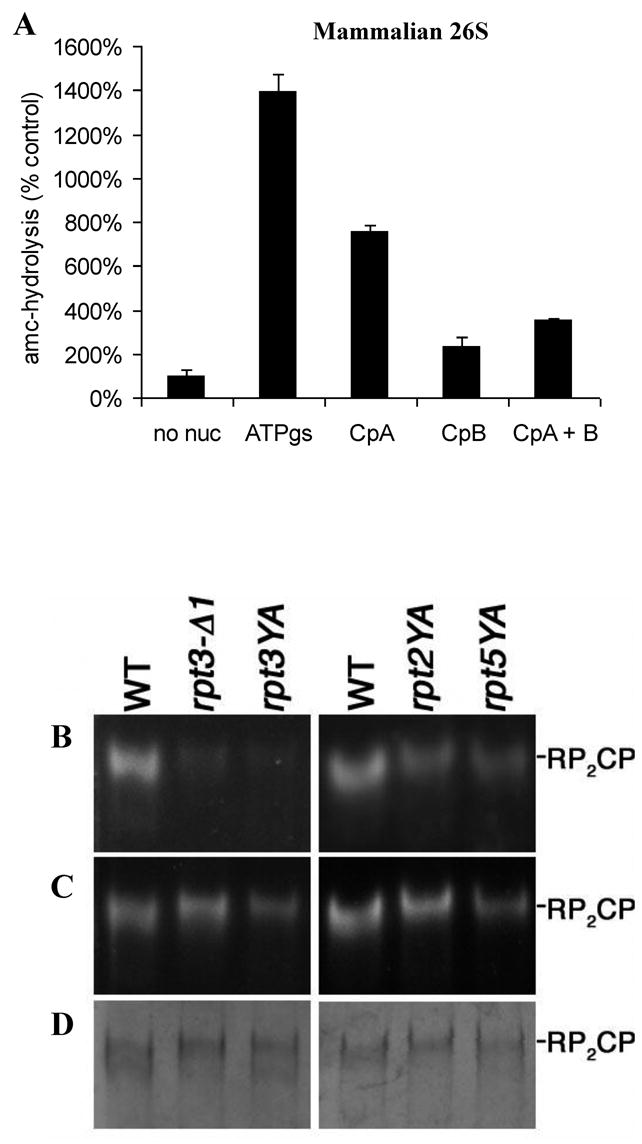
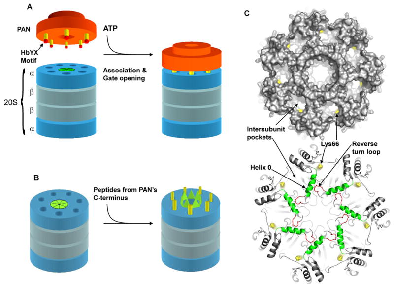
The 20S proteasome functions in protein degradation in eukaryotes together with the 19S ATPases or in archaea with the homologous PAN ATPase complex. These ATPases contain a conserved C-terminal hydrophobic-tyrosine-X motif (HbYX). We show that these residues are essential for PAN to associate with the 20S and open its gated channel for substrate entry. Upon ATP binding, these C-terminal residues bind to pockets between the 20S's alpha subunits. Seven-residue or longer peptides from PAN's C terminus containing the HbYX motif also bind to these sites and induce gate opening in the 20S. Gate opening could be induced by C-terminal peptides from the 19S ATPase subunits, Rpt2, and Rpt5, but not by ones from PA28/26, which lack the HbYX motif and cause gate opening by distinct mechanisms. C-terminal residues in the 19S ATPases were also shown to be critical for gating and stability of 26S proteasomes. Thus, the C termini of the proteasomal ATPases function like a "key in a lock" to induce gate opening and allow substrate entry.


Figures

Figure 1
Figure 1
A) Carboxypeptidase treatment of the proteasomal ATPase regulatory complexes’ eliminates their ability to stimulate the 20S for peptide hydrolysis. A) Two residues preceding the C-terminal arginine in PAN are conserved in the eukaryotic 19S proteasome-regulatory ATPases. B) A solution of PAN, 20S and the fluorescent peptide LFP (Mca-AKVYPYPME-Dpa-amide) was pre-incubated with Carboxypeptidase A (CpA), B (CpB) or without either carboxypeptidase (control and ATPγS) for 5 minutes followed by addition of a general Carboxypeptidase inhibitor from potato tuber, (0.01mg/ml). H2O (Control), or ATPγS (ATPγS, CpA, CpB) was added, and the rate of LFP hydrolysis monitored in real-time. Both CpA and CpB were used at a final concentration of 0.08 Units/ml. C) Same as in A except ATPγS was added prior to addition of CpB. Values are mean’s of three independent experiments, error bars indicate standard deviations and all of these experiment were performed at least three times with similar results.
Figure 2
Figure 2
The fluorescent spectrum of PAN R430W and PAN L428W mutants in solution and upon binding the 20S. A) Fluorescent emission spectra of the various PAN mutants. B) Fluorescent emission spectra of PAN R430W in the presence of 20S proteasomes without and with ATPγS. λem-max is indicated. C) Polarization of PAN’s (R430W) tryptophan in the presence of ATPγS upon addition of increasing amount of 20S proteasomes. PAN’s MW was taken as a 585kDa (2-hexameric rings) and 20S as 674kDa. The final concentration of PAN6 was 340nM the 20S concentration is shown. The fit line was calculated from regression analysis of the simple ligand binding equation with one site saturation, the Kd was calculated to be 34 +/−12 nM, and the maximum change in polarization (Bmax) was 45mP. D) A difference plot showing the changes that occur in the fluorescent spectrum of the tryptophan in PAN R430W that is induced by addition of WT-20S or by addition of K66A-20S. The plot was generated by subtracting the spectrum of PAN R430W with the 20S from it’s plot without the 20S, a flat line indicates no change in the spectrum. These experiment were performed at least three times with similar results.
Figure 2
Figure 2
The fluorescent spectrum of PAN R430W and PAN L428W mutants in solution and upon binding the 20S. A) Fluorescent emission spectra of the various PAN mutants. B) Fluorescent emission spectra of PAN R430W in the presence of 20S proteasomes without and with ATPγS. λem-max is indicated. C) Polarization of PAN’s (R430W) tryptophan in the presence of ATPγS upon addition of increasing amount of 20S proteasomes. PAN’s MW was taken as a 585kDa (2-hexameric rings) and 20S as 674kDa. The final concentration of PAN6 was 340nM the 20S concentration is shown. The fit line was calculated from regression analysis of the simple ligand binding equation with one site saturation, the Kd was calculated to be 34 +/−12 nM, and the maximum change in polarization (Bmax) was 45mP. D) A difference plot showing the changes that occur in the fluorescent spectrum of the tryptophan in PAN R430W that is induced by addition of WT-20S or by addition of K66A-20S. The plot was generated by subtracting the spectrum of PAN R430W with the 20S from it’s plot without the 20S, a flat line indicates no change in the spectrum. These experiment were performed at least three times with similar results.
Figure 3
Figure 3
Peptides derived from PAN’s extreme C-terminal sequence induce gate opening and inhibit PAN-20S complex assembly. A) Ability of peptides of different lengths to stimulate gate opening. Peptides (250μM) were incubated with 0.2 μg 20S proteasomes and LFP. In each case, a correction was made because the added peptide, in addition to causing gate opening, competed with LFP at the active sites. To determine the actual percentage stimulation of LFP hydrolysis due to gate-opening, the ability of the various peptides to inhibit LFP hydrolysis by the gateless (Δα2–12) 20S proteasome was also measured, and the values used to normalize data on LFP hydrolysis by WT 20S proteasomes. Without normalization, 7-8 residue peptides still stimulated 20S proteasomes 3–4 fold (see also B). Values in A, C, D and E are the means of three independent experiments +/− SD’s. B) The 7 residue peptide from PAN’s C-terminus (HLDVLYR) induces gate opening in the Thermoplasma 20S at much lower concentrations than the 10 residue peptide from PAN’s C-terminus (EPAHLDVLYR). This experiment was performed as in A) using the concentrations of the peptide shown, except that no adjustment for competition with the fluorogenic substrate was calculated, since such corrections become unreliable at the high peptide concentrations (>500 μM). The peptide HLDVLYR inhibits cleavage of LFP by Δα(2–12)20S at ≥50μM. C) Replacement of the Hb (hydrophobic) and Y residues in the 8-residue peptides prevented gate-opening. Proteasome activity was measured as in A) in the presence of the indicated peptides. Also shown is the inability of the WT peptide (250μM) to induce gate opening in the 20S K66A, and the inability of the peptide corresponding to PA26’s C-termini (500 SM)* to induce gate opening in wild-type 20S. D) Peptides that activate gate-opening prevent association of the 20S and PAN, as does the peptide corresponding to the C-terminus of PA26. Each peptide was pre-incubated with 20S and PAN for five minutes followed by addition of ATPγS. Polarization was measured before and after nucleotide addition. No peptide (control) was taken as 100% polarization and was similar to that shown in Fig. 2. The values are means +/− SD’s from at least three experiments. Peptide concentrations were used as in C). E) A similar experiment as in A (using peptides corresponding to PAN’s c-terminus) was carried out using purified rabbit muscle 20S proteasomes and GGL-amc as the fluorogenic substrate (Smith et al., 2005). F) A similar experiment as in C was carried out using purified rabbit muscle 20S but with peptides that corresponded to the C-termini of the mammalian 19S ATPases: Rpt1, Rpt2, Rpt3, Rpt4, Rpt5, and Rpt6. The cleavage rates of fluorogenic substrates specific to its three peptidase sites were monitored (see text). The peptide corresponding to Rpt1 stimulated gate opening weakly under some conditions (e.g. no glycerol but with 60mM KCl). The sequences for each of the C-terminal peptides are shown. All values are the Means +/− SD of three independent experiments.
Figure 3
Figure 3
Peptides derived from PAN’s extreme C-terminal sequence induce gate opening and inhibit PAN-20S complex assembly. A) Ability of peptides of different lengths to stimulate gate opening. Peptides (250μM) were incubated with 0.2 μg 20S proteasomes and LFP. In each case, a correction was made because the added peptide, in addition to causing gate opening, competed with LFP at the active sites. To determine the actual percentage stimulation of LFP hydrolysis due to gate-opening, the ability of the various peptides to inhibit LFP hydrolysis by the gateless (Δα2–12) 20S proteasome was also measured, and the values used to normalize data on LFP hydrolysis by WT 20S proteasomes. Without normalization, 7-8 residue peptides still stimulated 20S proteasomes 3–4 fold (see also B). Values in A, C, D and E are the means of three independent experiments +/− SD’s. B) The 7 residue peptide from PAN’s C-terminus (HLDVLYR) induces gate opening in the Thermoplasma 20S at much lower concentrations than the 10 residue peptide from PAN’s C-terminus (EPAHLDVLYR). This experiment was performed as in A) using the concentrations of the peptide shown, except that no adjustment for competition with the fluorogenic substrate was calculated, since such corrections become unreliable at the high peptide concentrations (>500 μM). The peptide HLDVLYR inhibits cleavage of LFP by Δα(2–12)20S at ≥50μM. C) Replacement of the Hb (hydrophobic) and Y residues in the 8-residue peptides prevented gate-opening. Proteasome activity was measured as in A) in the presence of the indicated peptides. Also shown is the inability of the WT peptide (250μM) to induce gate opening in the 20S K66A, and the inability of the peptide corresponding to PA26’s C-termini (500 SM)* to induce gate opening in wild-type 20S. D) Peptides that activate gate-opening prevent association of the 20S and PAN, as does the peptide corresponding to the C-terminus of PA26. Each peptide was pre-incubated with 20S and PAN for five minutes followed by addition of ATPγS. Polarization was measured before and after nucleotide addition. No peptide (control) was taken as 100% polarization and was similar to that shown in Fig. 2. The values are means +/− SD’s from at least three experiments. Peptide concentrations were used as in C). E) A similar experiment as in A (using peptides corresponding to PAN’s c-terminus) was carried out using purified rabbit muscle 20S proteasomes and GGL-amc as the fluorogenic substrate (Smith et al., 2005). F) A similar experiment as in C was carried out using purified rabbit muscle 20S but with peptides that corresponded to the C-termini of the mammalian 19S ATPases: Rpt1, Rpt2, Rpt3, Rpt4, Rpt5, and Rpt6. The cleavage rates of fluorogenic substrates specific to its three peptidase sites were monitored (see text). The peptide corresponding to Rpt1 stimulated gate opening weakly under some conditions (e.g. no glycerol but with 60mM KCl). The sequences for each of the C-terminal peptides are shown. All values are the Means +/− SD of three independent experiments.
Figure 4
Figure 4
Carboxypeptidase induced truncation and mutations within the HbYX motif result in 26S instability and gating defects. A) Carboxypeptidase treatment of free mammalian 19S/PA700 inhibits its ability to stimulate peptide hydrolysis by the mammalian 20S in the presence of ATPγS. The 19S particles (1μg/reaction) were preincubated with or without 0.1 U of carboxypeptidase A or B for 10 min. prior to addition of the 20S (0.1μg/reaction) and ATPγS (0.1mM). The assembly reaction mix was diluted 5 fold in the reaction buffer with Ac-nLPnLD-amc and the rate of hydrolysis was monitored. B) Purified proteasomes (3μg) from wild-type (WT) and rpt mutant strains were resolved on 3.5% nondenaturing gels, and proteasome activity was visualized by staining with the fluorogenic substrate suc-LLVY-AMC. The YA mutants are single-residue substitutions of the penultimate Tyr of the indicated Rpt protein. rpt3-Δ1 is a deletion of the C-terminal lysine of Rpt3. C) After the initial peptidase assay in (A), the assay was repeated in the presence of 0.02% SDS to cause gate opening. D) The native gels were stained with Coomassie Brilliant Blue to assess the amount of protein in each sample.
Figure 5
Figure 5
A) Model depicting the association of PAN with the α-ring of the 20S proteasome, in which the C-termini (yellow) of PAN (orange) dock into the intersubunit pockets in the top of the 20S. B) Schematic model for gate opening in the 20S upon binding of peptides derived form PAN’s C-terminus to the intersubunit pockets in the 20S. C) Top: top view of the 20S α-ring with a surface rendering to demonstrate the intersubunit pockets and location of lys66 (yellow). Bottom: Ribbon representation of the 20S α-ring. Peptide binding to lys66 (yellow) adjacent to Helix 0 (green) may cause a conformational change in Helix 0 that propagates to the reverse turn loop (red) to induce gate opening. The N-terminal gating residues are not resolved in this crystal structure (1PMA). Images in C) were rendered with Pymol.

Comment in

  • Unlocking the proteasome door.
    Saeki Y, Tanaka K. Saeki Y, et al. Mol Cell. 2007 Sep 21;27(6):865-7. doi: 10.1016/j.molcel.2007.09.001. Mol Cell. 2007. PMID: 17889660

References

    1. Adams J. Proteasome inhibitors in cancer therapy. Totowa, N.J: Humana Press; 2004.
    1. Ames BN. Assay of inorganic Phosphate, Total Phosphate and Phosphatases. Methods in Enzymology. 1966;8:115–118.
    1. Benaroudj N, Smith DM, Goldberg AL. What the archaeal PAN-proteasome complex and bacterial ATPdependent proteases can teach us about the 26S proteasome. Vol. 2. The Ubiquitin-Proteasome System; Wiley-VCH: 2005.
    1. Benaroudj N, Zwickl P, Seemuller E, Baumeister W, Goldberg AL. ATP hydrolysis by the proteasome regulatory complex PAN serves multiple functions in protein degradation. Mol Cell. 2003;11:69–78. - PubMed
    1. Bochtler M, Hartmann C, Song HK, Bourenkov GP, Bartunik HD, Huber R. The structures of HsIU and the ATP-dependent protease HsIU-HsIV. Nature. 2000;403:800–805. - PubMed

Publication types

MeSH terms

Substances