Review
. 2007 Nov 10;59(13):1351-65.
doi: 10.1016/j.addr.2007.08.008. Epub 2007 Aug 14.

Hyaluronan-dependent pericellular matrix

Affiliations
Review

Hyaluronan-dependent pericellular matrix

Stephen P Evanko et al. Adv Drug Deliv Rev. .

Abstract

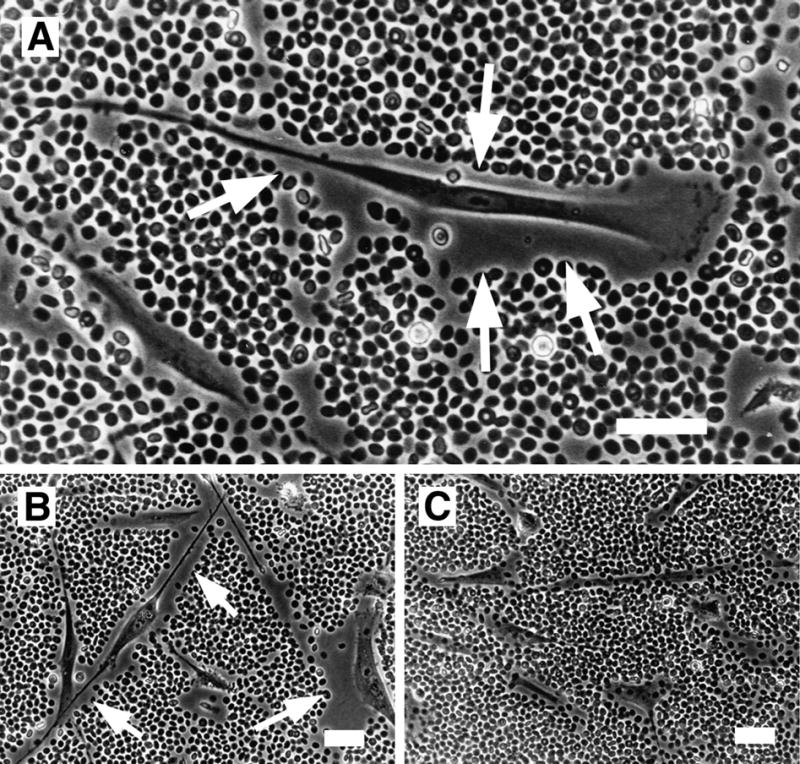
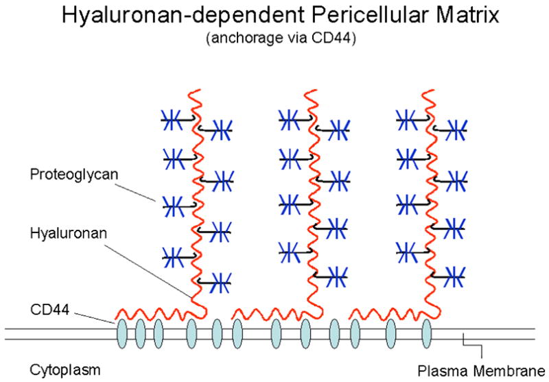
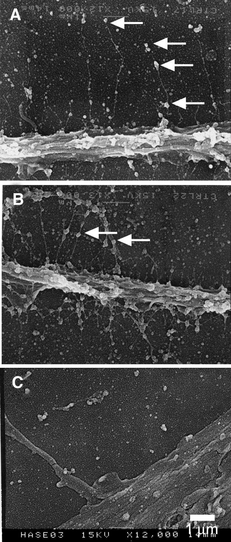
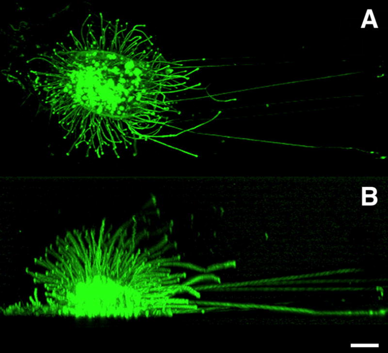
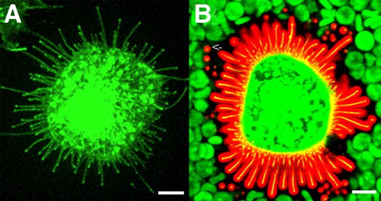
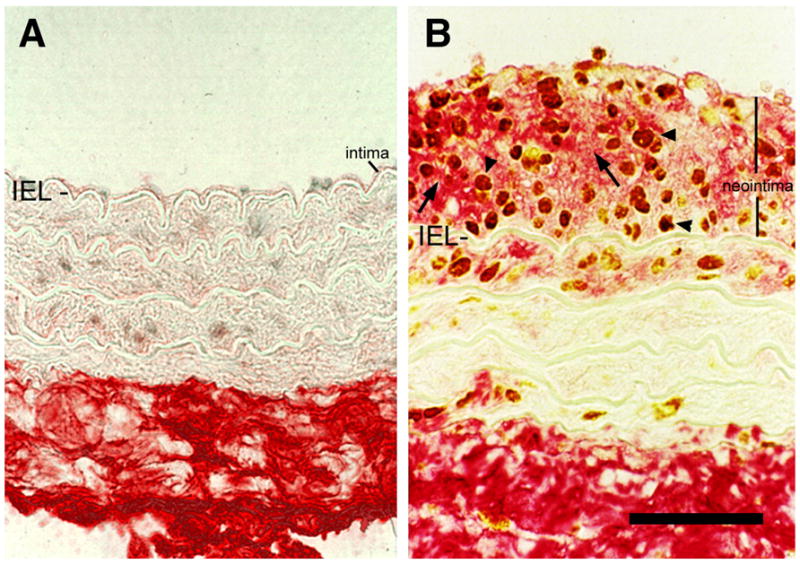
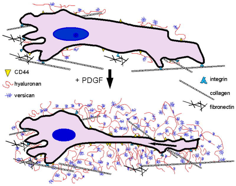
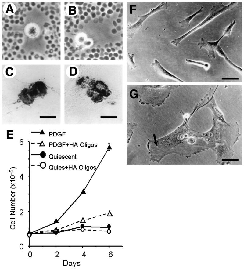
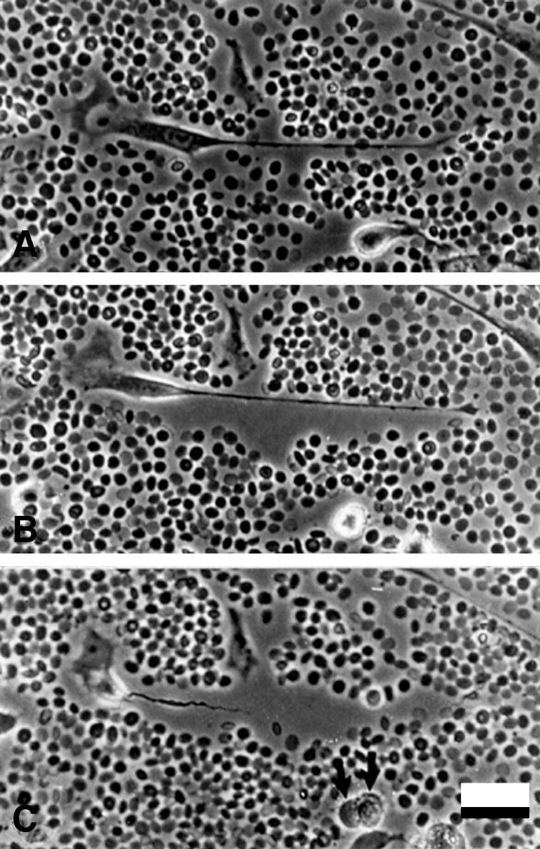
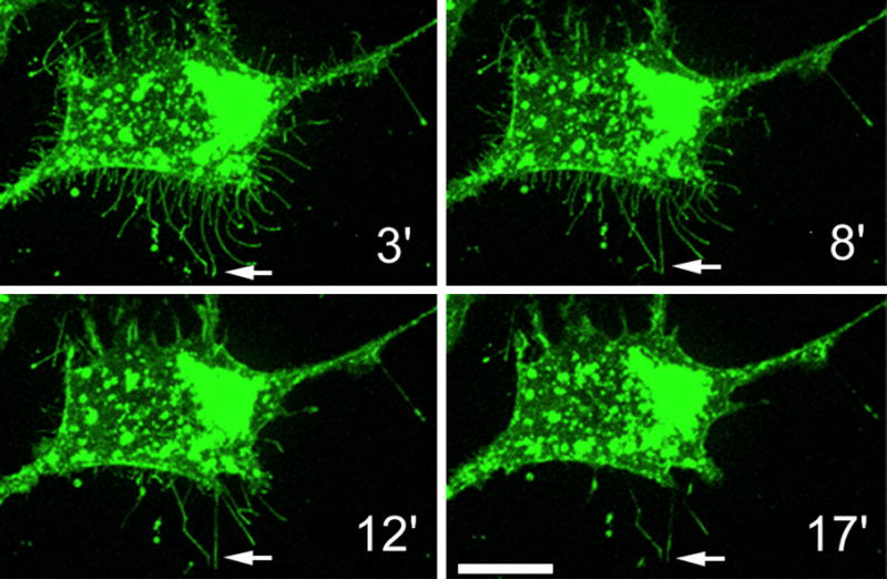
Hyaluronan is a multifunctional glycosaminoglycan that forms the structural basis of the pericellular matrix. Hyaluronan is extruded directly through the plasma membrane by one of three hyaluronan synthases and anchored to the cell surface by the synthase or cell surface receptors such as CD44 or RHAMM. Aggregating proteoglycans and other hyaluronan-binding proteins, contribute to the material and biological properties of the matrix and regulate cell and tissue function. The pericellular matrix plays multiple complex roles in cell adhesion/de-adhesion, and cell shape changes associated with proliferation and locomotion. Time-lapse studies show that pericellular matrix formation facilitates cell detachment and mitotic cell rounding. Hyaluronan crosslinking occurs through various proteins, such as tenascin, TSG-6, inter-alpha-trypsin inhibitor, pentraxin and TSP-1. This creates higher order levels of structured hyaluronan that may regulate inflammation and other biological processes. Microvillous or filopodial membrane protrusions are created by active hyaluronan synthesis, and form the scaffold of hyaluronan coats in certain cells. The importance of the pericellular matrix in cellular mechanotransduction and the response to mechanical strain are also discussed.


Figures

Figure 1
Figure 1
Hyaluronan-dependent pericellular matrix in human smooth muscle cells visualized using the particle exclusion assay. The cell coat excludes the fixed erythrocytes and is seen as a clear zone surrounding the cell (arrows). A. A typical locomoting cell with a small amount of pericellular matrix at the lammellipodium in front and more abundant matrix along the cell flanks and trailing uropod. B, C. Pericellular matrices were visualized before, B, or after, C, digestion with Streptomyces hyaluronidase. Bars equal 50 μm. Panel A originally published in: S. Evanko, J. Angello, T. Wight, Formation of hyaluronan and versican rich pericellular matrix is required for proliferation and migration of vascular smooth muscle cells. Arterioscler. Thromb. Vasc. Biol., 1999, 19(4):1004–1013. Used with permission.
Figure 2
Figure 2
Model depicting the pericellular matrix with the hyaluronan chains anchored to the cell surface via CD44 and the associated aggregating proteoglycans. Adapted by permission from Macmillan Publishers LTD: Nature Reviews Cancer, Toole, B.P., “Hyaluronan: from extracellular glue to pericellular cue,” 4:528 (2004).
Figure 3
Figure 3
Scanning electron microscopy of the pericellular matrix. Smooth muscle cells were fixed in the presence of ruthenium red, air-dried and coated for electron microscopy. A, Individual hyaluronan chains, several micrometers long extend perpendicularly from the surface of a trailing uropod of a locomoting cell. Proteoglycans are seen as large granules periodically decorating the hyaluronan filaments. B. An example of pericellular matrix that is more tangled with more condensed clusters of proteoglycan granules. C. Hyaluronidase digestion removes the hyaluronan strands and granules from the pericellular matrix and cell surface. Bar equals 1 micrometer. Panels A, B, and C were originally published in: S. Evanko, J. Angello, T. Wight, Formation of hyaluronan and versican rich pericellular matrix is required for proliferation and migration of vascular smooth muscle cells. Arterioscler. Thromb. Vasc. Biol., 1999, 19(4):1004–1013. Used with permission.
Figure 4
Figure 4
Microvillous plasma membrane protrusions induced by hyaluronan synthesis. GFP- labeled Has3 was transfected into LP-9 cells. The next day, a live cell was studied at multiple horizontal optical sections by confocal microscope. The stack of the optical sections was combined for a downwards (A) and sideways view (B). Bar equals 20 μm.
Figure 5
Figure 5
Hyaluronan coat on MCF-7 cells expressing GFP-HAS3. The microvilli (with green GFP-Has3), shown alone in (A), are actually covered by a 0.5–2 μm layer of hyaluronan (B), visualized by a probe made of aggrecan G1 domain and link protein tagged with Alexa Fluor 594® (red). Note that the hyaluronan coat visualized by red blood cells (green) corresponds to the space occupied by the microvilli (yellow) and their hyaluronan cover. The image represents a single confocal optical section; many of the microvilli are shown in cross section (arrow). Bar equals 10 μm.
Figure 6
Figure 6
Hyaluronan is increased in arterial neointima following balloon injury. Hyaluronan staining of arterial tissue using biotinylated-HABP (red, arrows) and PCNA staining to localize proliferating cells (brown, arrowheads) in uninjured, A, and injured B, rat carotid arteries. Note the abundance of hyaluronan surrounding the proliferating cells in the neointima of the injured vessel. IEL, internal elastic lamina. Bar equals 50 μm. Panels A and B were originally published in: R. Riessen, T.N. Wight, C. Pastore, C. Henley, J.M. Isner, Distribution of hyaluronan during extracellular matrix remodeling in human restenotic arteries and balloon-injured rat carotid arteries. Circulation, 1996, 93(6):1141–1147. Used with permission.
Figure 7
Figure 7
Model of pericellular matrix expansion and cell shape change following stimulation with PDGF.
Figure 8
Figure 8
Hyaluronan-dependent pericellular matrix regulates proliferation and cell shape. A, B, Particle exclusion assay showing cell coats surrounding mitotic smooth muscle cells in different stages of division. Mitotic cells stained for hyaluronan, C, and versican D, show concentrated deposits of these components. E, treatment of human smooth muscle cells with oligosaccharides of hyaluronan (20 μg/ml) inhibits PDGF-induced proliferation. Oligosaccharides also stimulated flattening of SMC. F, untreated cells. G, cells treated with oligosaccharides. Bar equals 50 μm. Portions of this figure were originally published in: S. Evanko, J. Angello, T. Wight, Formation of hyaluronan and versican rich pericellular matrix is required for proliferation and migration of vascular smooth muscle cells. Arterioscler. Thromb. Vasc. Biol., 1999, 19(4):1004–1013. Used with permission.
Figure 9
Figure 9
Pericellular matrix formation accompanies cell detachment and mitotic cell rounding. Time-lapse microscopy was begun 24 h after PDGF treatment, when cells are actively moving and dividing. The formation and expansion of the pericellular matrix occurs mainly at the time cells detach from the tissue culture substrate. (A) At time zero (immediately after settling of the red blood cells), an elongated cell with a long, trailing process and relatively little hyaluronan-dependent matrix. (B) 40 min later, a distinct pericellular matrix has formed around the cell and trailing process, pushing the red blood cells away, while the main cell body is beginning to detach from the substrate. (C) At 80 min, the cell is more rounded and is retracting the trailing process through the sleeve of hyaluronan-rich matrix. Note also the rounded cell with a distinct pericellular matrix in the lower part of the field in A and which has completed mitosis in C (arrows). Bar equals 50 μm.
Figure 10
Figure 10
The maintenance of the microvilli is dependent on the hyaluronan coat. Streptomyces hyaluronidase was introduced in a MCF-7 cell culture overexpressing GFP-HAS3 to degrade cell surface hyaluronan. The microvilli gradually shrink and eventually disappear when the external support given by the Has-associated hyaluronan is lost. Note that those microvilli which have adhered to the substratum do not retract (arrows).

References

    1. Weigel PH, Hascall VC, Tammi M. Hyaluronan synthases. J Biol Chem. 1997;272:13997–14000. - PubMed
    1. Tammi MI, Day AJ, Turley EA. Hyaluronan and homeostasis: a balancing act. J Biol Chem. 2002;277:4581–4584. - PubMed
    1. Stern R, Csoka A. Mammalian Hyaluronidases. In: Hascall V, Yanagashita M, editors. Glycoforum/Science of Hyaluronan Today. Seikagaku, Corporation; Tokyo, Japan: 2000. http://www.glycoforum.gr.jp.
    1. Toole BP. Hyaluronan: from extracellular glue to pericellular cue. Nat Rev Cancer. 2004;4:528–539. - PubMed
    1. Cowman MK, Spagnoli C, Kudasheva D, Li M, Dyal A, Kanai S, Balazs EA. Extended, relaxed, and condensed conformations of hyaluronan observed by atomic force microscopy. Biophys J. 2005;88:590–602. - PMC - PubMed

Publication types

MeSH terms