. 2007 Nov;36(3):332-42.
doi: 10.1016/j.mcn.2007.07.004. Epub 2007 Jul 24.

ARHGAP4 is a novel RhoGAP that mediates inhibition of cell motility and axon outgrowth

Affiliations

ARHGAP4 is a novel RhoGAP that mediates inhibition of cell motility and axon outgrowth

D L Vogt et al. Mol Cell Neurosci. 2007 Nov.

Abstract

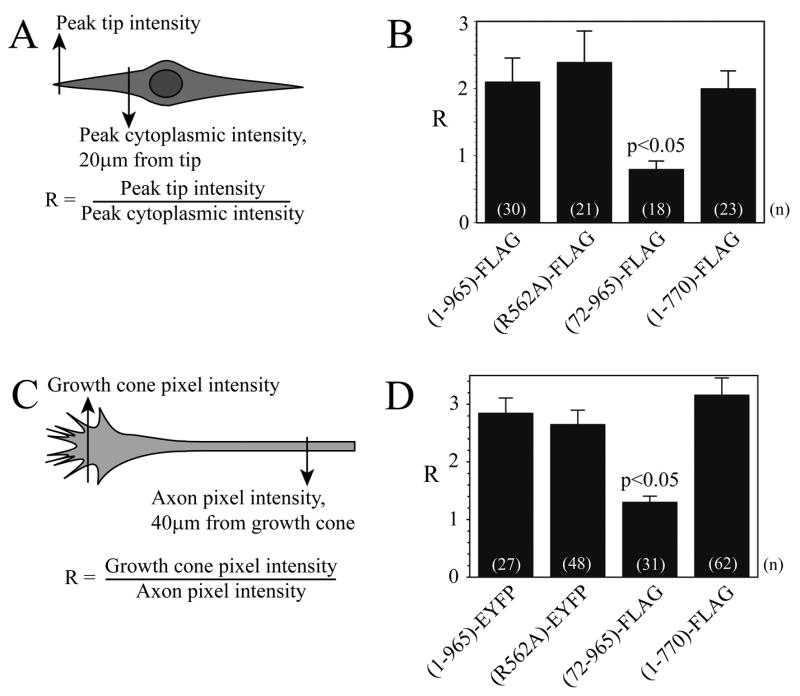
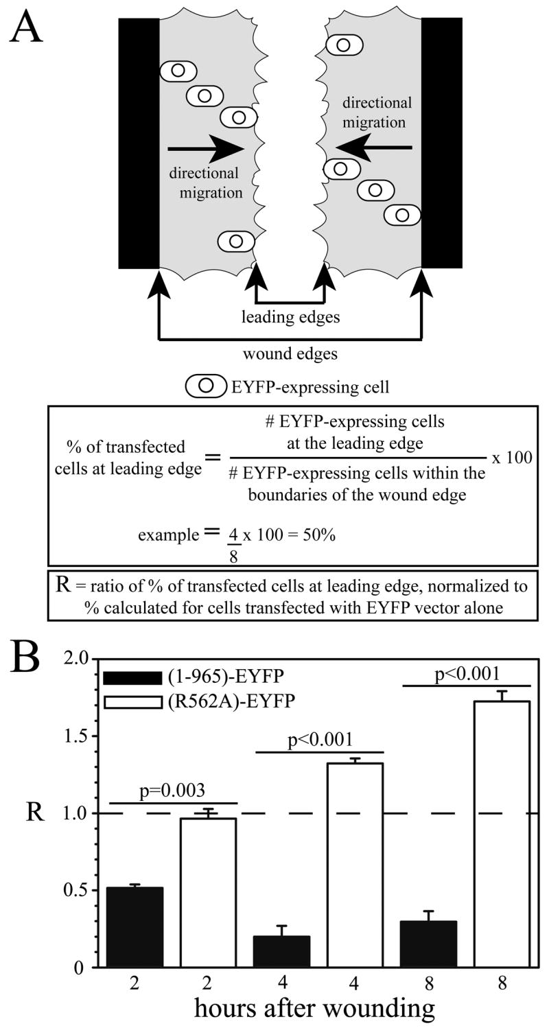
This report examines the structure and function of ARHGAP4, a novel RhoGAP whose structural features make it ideally suited to regulate the cytoskeletal dynamics that control cell motility and axon outgrowth. Our studies show that ARHGAP4 inhibits the migration of NIH/3T3 cells and the outgrowth of hippocampal axons. ARHGAP4 contains an N-terminal FCH domain, a central GTPase activating (GAP) domain and a C-terminal SH3 domain. Our structure/function analyses show that the FCH domain appears to be important for spatially localizing ARHGAP4 to the leading edges of migrating NIH/3T3 cells and to axon growth cones. Our analyses also show that the GAP domain and C-terminus are necessary for ARHGAP4-mediated inhibition of cell and axon motility. These observations suggest that ARHGAP4 can act as a potent inhibitor of cell and axon motility when it is localized to the leading edge of motile cells and axons.


Figures

Figure 1
Figure 1. Structural domains of the ARHGAP4 protein
ARHGAP4 contains N-terminal FCH (Fps/Fes/Fer/CIP4 homology), coiled-coil (CC) and ARNEY domains. The GAP domain is centrally located and the C-terminal contains a SH3 (Src homology 3) domain.
Figure 2
Figure 2. Endogenous ARHGAP4 protein is present in the peripheral zone of NIH/3T3 cells and growth cones
NIH/3T3 cells (A–C) and growth cones from dissociated dentate granule cell cultures (D–F) were immunostained for endogenous ARHGAP4 using a rabbit antibody (Foletta et al, 2002) and a mouse monoclonal antibody against β-tubulin III to label axons and the central region of growth cones. Fluorescence was visualized using biotinylated secondary antibodies labeled with avidin-Alexa 594 (red) and avidin-Alexa 488 (green). Images were viewed using a Leica confocal laser scanning microscope. Yellow indicates regions of overlapping immunoreactivity. Panels A–C: NIH/3T3 cells, Panels D–F: MF growth cones. The leading edge of the NIH/3T3 cell and the distal tips of filopodia of dentate granule cells are indicated by arrows in these representative images (n =3). Calibration = 5μm.
Figure 3
Figure 3. The ARHGAP4 N-terminus is required for normal protein localization in NIH/3T3 cells and axon terminals
A,B) Localization of FLAG-tagged full-length ARHGAP4 protein was compared to FLAG-tagged N-terminal and C-terminal truncation mutants in NIH/3T3 cells transiently transfected using Lipofectamine 2000 (Invitrogen), as described in Methods. Anti-FLAG immunohistochemistry was used to label mutant proteins using a biotinylated secondary antibody and avidin-Alexa 488. A)Fluorescence intensity was analyzed in a semi-quantitative fashion using NIH Image software. The ratio (R) of fluorescence intensity at the tip of the leading edge to the intensity 20μm from the tip was calculated. B)Results of the analysis are shown as mean ± SEM, and ANOVA was performed as described in Methods. Supplemental Figure 1 shows Western analysis of cells transfected to express each protein using the M2 anti-FLAG antibody. C,D) Localization of ARHGAP4 in axons and growth cones. C) The distribution of full-length ARHGAP4 and of 3 mutant proteins was quantified by dividing the pixel intensity of the respective tags at the growth cone by the pixel intensity at the axon 40μm from the growth cone, similar to the method used for NIH/3T3 cells. B)Results are shown as mean ± SEM, and ANOVA was performed using SigmaStat 3.1 software.
Figure 4
Figure 4. Effects of actin and microtubule destabilizing drugs on the localization of full-length ARHGAP4 (1-965-FLAG)
Wound assay experiments were performed on NIH/3T3 cells that expressed full length FLAG-tagged ARHGAP4 (1-965-FLAG). Three hours after wounding, the cells were treated with vehicle alone (DMSO), nocodazole, or cytochalasin-D to disrupt MTs and actin filaments, respectively. Fluorescent images were merged with brightfield images (C,F,I) to show leading edge boundaries.
Figure 5
Figure 5. Effects of actin and microtubule destabilizing drugs on the localization of the C-terminal truncation mutant ARHGAP4 (1-770-FLAG)
Wound assay experiments were performed on NIH/3T3 cells that expressed C-terminal deletion mutant FLAG-tagged ARHGAP4 (1-770-FLAG). Three hours after wounding, the control cells were treated with vehicle alone (DMSO), nocodazole, or cytochalasin-D to disrupt MTs and actin filaments, respectively. Fluorescent images were merged with brightfield images (C,F,I) to show leading edge boundaries.
Figure 6
Figure 6. Effects of mutant ARHGAP4 protein expression on MF axon outgrowth
Dentate explants were transfected using ExGen 500 to express ARHGAP4 proteins as described in Methods. Representative images obtained using a SPOT CCD camera attached to a Nikon Optiphot-2 fluorescence microscope are shown. Explants were transfected with the FLAG parent vector as control (A1); full-length ARHGAP4 (1-965)-FLAG (A2); the GAP loss of function mutant (R562A)-FLAG (A3); the C-terminal truncation mutant (1-770)-FLAG (A4); or the N-terminal truncation mutant (72-965)-FLAG (A5). MF axons were visualized using an anti-β-tubulin III antibody and nuclei of granule cells seen at the edge of the explant were stained blue using DAPI. Mossy fiber axon outgrowth was analyzed as described in Methods. Results are shown as mean ± SEM, and ANOVA analysis was performed using SigmaStat 3.1 software (B). Astrocytes (immunopositive for GFAP) were visualized for outgrowth assays as described in Methods (not shown). Results of astrocyte outgrowth assays are shown as mean ± SEM, and ANOVA was performed as described in Methods (C).
Figure 7
Figure 7. Expression of the R562A GAP activity mutant in NIH/3T3 cells results in increased cell motility
To assess the importance of GAP activity on cell motility, the (R562A)-EYFP mutant protein was expressed in NIH/3T3 cells. The parent EYFP, full-length (1-965)-EYFP, or (R562A)-EYFP vectors were transfected into NIH 3T3 cells using Lipofectamine 2000. Twenty-four hours after transfection, a scratch/wound was made in a confluent area of the culture plate and migration of transfected cells was assessed 2, 4 and 8 hours after the wound. The migration of transfected cells was quantified by counting the number of EYFP-positive cells at the line that represents the leading edge of migrating cells in the wound (fastest cells), and dividing by the total number of EYFP-positive cells between the wound edges (Fig. 9A). All percentage values were normalized to control values (cells expressing EYFP alone) and the ratio for control cells is represented by the horizontal dashed line (R=1.0, panel B). When compared to control cells expressing EYFP vector alone, significantly fewer cells expressing the full-length protein (1-965)-EYFP are observed at the leading edge of migrating cells at 2, 4 and 8 hours (Fig. 9B, black bars), while significantly more cells are observed at the leading edge if they express the (R562A)-EYFP mutant at 4 and 8 hours (Fig. 9B, white bars) (p ≤ 0.001). Importantly, there is a dramatic difference between the migration of cells expressing the full-length versus the R562A mutant proteins (p values shown in Fig. 9B). These observations suggest that the GAP function of ARHGAP4 is extremely important for regulation of cell motility. ANOVA analysis was performed using SigmaStat 3.1 software; n=3–5.
Figure 8
Figure 8. Knockdown of endogenous ARHGAP4 increased motility of NIH/3T3 cells
NIH/3T3 cells were co-transfected with the indicated siRNAs and an EYFP expression vector to identify transfected cells as previously described (Kanai et al., 2004). A: Cell motility was analyzed as described in Figure 7, in cells transfected with Control siRNA or ARHGAP4 siRNA. Results are shown as mean ± SEM, and statistical analysis was performed using SigmaStat software; n = 3. B: Western analysis (IB) was performed on cell lysates. Densitometric analysis of immunoreactive ARHGAP4 and actin bands was used to normalize endogenous ARHGAP4 expression, which corresponded to a 40% reduction in response to ARHGAP4 siRNA.

References

    1. Aspenstrom P. A Cdc42 target protein with homology to the non-kinase domain of FER has a potential role in regulating the actin cytoskeleton. Curr Biol. 1997;7:479–487. - PubMed
    1. Brouns MR, Matheson SF, Settleman J. p190 RhoGAP is the principal Src substrate in brain and regulates axon outgrowth, guidance and fasciculation. Nat Cell Biol. 2001;3:361–367. - PubMed
    1. Butler CD, Schnetz SA, Yu EY, Davis JB, Temple K, Silver J, Malouf AT. Keratan sulfate proteoglycan phosphacan regulates mossy fiber outgrowth and regeneration. J Neurosci. 2004;24:462–473. - PMC - PubMed
    1. Chen X, Dai JC, Orellana SA, Greenfield EM. Endogenous protein kinase inhibitor gamma terminates immediate-early gene expression induced by cAMP-dependent protein kinase (PKA) signaling: termination depends on PKA inactivation rather than PKA export from the nucleus. J Biol Chem. 2005;280:2700–2707. - PubMed
    1. Dent EW, Gertler FB. Cytoskeletal dynamics and transport in growth cone motility and axon guidance. Neuron. 2003;40:209–227. - PubMed

Publication types

MeSH terms