

doi: 10.1128/JVI.01083-07.
Epub 2007 Sep 5.
Human papillomavirus E6 regulates the cytoskeleton dynamics of keratinocytes through targeted degradation of p53
Affiliations
- PMID: 17804489
- PMCID: PMC2168984
- DOI: 10.1128/JVI.01083-07
Item in Clipboard
Human papillomavirus E6 regulates the cytoskeleton dynamics of keratinocytes through targeted degradation of p53
J Virol.
2007 Nov.
Abstract
The attachment and spreading of keratinocyte cells result from interactions between integrins and immobilized extracellular matrix molecules. Human papillomavirus type 16 (HPV-16) E6 augmented the kinetics of cell spreading, while E6 genes from HPV-11 or bovine papillomavirus type 1 did not. The ability of E6 to interact with the E6AP ubiquitin ligase and target p53 degradation was required to augment cell-spreading kinetics; dominant negative p53 alleles also enhanced the kinetics of cell spreading and the level of attachment of cells to hydrophobic surfaces. The targeted degradation of p53 by E6 may contribute to the invasive phenotype exhibited by cervical cells that contain high-risk HPV types.
Figures

HPV-16 E6 enhances early cell spreading after attachment of the cells to matrix-coated surfaces. (A) Cell spreading is altered by cancer-associated HPV E6. Only HPV-16 E6 enhanced early cell spreading after attachment. The values shown are the averages of the results of three assays described in the text. (B) 16E6 enhances early cell spreading. The cells shown were fixed and stained with formalin as described in the text. (C) Quantitation of the enhancement of cell spreading by 16E6. The total cell surface area was quantitated and compared by Image analysis software (NIH Image) 20 min after plating. Student's t test was used to determine the P value for a 300-cell population of each cell type. The error bars indicate the standard errors of the means.
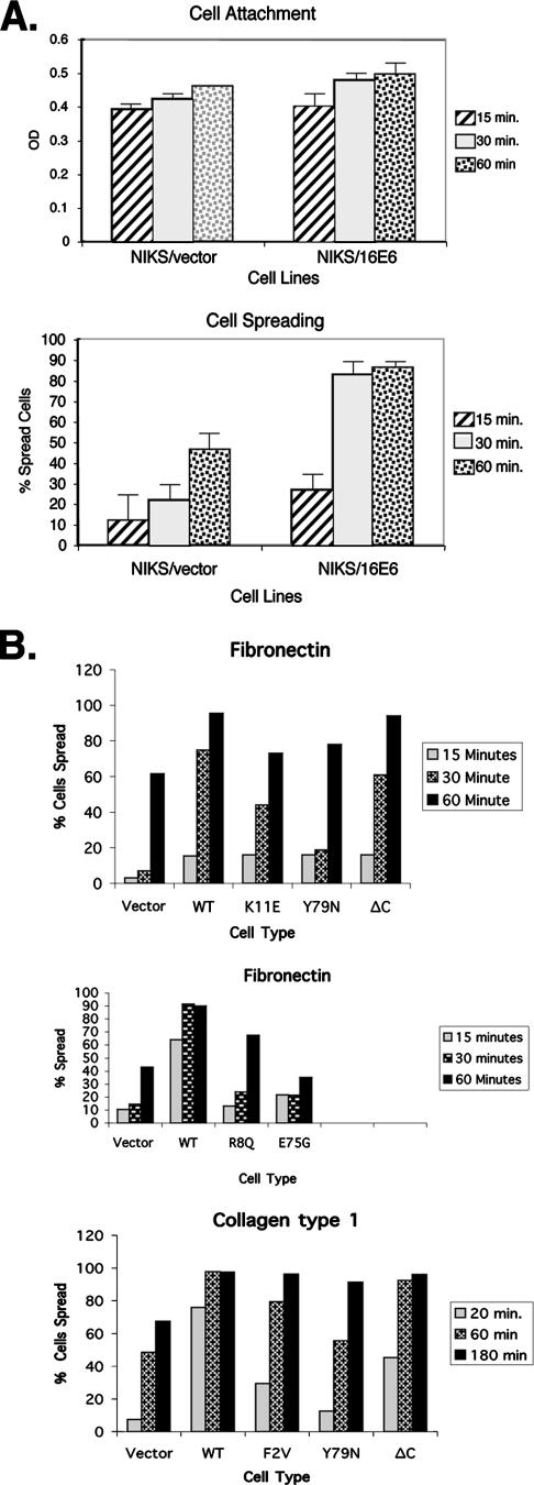
The effects of matrix and E6 mutations on cell spreading. (A) 16E6 augments cell spreading but not cell attachment. NIKS cells transduced with either empty retroviral vectors or 16E6 were assayed to determine the levels of cell attachment (top panel) and cell spreading (bottom panel) 15, 30, or 60 min after being plated on fibronectin-coated plates (10 μg/ml) as previously described (28). OD, optical density. (B) Enhanced spreading is independent of the matrix type and requires amino-terminal sequences of 16E6. NIKS cells transduced with empty retroviral vectors, 16E6 (shown as WT), or the indicated 16E6 mutants were seeded on plastic plates coated with either collagen type 1 or fibronectin for 20, 60, or 180 min. At least 300 cells were counted at each time point in three representative experiments.
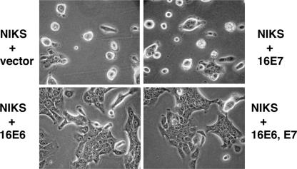
16E6 augments cell spreading on hydrophobic surfaces. (A) Spread of NIKS cells on bacterial plates 24 h after plating. NIKS cells were stably transduced with vectors or the indicated oncogenes, seeded on bacterial plates, and photographed 24 h later.

p53 regulates spreading on hydrophobic surfaces. (A) NIKS cells transduced with the indicated 16E6 genes or a dominant negative p53 gene (p53 DD) (25) were photographed 48 h after being plated on bacterial plates. (B) Expression levels of 16E6 and 16E6 mutants in NIKS cells. NIKS cells transduced with retrovirus vectors and lysed in sodium dodecyl sulfate were analyzed by Western blotting, using monoclonal antibodies (MAb) to 16E6 (11). Monoclonal antibody 3F8 recognizes an epitope common to 16E6 and all the mutants, while monoclonal antibody 6F4 recognizes the amino terminus of 16E6; this epitope is disrupted by the 16E6_F2V mutation.
Similar articles
-
Multiple regions of E6AP (UBE3A) contribute to interaction with papillomavirus E6 proteins and the activation of ubiquitin ligase activity.PLoS Pathog. 2020 Jan 23;16(1):e1008295. doi: 10.1371/journal.ppat.1008295. eCollection 2020 Jan. PLoS Pathog. 2020. PMID: 31971989 Free PMC article.
-
Structure of the E6/E6AP/p53 complex required for HPV-mediated degradation of p53.Nature. 2016 Jan 28;529(7587):541-5. doi: 10.1038/nature16481. Epub 2016 Jan 20. Nature. 2016. PMID: 26789255 Free PMC article.
-
Regulation of human papillomavirus E6 oncoprotein function via a novel ubiquitin ligase FBXO4.mBio. 2025 Feb 5;16(2):e0278324. doi: 10.1128/mbio.02783-24. Epub 2024 Dec 17. mBio. 2025. PMID: 39688415 Free PMC article.
-
The role of TP53 in Cervical carcinogenesis.Hum Mutat. 2003 Mar;21(3):307-12. doi: 10.1002/humu.10178. Hum Mutat. 2003. PMID: 12619117 Review.
-
HPV E6, E6AP and cervical cancer.BMC Biochem. 2008 Oct 21;9 Suppl 1(Suppl 1):S4. doi: 10.1186/1471-2091-9-S1-S4. BMC Biochem. 2008. PMID: 19007434 Free PMC article. Review.
Cited by
-
Human papillomavirus type 16 E6 induces cell competition.PLoS Pathog. 2022 Mar 23;18(3):e1010431. doi: 10.1371/journal.ppat.1010431. eCollection 2022 Mar. PLoS Pathog. 2022. PMID: 35320322 Free PMC article.
-
Cellular binding partners of the human papillomavirus E6 protein.Arch Virol. 2008;153(3):397-408. doi: 10.1007/s00705-007-0022-5. Epub 2008 Jan 3. Arch Virol. 2008. PMID: 18172569 Free PMC article. Review.
-
Human papillomavirus E6 and E7 oncoproteins as risk factors for tumorigenesis.J Biosci. 2009 Mar;34(1):113-23. doi: 10.1007/s12038-009-0013-7. J Biosci. 2009. PMID: 19430123 Review.
-
Papillomavirus E6 PDZ interactions can be replaced by repression of p53 to promote episomal human papillomavirus genome maintenance.J Virol. 2014 Mar;88(5):3027-30. doi: 10.1128/JVI.02360-13. Epub 2013 Dec 18. J Virol. 2014. PMID: 24352452 Free PMC article.
-
Expression of E6, p53 and p21 proteins and physical state of HPV16 in cervical cytologies with and without low grade lesions.Int J Clin Exp Med. 2014 Jan 15;7(1):186-93. eCollection 2014. Int J Clin Exp Med. 2014. PMID: 24482706 Free PMC article.
References
-
- Allen-Hoffmann, B. L., S. J. Schlosser, C. A. Ivarie, C. A. Sattler, L. F. Meisner, and S. L. O'Connor. 2000. Normal growth and differentiation in a spontaneously immortalized near-diploid human keratinocyte cell line, NIKS. J. Investig. Dermatol. 114:444-455. - PubMed
-
- Cooper, B., S. Schneider, J. Bohl, Y. Jiang, A. Beaudet, and S. Vande Pol. 2003. Requirement of E6AP and the features of human papillomavirus E6 necessary to support degradation of p53. Virology 306:87-99. - PubMed
-
- Grm, H. S., and L. Banks. 2004. Degradation of hDlg and MAGIs by human papillomavirus E6 is E6-AP-independent. J. Gen. Virol. 85:2815-2819. - PubMed
Publication types
MeSH terms
Substances
Grants and funding
LinkOut - more resources
Full Text Sources
Research Materials
Miscellaneous
