. 2007 Nov;81(22):12472-84.
doi: 10.1128/JVI.01123-07. Epub 2007 Sep 5.

Characterization of the intracellular deproteinized relaxed circular DNA of hepatitis B virus: an intermediate of covalently closed circular DNA formation

Affiliations

Characterization of the intracellular deproteinized relaxed circular DNA of hepatitis B virus: an intermediate of covalently closed circular DNA formation

Haitao Guo et al. J Virol. 2007 Nov.

Abstract

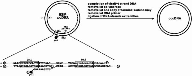
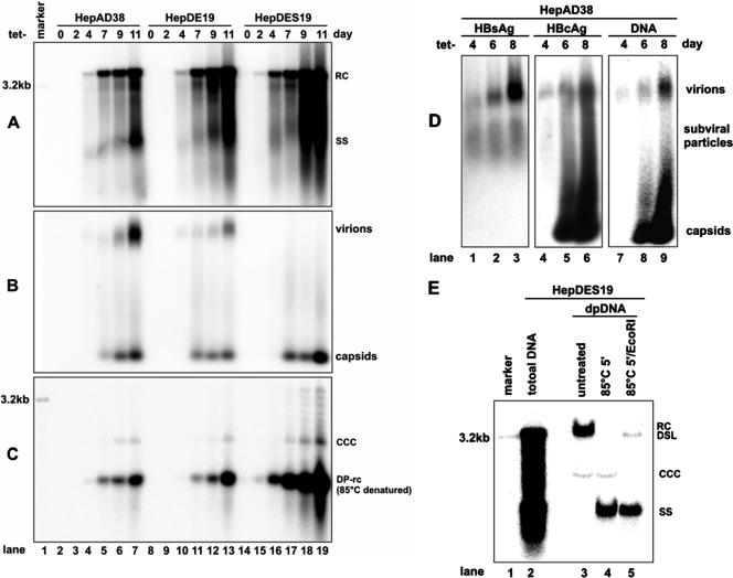
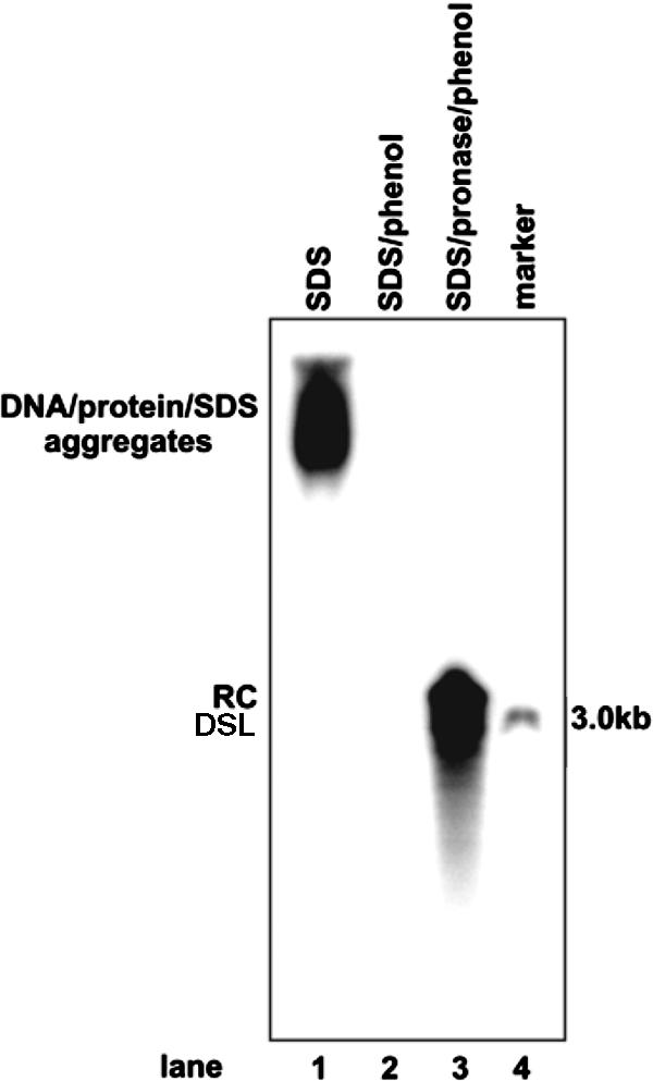
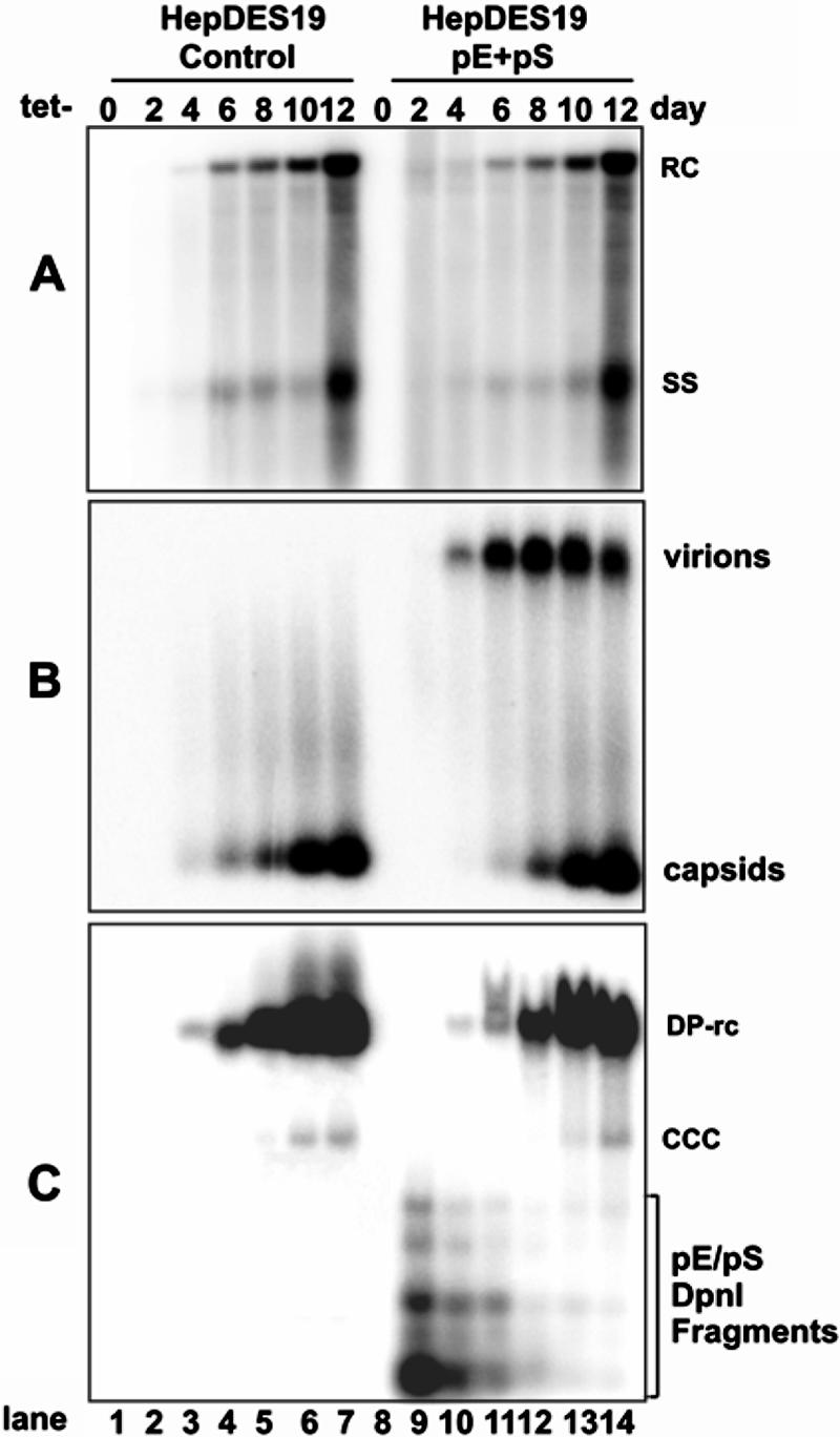
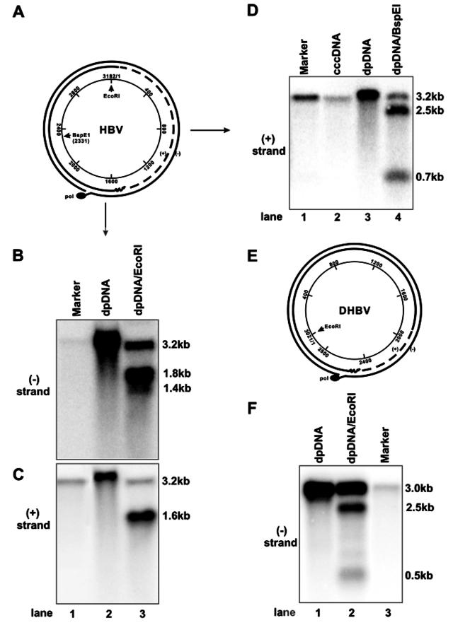
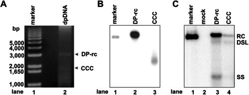
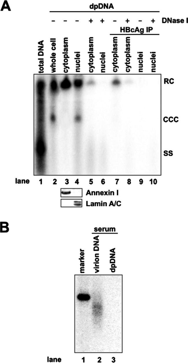
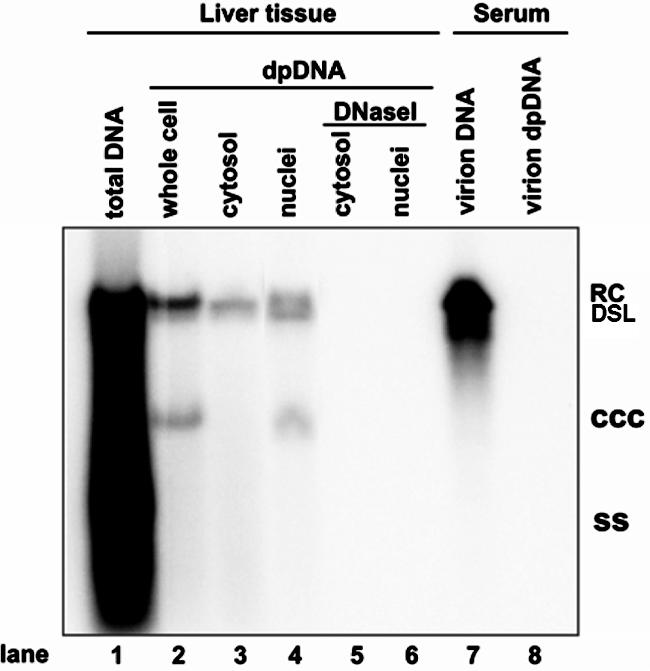
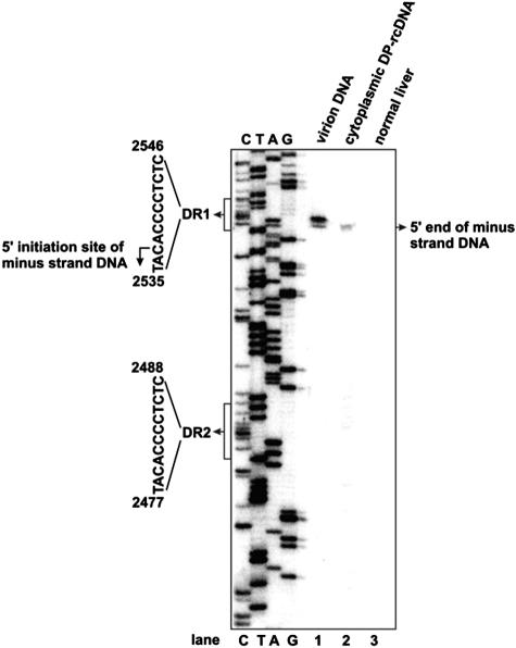
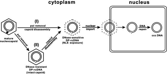
Covalently closed circular DNA (cccDNA) of hepatitis B virus (HBV) is formed by conversion of capsid-associated relaxed circular DNA (rcDNA) via unknown mechanisms and exists in the nucleus of the infected hepatocyte as a minichromosome that serves as the transcription template for viral RNAs. To study the molecular pathway of cccDNA formation and its regulation by viral and cellular factors, we have established a cell line that supports the replication of an envelope protein-deficient HBV genome in a tetracycline-inducible manner. Following induction of HBV replication, the cells accumulate higher levels of cccDNA as well as larger amounts of deproteinized rcDNA (DP-rcDNA) than cells that replicate wild-type HBV genomes. These results indicate that HBV envelope proteins negatively regulate cccDNA formation, and conversion of DP-rcDNA into cccDNA is a rate-limiting step of cccDNA formation in HepG2 cells. Detailed analyses reveal the following: (i) DP-rcDNA exists in both cytoplasm and nucleus; (ii) while nuclear DP-rcDNA is sensitive to DNase I digestion, a small fraction of cytoplasmic DP-rcDNA is DNase I resistant; (iii) both DNase I-sensitive and -resistant cytoplasmic DP-rcDNAs cosediment with capsids and can be immunoprecipitated with HBV core antibody; and (iv) a primer extension assay maps the 5' end of the minus strand of DP-rcDNA at the authentic end of virion rcDNA. Hence, our results favor a hypothesis that the removal of viral polymerase protein covalently linked to the 5' end of the minus-strand DNA occurs inside the capsid in the cytoplasm and most possibly via a reaction that cleaves the phosphodiester bond between the tyrosine of the polymerase and the 5' phosphoryl group of minus-strand DNA.


Figures

FIG. 1.
FIG. 1.
Schematic illustration of rcDNA structural features and requirement for the conversion of rcDNA into cccDNA. Incompletely synthesized plus-strand DNA is indicated with dashed line. RNA primer for plus-strand DNA synthesis is shown as curved line, and sequence is shown in lowercase letters. Polymerase (pol) covalently linked to the 5′ end of minus-strand rcDNA is highlighted with a filled circle. Direct repeat (DR) sequences 1 and 2 are indicated.
FIG. 2.
FIG. 2.
Kinetics of HBV DNA synthesis, cccDNA formation, and virion secretion in HepAD38, HepDE19, and HepDES19 cell lines upon induction. Cells were seeded in 35-mm dishes and cultured in the presence of tetracycline (1 μg/ml) until cells became confluent; cells continued to be cultured in tetracycline-free medium for 11 days. Cells and culture fluids were harvested at the indicated time points before and after tetracycline removal. Cytoplasmic core DNA (A), secreted viral particle-associated DNA (B), and Hirt DNA (C) were analyzed by Southern blot hybridization and particle gel assay with [α-32P]UTP-labeled minus-strand-specific HBV riboprobe. The Hirt DNA samples were denatured at 85°C for 5 min before loading to improve the detection sensitivity of cccDNA. As a consequence, the protein-free rcDNA (labeled DP-rcDNA) migrates at the position of the single-strand DNA. (D) Culture medium was harvested from HepAD38 cells 4, 6, and 8 days after removal of tetracycline, and viral particles were pelleted with PEG 8000 and separated in a 1% agarose gel and transferred onto nitrocellulose membrane as described in Materials and Methods. The membrane was probed sequentially with anti-HBs (left panel) and anti-HBc (middle panel) antibodies and hybridized with minus-strand-specific full-length HBV riboprobe (right panel). (E) Hirt DNA preparations made from HepDES19 cells cultured in the absence of tetracycline for 8 days were separated in a 1.5% agarose gel without treatment (lane 3), after denaturalization at 85°C for 5 min (lane 4), or digestion with EcoRI after denaturalization at 85°C for 5 min (lane 5). Core DNA prepared from the same culture (lane 2) served as a control. Unit-length linear HBV DNA (50 pg; lane marker) served as a hybridization standard for estimating the copy number of viral DNA. RC, rcDNA; ccc, cccDNA; SS, single-strand DNA; DSL, double-strand linear DNA.
FIG. 3.
FIG. 3.
Hirt DNA extraction procedure extracts only protein-free viral DNA. DHBV viral particles were purified from 1 ml of DHBV-positive duck serum by centrifugation through a 10 to 20% sucrose cushion and lysed in a 60-μl solution containing 0.1% SDS, 10 mM Tris-HCl (pH 7.5), and 10 mM EDTA at 37°C for 1 h. Viral DNA was extracted from the lysates by direct phenol extraction (Hirt procedure; lane 2) or by phenol extraction after digestion with 1 mg/ml pronase at 37°C for 1 h (core DNA procedure; lane 3). The lysate of viral particles (lane 1) and purified viral DNA were resolved on a 1.2% agarose gel. Viral DNA was detected by Southern blot hybridization with [α-32P]UTP-labeled full-length minus-strand-specific DHBV riboprobe. Fifty picograms of unit-length DHBV DNA served as a hybridization standard (lane 4). DSL, double-strand linear DNA.
FIG. 4.
FIG. 4.
HBV envelope proteins regulate viral cccDNA accumulation in cell culture. HepDES19 cells were seeded in 35-mm dishes at a density of 1 × 10⁶. Six hours later, cells were transfected with a total of 4 μg of plasmid pE (2 μg) and pS (2 μg) or 4 μg of control vector plasmid pUC19. After transfection, cells were cultured in the absence of tetracycline (Tet−). Cells and culture medium were harvested at the indicated time points. Intracellular HBV core DNA (A), viral particle-associated DNA (B), and protein-free viral DNA (C) were subjected to analysis as described in the legend of Fig. 2. Protein-free DNA samples were treated with DpnI prior to loading to digest input plasmid DNA. Fragments from the DpnI-digested plasmids are indicated. RC, rcDNA; CCC, cccDNA; SS, single-strand DNA.
FIG. 5.
FIG. 5.
Protein-free HBV and DHBV rcDNA species are not derived from nicked cccDNA. (A and E) Schematic representation of the HBV and DHBV rcDNA structures, respectively. EcoRI and BspEI restriction sites are indicated. (B, C, and D) Hirt DNA preparations from HepDES19 cells cultured in the absence of tetracycline for 10 days were left untreated (B and C, lane 2; D, lane 3) or digested with EcoRI (B and C, lane 3) or BspEI (D, lane 4) and then resolved in a 1.5% alkaline agarose gel. Purified HBV cccDNA (D, lane 2) and unit-length linear HBV DNA (lane 1) served as controls. DNAs were transferred onto nylon membrane and hybridized with [α-32P]UTP-labeled full-length minus-strand-specific (B) or plus-strand-specific (C and D) HBV riboprobe. (F) Hirt DNA preparations from dstet5 cells cultured in the absence of tetracycline for 7 days were left untreated (lane 1) or digested with EcoRI (lane 2) and then resolved in a 1.5% alkaline agarose gel. Unit-length linear DHBV DNA served as a hybridization standard (lane 3). DNAs were transferred onto nylon membrane and hybridized with [α-32P]UTP-labeled full-length minus-strand-specific DHBV riboprobe.
FIG. 6.
FIG. 6.
Transfection of DHBV DP-rcDNA into LMH cells initiates viral DNA replication. (A) Hirt DNA preparations from dstet5 cells cultured in the absence of tetracycline for 7 days were resolved in a 1.5% agarose gel, and DHBV cccDNA and DP-rcDNA were visualized by ethidium bromide staining. (B) Recovered HBV cccDNA and DP-rcDNA were resolved on agarose gel and detected by Southern blot hybridization. (C) LMH cells were transfected with approximately 5 ng of gel-purified DHBV cccDNA and DP-rcDNA mixed with 2 μg of plasmid pUC19 or with 2 μg of pUC19 alone (mock) by using Lipofectamine 2000. Cells were harvested at day 7 posttransfection, and capsid-associated viral DNA was extracted and determined by Southern blot hybridization with [α-32P]UTP-labeled full-length minus-strand-specific DHBV riboprobe. Fifty picograms of unit-length DHBV DNA served as a hybridization standard. RC, rcDNA; CCC, cccDNA; SS, single-strand DNA; DP-rc, DP-rcDNA; DSL, double-strand linear DNA.
FIG. 7.
FIG. 7.
Subcellular distribution of HBV DP-rcDNA in HepDES19 cells. (A) HepDES19 cells were cultured in the absence of tetracycline for 10 days. Intracellular HBV capsid DNA (lane 1) and Hirt DNA (lanes 2 to 10) extracted from the lysates of whole cell, cytoplasm, and nuclei of HepDES19 cells with or without prior DNase I digestions were analyzed by Southern blot hybridization. Lane 1 was loaded with one-quarter of the capsid DNA extracted from a 35-mm dish. Lane 2 was loaded with one-half of total Hirt DNA from a 35-mm dish. Lanes 3 to 10 were each loaded with Hirt DNA prepared from the cytoplasmic or nuclear fraction from one 35-mm dish with the treatments described below. Hirt DNAs were extracted from both fractions without (lanes 3 and 4) or with (lanes 5 and 6) prior DNase I digestion or extracted after immunoprecipitation (IP) with a polyclonal antibody against HBV capsid (lanes 7 and 9) and digestion with DNase I (lanes 8 and 10). Total proteins of each fraction were resolved by SDS-polyacrylamide gel electrophoresis, transferred on polyvinylidene difluoride membrane, and probed with antibodies against annexin I and lamin A/C, respectively. Bound antibodies were revealed by HRP-labeled secondary antibodies and visualized with an enhanced chemiluminescence detection system (lower panel). (B) HBV DNAs extracted from 250 μl of HBV-positive human serum with (lane 2) or without (lane 3) prior protease digestion were resolved in a 1.5% agarose gel, transferred onto membrane, and hybridized with an HBV minus-strand DNA-specific riboprobe. Fifty picograms of unit-length HBV DNA served as a hybridization standard (lane 1). RC, rcDNA; CCC, cccDNA; SS, single-strand DNA.
FIG. 8.
FIG. 8.
Subcellular distribution of DHBV DP-rcDNA in the liver of virally infected duck. Total intracellular capsid DNA (lane 1), Hirt DNA extracted from total lysate (lane 2), or cytoplasmic and nuclear fractions of a DHBV-positive duck liver without (lanes 3 and 4) or with (lanes 5 and 6) prior DNase I digestion and DHBV DNA extracted from 250 μl of DHBV-positive duck serum with (lane 7) or without (lane 8) prior pronase digestion were resolved in a 1.5% agarose gel, transferred onto membrane, and hybridized with a DHBV minus-strand DNA-specific riboprobe. Total DNA loaded in lane 1 was prepared from 7 mg of duck liver tissue, and each Hirt DNA sample was prepared from lysate prepared from 15 mg of liver tissue. RC, rcDNA; CCC, cccDNA; SS, single-strand DNA; DSL, double-strand linear DNA.
FIG. 9.
FIG. 9.
HBV cytoplasmic DP DNA cosediments with nucleocapsids. Three milliliters of cytoplasmic fraction prepared from 2 × 10⁷ HepDES19 cells cultured in the absence of tetracycline for 10 days was overlaid on 33 ml of a 10 to 60% (wt/vol) sucrose gradient and centrifuged at 24,000 rpm for 16 h at 4°C using a Beckman SW28 rotor. Seventeen 2.25-ml fractions were collected from the bottom. HBV core antigen (A), total HBV DNA (B), and capsid DNA (C) in each fraction were assayed as described in Material and Methods. DP-rcDNA was extracted from each fraction without (D) or with (E) prior DNase I digestion. For the immunoprecipitation (IP) assay, each faction was mixed with Sepharose 4B-protein A beads preabsorbed with rabbit anti-HBc and incubated at 4°C overnight. Beads were washed three times with TNE buffer, and protein-free DNA was extracted without (F) or with (G) prior DNase I digestion. The Hirt DNAs were resolved in a 1.2% agarose gel and detected by Southern blot hybridization. RC, rcDNA; SS, single-strand DNA; EIA, enzyme immunoassay.
FIG. 10.
FIG. 10.
Mapping the 5′ end of the minus strand of DHBV DP-rcDNA. Virion DNA prepared from DHBV-positive duck serum (lane 1) and Hirt DNA prepared from the cytoplasmic fraction of DHBV-infected (lane 2) and normal (lane 3) duck liver tissues were annealed with a 5′ end-labeled oligonucleotide coordinating with nt 2375 to 2396 in the DHBV genome. The primer extension reaction was carried out as described in Materials and Methods. Products were resolved with a 6% acrylamide-8 M urea gel alongside a sequencing ladder (lanes C, T, A, and G) and visualized by a PhosphorImager (Bio-Rad).
FIG. 11.
FIG. 11.
Proposed model for the molecular pathway of hepadnavirus cccDNA formation. See text for a detailed explanation. pol, polymerase; NLS, nuclear localization signal.

References

    1. Barrasa, M. I., J. T. Guo, J. Saputelli, W. S. Mason, and C. Seeger. 2001. Does a cdc2 kinase-like recognition motif on the core protein of hepadnaviruses regulate assembly and disintegration of capsids? J. Virol. 75:2024-2028. - PMC - PubMed
    1. Barthelmes, H. U., M. Habermeyer, M. O. Christensen, C. Mielke, H. Interthal, J. J. Pouliot, F. Boege, and D. Marko. 2004. TDP1 overexpression in human cells counteracts DNA damage mediated by topoisomerases I and II. J. Biol. Chem. 279:55618-55625. - PubMed
    1. Beck, J., and M. Nassal. 2007. Hepatitis B virus replication. World J. Gastroenterol. 13:48-64. - PMC - PubMed
    1. Connelly, J. C., and D. R. Leach. 2004. Repair of DNA covalently linked to protein. Mol. Cell 13:307-316. - PubMed
    1. Dixon, K., and E. Kopras. 2004. Genetic alterations and DNA repair in human carcinogenesis. Semin. Cancer Biol. 14:441-448. - PubMed

Publication types