. 2007 Nov;81(22):12135-44.
doi: 10.1128/JVI.01296-07. Epub 2007 Sep 5.

High fidelity of murine hepatitis virus replication is decreased in nsp14 exoribonuclease mutants

Affiliations

High fidelity of murine hepatitis virus replication is decreased in nsp14 exoribonuclease mutants

Lance D Eckerle et al. J Virol. 2007 Nov.

Abstract

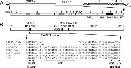
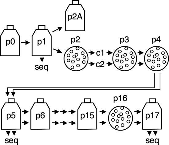
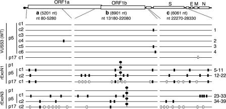
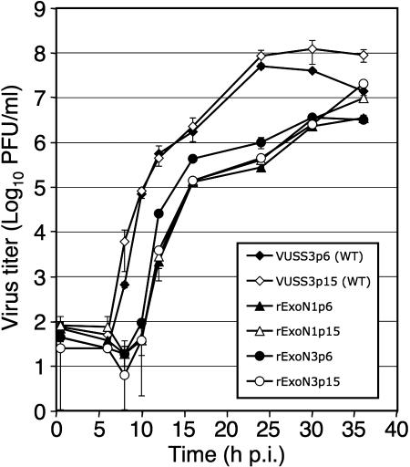
Replication fidelity of RNA virus genomes is constrained by the opposing necessities of generating sufficient diversity for adaptation and maintaining genetic stability, but it is unclear how the largest viral RNA genomes have evolved and are maintained under these constraints. A coronavirus (CoV) nonstructural protein, nsp14, contains conserved active-site motifs of cellular exonucleases, including DNA proofreading enzymes, and the severe acute respiratory syndrome CoV (SARS-CoV) nsp14 has 3'-to-5' exoribonuclease (ExoN) activity in vitro. Here, we show that nsp14 ExoN remarkably increases replication fidelity of the CoV murine hepatitis virus (MHV). Replacement of conserved MHV ExoN active-site residues with alanines resulted in viable mutant viruses with growth and RNA synthesis defects that during passage accumulated 15-fold more mutations than wild-type virus without changes in growth fitness. The estimated mutation rate for ExoN mutants was similar to that reported for other RNA viruses, whereas that of wild-type MHV was less than the established rates for RNA viruses in general, suggesting that CoVs with intact ExoN replicate with unusually high fidelity. Our results indicate that nsp14 ExoN plays a critical role in prevention or repair of nucleotide incorporation errors during genome replication. The established mutants are unique tools to test the hypothesis that high replication fidelity is required for the evolution and stability of large RNA genomes.


Figures

FIG. 1.
FIG. 1.
MHV genome organization and nsp14 exoribonuclease motifs. (A) MHV genome organization and ORF1a/b replicase polyprotein expression. The MHV genome is a 31.3-kb positive-sense RNA molecule that is capped (dark circle) and polyadenylated. The genes are indicated for the replicase (ORF1a and ORF1b), spike (S), envelope (E), membrane (M), and nucleocapsid (N) proteins. ORF1b is accessed by ribosomal frameshift. The ORF1a/b polyprotein is translated from input genome RNA and processed into 16 mature nsps by three virus-encoded proteinases (gray). nsps 12 to 16 have predicted or demonstrated activities as described in the text. Hel, helicase; Endo, endoribonuclease; MT, methyltransferase; (A)n, polyadenylate tract. (B) Organization of nsp14 and partial sequence alignment of representative CoV nsp14 sequences with the sequence of Saccharomyces cerevisiae PAN2 (SwissProt accession number P53010), a poly(A)-specific ExoN, and Escherichia coli DNA polymerase III epsilon subunit (DP3E), the proofreading exonuclease subunit of the replicative DNA polymerase (SwissProt P03007). GenBank accession numbers for the full-length CoV genomes are as follows: MHV-A59, AY910861; SARS-CoV (Urbani strain), AY278741; HCoV-229E, NC_002645; transmissible gastroenteritis virus (TGEV), NC_002306; infectious bronchitis virus (IBV)-Beaudette, NC_001451. Sequence alignment was adapted from Snijder et al. (34). Active-site residues of conserved motifs I to III of the DEDD superfamily (45) are indicated by bold text and amino acid position in MHV nsp14. A predicted zinc finger domain (ZnF) is located between motifs I and II in the viral sequences. Residues replaced with alanine are indicated by black circles for MHV mutants rExoN1 (ExoN motif I) and rExoN3 (ExoN motif III) (see Table 1 for details).
FIG. 2.
FIG. 2.
Replication and plaque morphology of rExoN1 and rExoN3 mutants. (A) Multicycle growth analysis of ExoN mutants. DBT-9 cells were infected with the indicated viruses at an MOI of 0.01 PFU/cell. Viruses are identified in the text. Supernatant samples were obtained at 0.5, 6, 8, 10, 12, 16, 24, 30, 36, 42, and 48 hpi, and virus titers were determined by plaque assay. Mean titers and standard deviations from duplicate samples are indicated. (B) Single-cycle growth analysis of rExoN1 mutants. DBT-9 cells were infected with the indicated viruses at an MOI of 3. Supernatant samples were obtained at 0.5, 4, 6, 8, 10, 12, 16, and 24 hpi, and virus titers were determined by plaque assay. Mean titers and standard deviations from duplicate samples are indicated. (C) Plaque morphology. p0 and p15 stocks of the indicated viruses were diluted and used to infect DBT-9 cells for 24 to 28 h, followed by fixation with 4% formaldehyde. Representative wells were scanned and images were prepared using Adobe Photoshop CS2.
FIG. 3.
FIG. 3.
Virus isolation and passage strategy. Flasks indicate population passage, and circles indicate plaque isolation. From p2 forward, two plaque clones were isolated and passed in parallel, indicated by double arrows. Passages where RNA from infected cells was purified and used for RT-PCR and determination of viral sequences are denoted by seq. Passages between p0 and p6 were conducted at an MOI of ≤0.1 PFU/cell. Passages between p6 and 15 were performed at unknown MOIs.
FIG. 4.
FIG. 4.
RNA synthesis from rExoN1 and rExoN3 mutant viruses. (A) Kinetics of viral RNA synthesis during multicycle growth. DBT-9 cells were mock infected or infected with the indicated viruses at an MOI of 0.1 PFU/cell. Viral RNA was metabolically labeled with [3H]uridine in the presence of ActD at 4-h intervals that began at 4, 8, 12, 16, 20, 24, 28, 32, 36, and 40 hpi. Total RNA was precipitated with 5% TCA, and incorporation of radiolabel was quantitated by scintillation counting. Mean values and standard deviations from duplicate samples obtained from duplicate infection series are indicated. (B) Kinetics of viral RNA synthesis during single-cycle growth. DBT-9 cells were mock infected or infected with the indicated viruses at an MOI of 3 PFU/cell. Viral RNA was metabolically labeled with [3H]uridine in the presence of ActD at 2-h intervals that began at 3, 5, 7, 9, 11, 13, and 15 hpi. (C) Electrophoretic analysis of RNA replication and transcription products. DBT-9 cells were mock infected (m) or infected with the indicated viruses at an MOI of 3 PFU/cell. Viral RNA was metabolically labeled with [3H]uridine in the presence of ActD from 11 to 13 hpi. Intracellular RNA was isolated, denatured, and resolved by electrophoresis in 1% agarose gels. Labeled RNA species were visualized by fluorography. Genomic RNA (R1) and sg mRNAs (R2 to R7) are indicated. The apparent band migrating between RNA3 and RNA4 in lanes 1, 3, 4, and 5 is most likely due to a physical effect of abundant 28S rRNA. The asterisk indicates that lane 1 received the same RNA sample as lane 5 but fivefold less. Note that all samples were electrophoresed on the same gel, but the image was cropped to remove extraneous lanes between lanes 4 and 5. (D) RT-PCR of MHV RNA2 (top) and cellular β-actin mRNA (bottom). The same RNA samples analyzed in panel C were subjected to RT-PCR, and the products were resolved in 1.2% (top) and 1.5% (bottom) agarose gels in the presence of ethidium bromide. The VUSS3 RNA sample was diluted fourfold prior to reverse transcription. Sizes (in kbp) of bands from a 100-bp DNA ladder are indicated. The predicted sizes of the RNA2-specific and actin-specific amplicons are 437 and 348 bp, respectively. Abbreviations: m, mock; con, negative control PCR for β-actin using water as a template. Images in panels C and D were prepared using Adobe Photoshop CS2.
FIG. 5.
FIG. 5.
Distribution of mutations in WT, rExoN1, and rExoN3 mutants. Schematic of MHV RNA genome shows the regions a, b, and c that were amplified by RT-PCR and sequenced for each WT and ExoN mutant virus. The 5′ and 3′ terminal nucleotides of the regions are indicated, as is the length in nucleotides. Lollipops indicate locations of engineered ExoN mutations. Black rectangles on each genome indicate additional mutations identified in p5 viruses. White rectangles indicate mutations identified in p17 but not in p5 viruses. Numbers at right indicate mutations for which details are shown in Table 3. VUSS3p17c1, rExoN1p17c1, and rExoN3p17c2 were derived from VUSS3p5c1, rExoN1p5c1, and rExoN3p5c2, respectively. S, spike; E, envelope; M, membrane; N, nucleocapsid protein.
FIG. 6.
FIG. 6.
Replication of p6 and p15 viruses. DBT-9 cells were infected with the indicated viruses at an MOI of 0.05 PFU/cell. p6 and p15 viruses of VUSS3, rExoN1, and rExoN3 were derived from VUSS3p5c1, rExoN1p5c1, and rExoN3p5c2, respectively. Supernatant samples were obtained at 0.5, 6, 8, 10, 12, 16, 24, 30, and 36 hpi, and virus titers were determined by plaque assay. Mean titers and standard deviations from duplicate experiments are indicated.
FIG. 7.
FIG. 7.
Model for generation of intergenic region mutations in rExoN3p5c1. (A) Single template switch during normal synthesis of sg mRNA4. The WT sequences of a portion of the intergenic region (IGR) between the spike gene and ORF4 (first line) and leader regions (bold text, second line) of genome RNA (RNA1) are shown. The conserved consensus sequence in the RNA4 transcription-regulating sequence and at the 3′ end of leader is boxed. The line arrow indicates the replicase-transcriptase template switch from the intergenic region to the leader region to produce negative-sense (−) RNA4, which is then copied to yield positive-sense RNA4. Lowercase text represents negative-sense sequence. (B) Proposed double template switch to generate the intergenic region mutations in rExoN3p5c1. The three clustered nucleotide substitutions (circles) identified in the intergenic region between the spike gene and ORF4 in rExoN3p5c1 (Mut) could result from a single step in which the replicase-transcriptase switches from copying the intergenic region to copying 4 to 5 nt of leader and then back to copying the intergenic region.

Similar articles

Cited by

References

    1. Almazán, F., M. L. Dediego, C. Galán, D. Escors, E. Álvarez, J. Ortego, I. Sola, S. Zuñiga, S. Alonso, J. L. Moreno, A. Nogales, C. Capiscol, and L. Enjuanes. 2006. Construction of a severe acute respiratory syndrome coronavirus infectious cDNA clone and a replicon to study coronavirus RNA synthesis. J. Virol. 80:10900-10906. - PMC - PubMed
    1. Baric, R. S., B. Yount, L. Hensley, S. A. Peel, and W. Chen. 1997. Episodic evolution mediates interspecies transfer of a murine coronavirus. J. Virol. 71:1946-1955. - PMC - PubMed
    1. Bernad, A., L. Blanco, J. M. Lazaro, G. Martin, and M. Salas. 1989. A conserved 3′→5′ exonuclease active site in prokaryotic and eukaryotic DNA polymerases. Cell 59:219-228. - PubMed
    1. Bhardwaj, K., L. Guarino, and C. C. Kao. 2004. The severe acute respiratory syndrome coronavirus Nsp15 protein is an endoribonuclease that prefers manganese as a cofactor. J. Virol. 78:12218-12224. - PMC - PubMed
    1. Bull, J. J., R. Sanjuan, and C. O. Wilke. 2007. Theory of lethal mutagenesis for viruses. J. Virol. 81:2930-2939. - PMC - PubMed

Publication types