. 2007 Sep 5;27(36):9545-59.
doi: 10.1523/JNEUROSCI.1930-07.2007.

Skin-derived precursors generate myelinating Schwann cells that promote remyelination and functional recovery after contusion spinal cord injury

Affiliations

Skin-derived precursors generate myelinating Schwann cells that promote remyelination and functional recovery after contusion spinal cord injury

Jeff Biernaskie et al. J Neurosci. .

Abstract

Transplantation of exogenous cells is one approach to spinal cord repair that could potentially enhance the growth and myelination of endogenous axons. Here, we asked whether skin-derived precursors (SKPs), a neural crest-like precursor that can be isolated and expanded from mammalian skin, could be used to repair the injured rat spinal cord. To ask this question, we isolated and expanded genetically tagged murine SKPs and either transplanted them directly into the contused rat spinal cord or differentiated them into Schwann cells (SCs), and performed similar transplantations with the isolated, expanded SKP-derived SCs. Neuroanatomical analysis of these transplants 12 weeks after transplantation revealed that both cell types survived well within the injured spinal cord, reduced the size of the contusion cavity, myelinated endogenous host axons, and recruited endogenous SCs into the injured cord. However, SKP-derived SCs also provided a bridge across the lesion site, increased the size of the spared tissue rim, myelinated spared axons within the tissue rim, reduced reactive gliosis, and provided an environment that was highly conducive to axonal growth. Importantly, SKP-derived SCs provided enhanced locomotor recovery relative to both SKPs and forebrain subventricular zone neurospheres, and had no impact on mechanical or heat sensitivity thresholds. Thus, SKP-derived SCs provide an accessible, potentially autologous source of cells for transplantation into and treatment of the injured spinal cord.


Figures
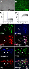
Figure 1.
Figure 1.
Characterization of SKP-derived SCs before transplantation. A, B, Photomicrographs of SKP-derived SCs generated from a mouse that expresses YFP in all cell types. Both panels show the same field photographed with phase illumination (A) and with fluorescence illumination (B). C, D, Flow cytometry with SKP-derived SCs for the genetic YFP tag (C; GFP) and for p75NTR (D; p75), a cell surface marker for SCs. Note that >97% of SKP-derived SCs express both of these proteins. E, Photomicrographs of SKP-derived SCs triple-labeled for the genetic YFP tag, and the SC markers S100β and p75NTR (p75). The bottom right panel is the merge. The arrows indicate a triple-labeled cell. Note that virtually all of the cells express all three proteins. F, Photomicrographs of SKP-derived SCs double-labeled for the genetic YFP tag, and the peripheral myelin protein P0. The top left panel is Hoechst nuclear staining to show all of the cells in the field, and the bottom right panel is the merge. The arrows indicate double-labeled cells.
Figure 2.
Figure 2.
Cavity size and cell survival in the contused rat spinal cord after transplantation of SKPs versus SKP-derived SCs. A–D, Fluorescence, low-magnification (10×) photomicrographs of longitudinal sections through the spinal cord of rats that were transplanted 11 weeks earlier with medium alone (A), YFP-expressing neonatal forebrain SVZ neurospheres (B), YFP-expressing neonatal SKPs (C), or YFP-expressing SKP-derived SCs (D). E–G, Note that only SKP-derived SCs formed bridges across the longitudinal plane of the cavity with good rostrocaudal integration. Quantification of sections similar to those shown in A–D to obtain the mean cavity size (E), the amount of spared tissue (F), and the transplant size (G). *p < 0.05 relative to medium alone (E, F) and/or to neurospheres (F, G). Note that naive SKPs significantly reduced the cavity size and demonstrated the largest transplant volumes, whereas SKP-derived SCs significantly spared the tissue rim. H, Number of surviving, YFP-positive cells 12 weeks after transplantation. Note that, of the three groups, SKPs had the highest survival, followed by SKP-derived SCs, both showing significantly greater survival than neurospheres. *p < 0.05. In E–H, results represent mean ± SEM.
Figure 3.
Figure 3.
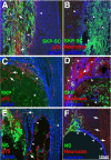
Transplanted SKPs and SKP-derived SCs modify the extracellular environment surrounding the lesion. A–F, Fluorescence photomicrographs of longitudinal sections through the spinal cord of rats that were transplanted 11 weeks earlier with neonatal YFP-expressing SKP-derived SCs (A, B), YFP-expressing SKPs (C, D), or YFP-expressing forebrain SVZ neurospheres (NS) (E, F), showing both the transplant and the adjacent tissue rim. A, C, E, Immunocytochemistry for YFP expressed by the transplanted cells (green) for rat-specific p75 neurotrophin receptor (red), which is specific for host SCs, and for GFAP (blue), which, in the absence of coexpression of p75NTR, is specific for astrocytes. The arrowheads denote GFAP-positive astrocytes, and the arrows denote host SCs. Note that SKP-derived SCs (green) allow infiltration into the transplant of both astrocytes (blue) and SCs (red) from the host tissue. The same occurs, albeit to a significantly lesser extent, for transplanted SKPs. B, D, F, Immunocytochemistry for YFP expressed by the transplanted cells (green), for neurocan (red), an inhibitory ECM molecule, and for laminin (blue). The arrows and arrowheads denote regions of neurocan and laminin expression, respectively. Note that the expression of neurocan (red) is greatly reduced in the spinal cord tissue surrounding the SKP-derived SC transplant relative to transplants of either SKPs or neurospheres. Moreover, laminin (blue) is expressed throughout the transplant and to some extent in the intact rim in spinal cords receiving SKP-derived SCs or SKPs, but is only found in the border zone lining the mostly empty cavities of neurosphere transplants.
Figure 4.
Figure 4.
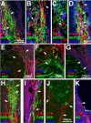
SKP-derived SCs promote axonal growth and sprouting into the transplant region. A–K, Fluorescence photomicrographs of longitudinal sections through the spinal cord of rats that were transplanted 11 weeks earlier with neonatal, YFP-expressing SKP-derived SCs (A–D, H); neonatal, YFP-expressing SKPs (E–G, I, J); or neonatal, YFP-expressing forebrain SVZ neurospheres (NS) (K). In all cases, sections were immunostained for GFAP (blue). A, B, Immunocytochemistry for the YFP tag in the transplanted SKP-derived SCs (green) and for TH (red), a marker for descending noradrenergic fibers. B is a higher magnification image, and the arrows indicate TH-positive axons coursing through the SKP-derived SC bridge in the center of the contusion lesion. C, D, Immunocytochemistry for the YFP tag in the transplanted SKP-derived SCs (green) and either serotonin (C; 5-HT, red), a marker for descending serotonergic axons, or CGRP (D; red), a marker for sensory axons. The arrows indicate serotonin- or CGRP-positive fibers. E–G, Immunocytochemistry for the YFP tag in transplanted SKPs (green), and for tyrosine hydroxylase (E), serotonin (F; 5-HT), or CGRP (G) (red in all cases). F is a higher magnification image. The arrows denote positive axons. Note that, unlike SKP-derived SCs, the axons do not course through the transplant but instead are primarily limited to the border of the transplant. H–K, Ascending sensory axons were traced with the anterograde tracer CTB at 10 weeks after lesion, animals were killed 2 weeks later, and immunocytochemistry was performed for the YFP tag in the transplanted cells (green), and for cholera toxin B (red). Photomicrographs of longitudinal sections through the spinal cords of these animals demonstrate that ascending, CTB-positive axons grew at least 1 mm into transplants of SKP-derived SCs (H), but not into transplants of naive SKPs (I, J) or of neurospheres (K). The arrows in H, I, and J indicate the CTB-positive axons, and those in K indicate terminal bulbs of CTB-positive axons.
Figure 5.
Figure 5.
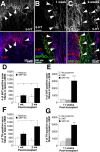
SKP-SCs promote robust axonal growth in the injured spinal cord. A, Top panel shows 5-HT-immunolabeled axons 2 weeks after medium-only injection into the contusion site. 5-HT-positive axon fibers (red; arrowheads) are mostly limited to the areas of intact CNS tissue rich with GFAP-positive astrocytes (blue; bottom panel). B, One week after SKP-derived SCs transplant, 5-HT-positive axons (top panel) are primarily limited to intact host tissue (GFAP, blue; bottom panel). C, In contrast, after 2 weeks, many more 5-HT-positive axons (top panel; arrowheads) are observed within SKP-derived SC transplants, likely an indication of axonal preservation or enhanced sprouting. Note that 5-HT fibers are observed even in areas that do not contain GFAP-positive astrocytes (blue; bottom panel). D, Quantification of 5-HT-positive axons within transplants of SKP-SCs at 1 and 2 weeks after injury. No differences in axon number were observed after 1 week; however, by 2 weeks after transplant, SKP-derived SCs supported a twofold increase in axon number relative to medium-alone controls. *p < 0.05. E, Quantification of 5-HT-positive axons in transplants of neurospheres, SKPs, or SKP-derived SCs (as shown in Fig. 4) 11 weeks after transplantation. Treatment with SKP-derived SCs resulted in a fourfold increase in 5-HT axon numbers relative to both SKPs and neurospheres. ***p < 0.001. F, Quantification of TH-positive axons within the transplants of SKP-derived SCs at 1 and 2 weeks after injury. There was no difference in TH-positive axon numbers after 1 week; however, by 2 weeks after transplant, SKP-derived SCs supported a threefold increase compared with medium-alone controls and a significant increase compared with SKP-derived SC transplants at 1 week. *p < 0.05. G, Quantification of TH-positive axons in transplants of neurospheres, SKPs, or SKP-derived SCs (as shown in Fig. 4) 11 weeks after transplantation. After 11 weeks, SKP-derived SC transplant supported a threefold increase in axon number relative to SKP- or neurosphere-treated animals. **p < 0.01. All group analyses were n = 5 with the exception of 1 week medium controls (n = 3). All data are group means ± SEM.
Figure 6.
Figure 6.
Naive SKPs and SKP-derived SCs myelinate axons in the injured spinal cord. Analysis of longitudinal sections of the spinal cord of animals transplanted with neonatal, YFP-expressing SKPs (SKP), SKP-derived SCs (SC), or neurospheres (NS) 11 weeks earlier. A, B, Double-label immunocytochemistry for the YFP tag in transplanted SKPs (green) and for the myelin protein MBP (red; the arrows indicate double-labeled cells). B is a higher magnification confocal image showing the bipolar morphology of the YFP-positive, MBP-positive cells. C, Triple-label immunocytochemistry for the YFP tag in transplanted SKP-SCs (green), for P0 (red), and for the axonal marker NFM (blue; the arrows indicate double-labeled P0-positive SKP-SCs). The inset shows a high magnification confocal image of the boxed area demonstrating a YFP-positive, P0-positive SC that is associated with an endogenous axon within the transplant. D, Low (left panel) and high (right panel) magnification confocal micrographs of a section double-labeled for the YFP tag in the transplanted SKP-SCs (green), and for the SC-specific myelin protein P0 (red). Note that, in the spared rim of the contused spinal cord (the boxed area in the left panel), there are P0-positive myelin sheaths. At least some of these patches of SC myelin derive from YFP-positive transplanted SKP-SCs, as shown in the right panel (arrows). E, Triple-label immunocytochemistry for the YFP tag in transplanted SKPs (green), for P0 (red), and for NFM (blue). The arrows denote transplanted, P0-positive cells. F, Transplanted neonatal forebrain SVZ neurospheres also occasionally myelinated host axons (arrows), as indicated by triple labeling for the YFP tag (green), the myelin-specific protein MBP (red), and the axonal marker NFM (blue). G, H, YFP-tagged SKPs (G) and SKP-SCs (H) induced nodes of Ranvier when they formed myelin sheaths on endogenous axons, as indicated by immunostaining for the paranodal, axonal potassium channel Kv1.2 (red; G; arrows) or contactin-associated protein Caspr (red; H; arrows). Quantification of the number (I) and percentage (J) of surviving, YFP-positive transplanted cells that coexpressed the peripheral myelin-specific protein P0 and associated with endogenous axons. Note that ∼35 and 15% of SKP-SCs and SKPs, respectively, made P0-positive myelin sheaths. *p < 0.05. Data represent mean ± SEM.
Figure 7.
Figure 7.
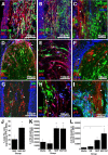
SKPs and SKP-derived SCs promote recruitment of endogenous SCs into the injured spinal cord. Analysis of longitudinal sections of the spinal cord of animals transplanted with neonatal YFP-tagged SKPs, SKP-derived SCs (SC), or forebrain SVZ neurospheres (NS) 11 weeks earlier. A–C, Fluorescence photomicrographs of spinal cord sections immunolabeled for the YFP tag in transplanted SKP-SCs (green), for GFAP (blue), and for either P0 (A) or rat p75NTR (B, C) (both red). C is a higher magnification image. D–F, Immunolabeling of SKP transplants for the YFP tag (green), the axonal protein NFM (NF; blue), and either P0 (D) or p75NTR (F). G–I, Immunolabeling of neurosphere transplants for the YFP tag (green), GFAP (G, I) or NFM (H) (both blue), and rat p75NTR (G, I) or P0 (H) (both red). In all panels, the arrows denote host SCs that are YFP-negative and P0-positive or YFP-negative and rat p75NTR-positive. J, Quantification of the percentage of P0-positive myelin sheaths double-labeled for the YFP tag present in transplanted SKPs or SKP-derived SCs. The other P0-positive myelin sheaths derive from endogenous SCs that have migrated into the injured spinal cord. K, L, Quantification of the total number of P0-positive myelin sheaths within the lesion site (K) and within the spared rim tissue (L) 11 weeks after transplantation with neurospheres, SKPs, SKP-derived SCs, or medium alone. Note that both SKPs and SKP-derived SCs recruit substantially more endogenous SCs than do transplants of neurospheres or medium alone. *p < 0.05; **p < 0.05 relative to both medium control and neurosphere-treated animals. In J–L, results represent mean ± SEM.
Figure 8.
Figure 8.
SKP-derived SCs improve locomotor function after a contusion injury of the spinal cord. A, Locomotor function 9 weeks after injury, and 8 weeks after transplantation of neonatal SKPs (n = 13), SKP-derived SCs (n = 16), or forebrain SVZ neurospheres (n = 11), as assessed by the BBB. Although all groups showed gradual improvement over the 9 weeks after injury, group comparisons showed that SKP-derived SCs showed a small but significant behavioral improvement relative to the other groups. B, The Basso locomotor subscore from the animals shown in A. SKP-derived SCs led to significantly enhanced locomotor behavior relative to transplants of either naive SKPs or neurospheres. C, Locomotor activity in the same group of animals as assessed by ladder rung walking. Animals receiving transplants of SKP-derived SCs showed a reduced number of hindlimb stepping and forelimb placement errors relative to those with transplants of SKPs or neurospheres, but this did not reach significance (p > 0.10). In all panels, results represent mean ± SEM. *p < 0.05 relative to SKPs; **p < 0.05 relative to all other groups. Group differences were ascertained using unpaired t tests.
Figure 9.
Figure 9.
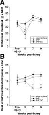
SKP-derived SCs do not reduce sensory thresholds when transplanted into the contused spinal cord. Animals were assessed for hindlimb sensory thresholds before their injury, and 8 weeks after transplantation of neonatal SKPs, SKP-derived SCs, or forebrain SVZ neurospheres. A, Results of the mechanical sensitivity test demonstrating that transplantation of SKP-derived SCs had no impact on mechanical pain thresholds. Interestingly, transplants of SKPs or neurospheres led to a transient reduction in mechanical thresholds that recovered to preinjury levels by 9 weeks after injury. B, Results of the thermal sensitivity. Analysis of the data by repeated-measures ANOVA revealed a significant week by treatment interaction (p < 0.03) and a main effect for treatment group (p < 0.01). Transplantation of SKP-derived SCs or neurospheres had no impact on sensitivity to heat, but transplantation of SKPs led to a significantly heightened sensitivity to heat at 7 weeks after injury that persisted at 9 weeks. In both panels, results represent mean ± SEM. *p < 0.05.

References

    1. Akiyama Y, Radtke C, Kocsis JD. Remyelination of the rat spinal cord by transplantation of identified bone marrow stromal cells. J Neurosci. 2002;22:6623–6630. - PMC - PubMed
    1. Azanchi R, Bernal G, Gupta R, Keirstead HS. Combined demyelination plus Schwann cell transplantation therapy increases spread of cells and axonal regeneration following contusion injury. J Neurotrauma. 2004;21:775–788. - PubMed
    1. Basso DM. Behavioral testing after spinal cord injury: congruities, complexities, and controversies. J Neurotrauma. 2004;21:395–404. - PubMed
    1. Basso DM, Beattie MS, Bresnahan JC. A sensitive and reliable locomotor rating scale for open field testing in rats. J Neurotrauma. 1995;12:1–21. - PubMed
    1. Biernaskie J, McKenzie IA, Toma JG, Miller FD. Isolation of skin-derived precursors (SKPs) and differentiation and enrichment of their Schwann cell progeny. Nat Protoc. 2006;1:2803–2812. - PubMed

Publication types

MeSH terms