

Spatial attention and the latency of neuronal responses in macaque area V4
- PMID: 17804623
- PMCID: PMC6672969
- DOI: 10.1523/JNEUROSCI.2734-07.2007
Spatial attention and the latency of neuronal responses in macaque area V4
Abstract
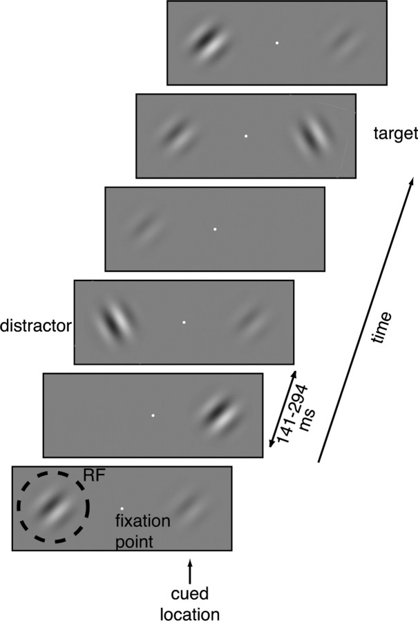
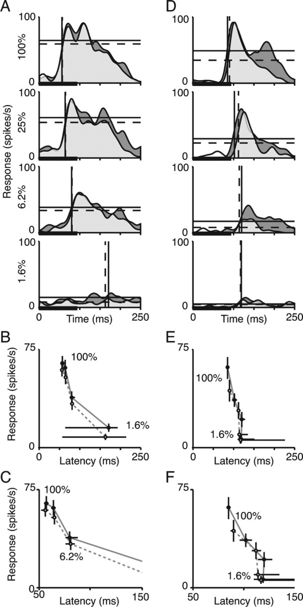
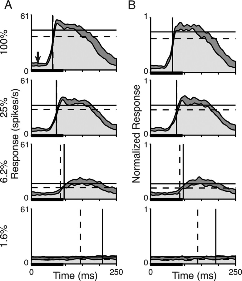
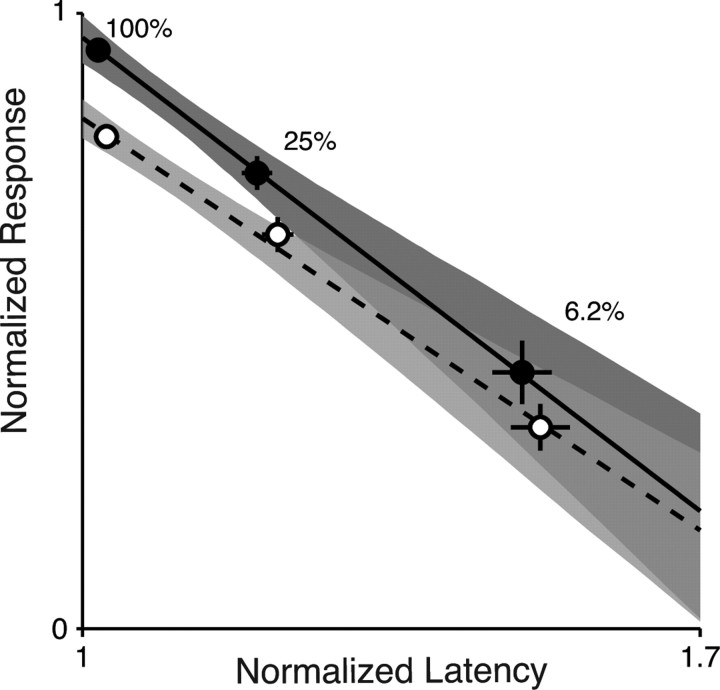
The effects of attention on neuronal responses in visual cortex have been likened to a change in stimulus contrast. Attention and stimulus contrast both modulate the magnitude of neuronal responses. However, changes in stimulus contrast also affect the latency of visual responses. Although many neurophysiological studies have examined how attention affects the strength of neuronal responses, few have considered whether attention affects neuronal latencies. To compare directly the effects of stimulus contrast and attention, we recorded responses from individual neurons in area V4 of macaque monkeys while they performed a task that independently controlled spatial attention and stimulus contrast. As expected, changes in stimulus contrast affected both the magnitude and latency of neuronal responses. Although attention had the expected effects on the magnitudes of neuronal responses, we did not detect statistically reliable changes in neuronal latency. A direct comparison of the effects of contrast and attention revealed a reliable difference. When a shift in spatial attention decreased response magnitude, response latency increased much less than when the same magnitude change was caused by reducing stimulus contrast. Thus, attention is distinct from contrast in the way it affects the relationship between neuronal response magnitude and latency.
Figures
Comment in
-
Turning on the spotlight: do attention and luminance contrast affect neuronal responses in the same way?J Neurosci. 2007 Nov 28;27(48):13043-4. doi: 10.1523/JNEUROSCI.4378-07.2007. J Neurosci. 2007. PMID: 18045898 Free PMC article. No abstract available.
Similar articles
-
Attention influences single unit and local field potential response latencies in visual cortical area V4.J Neurosci. 2012 Nov 7;32(45):16040-50. doi: 10.1523/JNEUROSCI.0489-12.2012. J Neurosci. 2012. PMID: 23136440 Free PMC article.
-
Spatial attention modulates center-surround interactions in macaque visual area v4.Neuron. 2009 Mar 26;61(6):952-63. doi: 10.1016/j.neuron.2009.02.023. Neuron. 2009. PMID: 19324003 Free PMC article.
-
Modulation of Neuronal Responses by Exogenous Attention in Macaque Primary Visual Cortex.J Neurosci. 2015 Sep 30;35(39):13419-29. doi: 10.1523/JNEUROSCI.0527-15.2015. J Neurosci. 2015. PMID: 26424888 Free PMC article.
-
Attention to stimulus features shifts spectral tuning of V4 neurons during natural vision.Neuron. 2008 Aug 14;59(3):509-21. doi: 10.1016/j.neuron.2008.07.001. Neuron. 2008. PMID: 18701075 Free PMC article.
-
Timing of response onset and offset in macaque V4: stimulus and task dependence.J Neurophysiol. 2020 Jun 1;123(6):2311-2325. doi: 10.1152/jn.00586.2019. Epub 2020 May 13. J Neurophysiol. 2020. PMID: 32401171 Free PMC article.
Cited by
-
The effect of attention on neuronal responses to high and low contrast stimuli.J Neurophysiol. 2010 Aug;104(2):960-71. doi: 10.1152/jn.01019.2009. Epub 2010 Jun 10. J Neurophysiol. 2010. PMID: 20538780 Free PMC article.
-
Temporal Dynamics and Response Modulation across the Human Visual System in a Spatial Attention Task: An ECoG Study.J Neurosci. 2019 Jan 9;39(2):333-352. doi: 10.1523/JNEUROSCI.1889-18.2018. Epub 2018 Nov 20. J Neurosci. 2019. PMID: 30459219 Free PMC article.
-
Turning on the spotlight: do attention and luminance contrast affect neuronal responses in the same way?J Neurosci. 2007 Nov 28;27(48):13043-4. doi: 10.1523/JNEUROSCI.4378-07.2007. J Neurosci. 2007. PMID: 18045898 Free PMC article. No abstract available.
-
Neuronal correlates of selective attention and effort in visual area V4 are invariant of motivational context.Sci Adv. 2022 Jun 10;8(23):eabc8812. doi: 10.1126/sciadv.abc8812. Epub 2022 Jun 10. Sci Adv. 2022. PMID: 35687684 Free PMC article.
-
Early dissociation between neural signatures of endogenous spatial attention and perceptual awareness during visual masking.Front Hum Neurosci. 2012 Feb 10;6:16. doi: 10.3389/fnhum.2012.00016. eCollection 2011 Dec 16. Front Hum Neurosci. 2012. PMID: 22363274 Free PMC article.
References
-
- Albrecht DG, Geisler WS, Frazor RA, Crane AM. Visual cortex neurons of monkeys and cats: temporal dynamics of the contrast response function. J Neurophysiol. 2002;88:888–913. - PubMed
-
- Gawne TJ. Temporal coding as a means of information transfer in the primate visual system. Crit Rev Neurobiol. 1999;13:83–101. - PubMed
Publication types
MeSH terms
Grants and funding
LinkOut - more resources
Full Text Sources
Research Materials