. 2007 Sep 5;27(36):9682-95.
doi: 10.1523/JNEUROSCI.2750-07.2007.

Delineation of multiple subpallial progenitor domains by the combinatorial expression of transcriptional codes

Affiliations

Delineation of multiple subpallial progenitor domains by the combinatorial expression of transcriptional codes

Nuria Flames et al. J Neurosci. .

Abstract

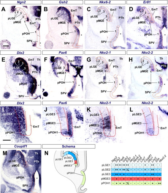
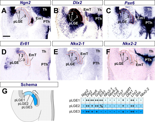
The mammalian telencephalon is considered the most complex of all biological structures. It comprises a large number of functionally and morphologically distinct types of neurons that coordinately control most aspects of cognition and behavior. The subpallium, for example, not only gives rise to multiple neuronal types that form the basal ganglia and parts of the amygdala and septum but also is the origin of an astonishing diversity of cortical interneurons. Despite our detailed knowledge on the molecular, morphological, and physiological properties of most of these neuronal populations, the mechanisms underlying their generation are still poorly understood. Here, we comprehensively analyzed the expression patterns of several transcription factors in the ventricular zone of the developing subpallium in the mouse to generate a detailed molecular map of the different progenitor domains present in this region. Our study demonstrates that the ventricular zone of the mouse subpallium contains at least 18 domains that are uniquely defined by the combinatorial expression of several transcription factors. Furthermore, the results of microtransplantation experiments in vivo corroborate that anatomically defined regions of the mouse subpallium, such as the medial ganglionic eminence, can be subdivided into functionally distinct domains.


Figures

Figure 1.
Figure 1.
Anatomical organization of the embryonic telencephalon. Dlx2 ventricular expression delineates the embryonic subpallial territory. A, Lateral view of the forebrain at E13.5 of development. The dotted lines represent the level and orientation of photographs in B–D and F–I. B–D, Horizontal sections of the E13.5 telencephalon showing the expression pattern of Dlx2 mRNA. E, Whole-mount in situ hybridization of Dlx2 at E13.5. The arrowhead points to the caudal end of the sulcus between the LGE and the MGE, which is typically referred as the rostral limit of the CGE. The dotted lines indicate the level and orientation of photographs (F–I). The crossed arrows mark the dorsoventral (D–V) and anteroposterior (A–P) axes. F–I, Coronal sections through the E13.5 telencephalon showing Dlx2 expression. The asterisks in B–D mark the lateral ventricle. AH, Anterior hypothalamus; dTh, dorsal thalamus; EmT, eminentia thalami; NCx, neocortex; ob, olfactory bulb; PTh, prethalamus; SCh, suprachiasmatic nucleus; SE, septum; SPV, supraoptic paraventricular region. Scale bars: A, 300 μm; B–D, 250 μm; E, 250 μm; F–I, 200 μm.
Figure 2.
Figure 2.
Molecular identification of distinct ventricular domains in the E13.5 rostral subpallium. A–M, Coronal sections through the rostral telencephalon at E13.5 showing the expression of Dlx2 (A), Pax6 (B), Gsh2 (C), Er81 (D), Nkx2-1 (E, I), Nkx6-2 (F, J), Lhx6 (G, K), Lhx7 (H, L), and Couptf1 (M) RNA. All sections were taken from the same level and plane of section. The red dashed lines delineate the VZ. Progenitor domains are named after the anatomical region they belong to, followed by a number given in dorsoventral order. The white or black dashed lines mark the limits between ventricular domains, as inferred from the combined information of the expression of all transcription factors shown in the figure. A, The dorsal limit of the Dlx2 mRNA expression in the VZ delineates the pallial/subpallial boundary. The most dorsal domain of the MGE has only scattered Dlx2 expression (pMGE1). B, Pax6 mRNA is expressed at high levels in the pallial VZ and in pLGE1, at low levels in the remaining of the LGE VZ (pLGE2, pLGE3, and pLGE4), and is not expressed in the MGE VZ. C, Gsh2 mRNA is expressed both in the LGE and MGE VZ. The most dorsal part of the LGE has stronger expression of Gsh2 (pLGE1 and pLGE2). As in the case for Dlx2, Gsh2 mRNA is only found in scattered cells in pMGE1. D, Er81 mRNA is expressed in the most dorsal part of the LGE, overlapping with the domain of strong Gsh2 expression (pLGE1 and pLGE2); it is also expressed in ventral parts of the pallium. E, Nkx2-1 mRNA expression defines the limit between pLGE4 and pMGE1. F, Nkx6-2 mRNA is expressed around the LGE/MGE boundary. The most ventral part of the LGE (pLGE4) expresses low levels of Nkx6-2, the dorsal part of the MGE (pMGE1) expresses high levels of Nkx6-2, and pMGE2 expresses low levels of Nkx6-2. G, Scattered cells expressing Lhx6 mRNA are found in the core of the MGE VZ (pMGE2, pMGE3, pMGE4), but are rare or not present in the most dorsal and ventral domains (pMGE1 and pMGE5). H, Lhx7 is also expressed by scattered cells in the ventricular zone of the ventral half of the MGE (pMGE4, pMGE5). I–L, Higher magnification of E–H, respectively. The arrowheads point the domain boundaries. M, Couptf1 is expressed at high levels in the ventricular zone of the LGE and in a dorsoventral gradient in the MGE but is not detected in pMGE5. N, Schematic summary depicting the anatomical disposition of the proliferative domains present at this level. The table summarizes the molecular identity of each domain: −, absent; +, present; ++, present at high levels; and +/s, scattered expression. pLGE, Progenitor domain from the LGE; pMGE, progenitor domain from the MGE; SE, septum. Scale bars: A–H, M, 140 μm; I–L, 70 μm.
Figure 3.
Figure 3.
Molecular identification of distinct ventricular domains in the E13.5 rostral-intermediate subpallium. A–L, Coronal sections through the rostral-intermediate telencephalon at E13.5 showing the expression of Dlx2 (A), Pax6 (B), Gsh2 (C), Er81 (D), Nkx2-1 (E), Nkx6-2 (F), Lhx6 (G), Couptf1 (H), Dbx1 (I), Lhx2 (J), Shh (K), and Nkx2-2 (L) mRNA. All sections were taken from the same level and plane of section. The red dashed lines delineate the VZ. Progenitor domains are named after the anatomical region they belong to, followed by a number given in dorsoventral order. The white or black dashed lines mark the limits between ventricular domains, as inferred from the combined information of the expression of all transcription factors shown in the figure. A, The ventricular domain of low Dlx2 expression (arrowheads) defines the caudal extension of the pMGE1. Dlx2 also shows scattered expression in the POA ventricular zone. B, Pax6 mRNA is expressed at high levels in the pallial VZ and pLGE1, and at low levels in pLGE2 and pLGE3. C, Gsh2 mRNA is expressed both in the LGE and MGE VZ but is absent from the POA VZ. D, Er81 mRNA is expressed in the VZ of the most dorsal parts of the basal ganglia (pLGE1 and pLGE2) as well as in the ventral MGE (pMGE5) and POA. Interestingly, the dorsal POA expresses higher levels of Er81 (pPOA1) than the ventral POA (pPOA2). E, Nkx2-1 is expressed in the ventral half of the basal ganglia (MGE and POA). As in more anterior levels, Pax6 and Nkx2-1 ventricular expression patterns are complementary. F, Nkx6-2 is expressed in three domains, the caudal pole of pLGE4, pMGE1, and the ventral part of the POA (pPOA2). G, Lhx6 is expressed by scatter cells in pMGE3 and pMGE5. H, Couptf1 mRNA is absent from the ventricular zone of the ventral MGE (pMGE5). I, Dbx1 mRNA is exclusively expressed in the ventral part of the POA (pPOA2). J, Lhx2 mRNA is expressed in a dorsoventral gradient in the LGE and MGE VZ, is absent from the dorsal POA (pPOA1), and is expressed at high levels in the ventral POA (pPOA2). K, Shh is expressed in the VZ of the dorsal and the ventral POA. L, Nkx2-2 is weakly expressed in the ventral half of the basal ganglia (MGE and POA). M, Summary of the figure. The schema depicts the anatomical disposition of the proliferative domains present at this level, and the table summarizes the molecular identity of each domain: −, absent; +, present; ++, present at high levels; and +/s, scatter expression. EmT, Eminentia thalami; pLGE, progenitor domain from the LGE; pMGE, progenitor domain from the MGE; pPOA, progenitor domain from the POA; Th, thalamus. Scale bar, 130 μm.
Figure 4.
Figure 4.
Molecular identification of distinct ventricular domains in the E13.5 caudal-intermediate subpallium. A–M, Coronal sections through the caudal-intermediate telencephalon at E13.5 showing the expression of Ngn2 (A), Gsh2 (B), Nkx6-2 (C), Er81 (D), Dlx2 (E, I), Pax6 (F, J), Nkx2-1 (G, K), Nkx2-2 (H, L), and Couptf1 (M) mRNA. All sections were taken from the same level and plane of section. The red dashed lines delineate the VZ. Progenitor domains are named after the anatomical region they belong to, followed by a number given in dorsoventral order. The white or black dashed lines mark the limits between ventricular domains, as inferred from the combined information of the expression of all transcription factors shown in the figure. At this caudal level, the VZ of the ganglionic eminences and the VZ of the telencephalic stalk are separated by the prethalamus (PTh). A, Ngn2 mRNA expression (a pallial and eminentia thalami marker) is absent from the subpallial ventricular zone including the ganglionic eminences and the telencephalic stalk. B, Gsh2 is expressed in all proliferative subpallial domains, indicating the absence of pPOA at this caudal level. As in anterior levels, Gsh2 expression is stronger in the most dorsal parts of the basal ganglia (pLGE1 and pLGE2). C, Nkx6-2 is not expressed in the telencephalon at this level, indicating the absence of pMGE1 or pPOA2. D, Er81 is expressed in pLGE1, pLGE2, and POH. E, Dlx2 mRNA is expressed in all proliferative subpallial domains; the absence of a domain with scattered Dlx2 expression suggests that pMGE1 is not present at this level. F, Pax6 expression defines the progenitor domains of the LGE (pLGE1, pLGE2, and pLGE3) and the POH (pPOH1), whereas Pax6 is absent from the progenitor domains of the MGE (pMGE3). G, Nkx2-1 ventricular expression shows the presence of MGE progenitor domains at this caudal level (pMGE3). H, Nkx2-2 mRNA is expressed at low levels in the MGE VZ (pMGE3) and at high levels in the POH VZ (pPOH1). I–L, Higher magnification of E and H, respectively, showing the presence of a small piece of the POH ventricle between the MGE and the EmT. The POH VZ expresses Dlx2, Pax6, and Nkx2-2 and lacks Nkx2-1 expression. M, Couptf1 mRNA is expressed in all proliferative subpallial domains at this level, suggesting that pMGE5 is absent at this posterior level. N, Summary of the figure. The schema depicts the anatomical disposition of the proliferative domains present at this level, and the table summarizes the molecular identity of each domain: −, absent; +, present; ++, present at high levels. EmT, eminentia thalami; pLGE, progenitor domain from the LGE; pMGE, progenitor domain from the MGE; pPOH, progenitor domain from the hypothalamic preoptic area; PTh, prethalamus; Th, thalamus; SPV, supraoptic paraventricular region; *, third ventricle. Scale bars: A–H, M, 150 μm; I–L, 75 μm.
Figure 5.
Figure 5.
Molecular identification of distinct ventricular domains in the E13.5 caudal subpallium. A–F, Coronal sections through the caudal telencephalon at E13.5 showing the expression of Ngn2 (A), Dlx2 (B), Pax6 (C), Er81 (D), Nkx2-1 (E), and Nkx2-2 (F) mRNA. All sections were taken from the same level and plane of section. The red dashed lines delineate the VZ. Progenitor domains are named after the anatomical region they belong to, followed by a number given in dorsoventral order. The white or black dashed lines mark the limits between ventricular domains, as inferred from the combined information of the expression of all transcription factors shown in the figure. A, Ngn2 expression, a pallial marker, is absent from the subpallium. B, Dlx2 is expressed in a complementary manner to Ngn2, labeling the VZ of the subpallium. C, Pax6 is expressed throughout the subpallial VZ. D, Er81 is expressed in the most dorsal domains of the subpallium (pLGE1 and pLGE2). E, Lack of Nkx2-1 mRNA expression indicates the absence of pMGE domains. F, Nkx2-2 is also absent from the VZ, suggesting the absence of POH. G, Summary of the figure. The schema depicts the anatomical disposition of the proliferative domains present at this level, and the table summarizes the molecular identity of each domain: −, absent; +, present. EmT, eminentia thalami; pLGE, progenitor domain from the LGE; PTh, prethalamus; Th, thalamus. Scale bar, 140 μm.
Figure 6.
Figure 6.
Molecular identification of distinct ventricular domains in the E13.5 subpallial septum. A–I, Horizontal sections through the telencephalon at E13.5 showing the expression of Dlx2 (A), Pax6 (B), Gsh2 (C), Er81 (D), Nkx2-1 (E), Nkx6-2 (F), Lhx5 (G), Shh (H), and Nkx2-2 (I) mRNA. All sections were taken from the same level and plane of section, except for F, which is more ventral. The horizontal dotted line marks the midline. The red dashed lines delineate the VZ. Progenitor domains are named after the anatomical region they belong to, followed by a number given in anterodorsal to caudoventral order. The white or black dashed lines mark the limits between ventricular domains, as inferred from the combined information of the expression of all transcription factors shown in the figure. A, Dlx2 mRNA is expressed in all septal domains except for pSe5, where only occasional cells expressing Dlx2 can be observed. B, Pax6 marks the LGE counterpart domains of the septum (pSe1, pSe2, and pSe3). C, Gsh2 is expressed in most parts of the septum but is absent from the most ventromedial part. D, Er81 is expressed throughout the septum. E, Nkx2.1 is expressed in the most ventromedial part of the septum (pSe4 to pSe6). F, Nkx6.2 is expressed in a small septal domain (pSe4). G, Lhx5 mRNA is exclusively expressed in the septum forming a medial-to-lateral gradient. H, Shh is expressed in a very small domain in the most medial part of the septum (pSe6), corresponding to the Dlx2- and Gsh2-negative domain. I, Nkx2-2 is expressed at low levels in two domains of the medial septum (pSe5 and pSe6). J, Summary of the figure. The schema depicts the anatomical disposition of the proliferative domains present at this level, and the table summarizes the molecular identity of each septal domain: −, absent; +, present; ++, present at high levels; +/s, scatter expression. Septal color codes match with their ganglionic eminences progenitor domain counterparts but are represented with a lined pattern to symbolize their singular characteristics. The white arrowheads in B and D indicate the possible postmitotic output of pLGE1, which is complementary to the postmitotic output of pLGE2 (D, black arrowhead). pLGE, Progenitor domain from the LGE; pMGE, progenitor domain from the MGE; pPOH, progenitor domain from the hypothalamic preoptic area; pSe, progenitor domains from the septum; *, lateral ventricle. Scale bar, 180 μm.
Figure 7.
Figure 7.
Characterization of ventricular zone cells at the intereminential (LGE/MGE) sulcus in the E13.5 subpallium. A–A″, C–C″, E–E″, Adjacent coronal sections through the rostral telencephalon at E13.5 showing the expression of Nkx2-1 (A, A″, B), Nkx6-2 (A′, A″, B, C′, C″, D, E′, E″, F), Olig2 (C, C″, D), and Lhx6 (E, E″, F) proteins. B, D, and F are high magnification confocal photographs of the areas boxed in A″, C″, and E″, respectively. The insets in B, D, and F show a high magnification of the boxed areas. The dashed lines delineate the VZ. Progenitor domains are named after the anatomical region they belong to, followed by a number given in dorsoventral order. The dotted lines mark the limits between ventricular domains pLGE4, pMGE1, and pMGE2. A, B, Expression of Nkx2-1 and Nkx6.2 at the LGE/MGE sulcus. Virtually every cell in the MGE expresses Nkx2-1. pMGE1 and pMGE2 contain cells that express both Nkx2-1 and Nkx6-2 (arrowheads). Double-labeled cells are more abundant in pMGE1 than in pMGE2. Only occasionally Nkx6-2+/Nkx2-1− cells are observed in pMGE1 (open arrows) or Nkx6-2+/Nkx2-1+ in pLGE4 (open arrowheads). C, D, Expression of Olig2 and Nkx6.2 at the LGE/MGE sulcus. Olig2 is expressed in many cells in pLGE4, pMGE1, and pMGE2; however, Olig2 is consistently expressed at higher levels in pMGE2 than in pMGE1 and pLGE4. pLGE4 contains cells that express Olig2 but not Nkx6-2 (arrows) and double-labeled cells (arrowheads). pMGE1 and pMGE2 contain cells that express Olig2 but not Nkx6-2 (open arrows) and double-labeled cells (arrowheads and open arrowheads). Olig2 levels are consistently higher in pMGE2 cells (open arrowheads) than in pMGE1 cells (arrowheads). E, F, Expression of Lhx6 and Nkx6.2 at the LGE/MGE sulcus. Lhx6 is expressed in very few cells in the VZ of the pMGE, but it is consistently absent from pMGE1 (E). Lhx6 (arrows) and Nkx6-2 (open arrows) are expressed in different VZ cells. Scale bars: A, C, E, 100 μm; B, D, F, 20 μm.
Figure 8.
Figure 8.
Fate of cortical interneurons derived from pMGE1 and pMGE4. A, Schematic diagram of the experimental design. Coronal slices were prepared from the telencephalon of E13.5 GFP+ donor embryos, and progenitor cells were isolated from a small cube of tissue approximately corresponding to pMGE1 at rostral-intermediate telencephalic levels. In parallel, the MGE was dissected from wild-type embryos and dissociated. Pooled donor GFP+ pMGE1 and MGE cells were then injected into the MGE of E13.5 host embryos. Host embryos were analyzed at P14. B, Coronal section obtained from a donor GFP+ slice showing the expression of Nkx6-2 mRNA (experiment T106). The asterisk indicates the original location of transplanted cells within the MGE (approximately pMGE1 at this level). The inset summarizes the molecular profiling of pMGE1. C, Coronal section through the somatosensory cortex of a transplanted P14 mouse showing two pMGE1-derived cells (arrowheads) after nuclear staining [4′,6′-diamidino-2-phenylindole (DAPI)] and immunohistochemistry for GFP (green). D, D′, The arrowhead points to a neuron expressing GFP and SST, but negative for PV. The open arrowhead points to a PV+ cell. E, Coronal section obtained from a donor GFP+ slice showing the expression of Lhx6 mRNA (experiment T129E). The asterisk indicates the original location of transplanted cells within the MGE (approximately pMGE4 at this level). The inset summarizes the molecular profiling of pMGE4. F, Coronal section through the somatosensory cortex of a transplanted P14 mouse showing two pMGE4-derived cells (arrowheads) after nuclear staining (DAPI) and immunohistochemistry for GFP (green). G, G′, The arrowhead points to a neuron expressing GFP and PV, but negative for SST. The open arrowhead points to a SST+ cell. H, Quantification of the percentage of SST+, PV+, or PV/SST− in the P14 cortex after whole MGE, pMGE1, and pMGE4 transplantation. GFP+/SST+ cells: 30.12 ± 2.27% (whole MGE), 63.88 ± 7.34% (pMGE1), and 7.24 ± 1.73% (pMGE4); GFP+/PV+ cells: 50.66 ± 1.26% (whole MGE), 21.96 ± 9.7% (pMGE1), and 48.96 ± 5.07% (pMGE4); GFP+/SST−/PV− cells: 19.2 ± 2.02% (whole MGE), 14.14 ± 2.52% (pMGE1), and 43.78 ± 5.7% (pMGE4). Error bars indicate SEM. ***p < 0.001, χ2 tests between whole MGE and pMGE1 or whole MGE and pMGE4. Scale bars: B, E, 200 μm; C, F, 300 μm; D, D′, G, G′, 10 μm.
Figure 9.
Figure 9.
A model of organization for the ventricular zone of the mouse subpallium. A, Whole-mount Nkx2-1 expression. Nkx2-1 is expressed in the MGE, POA, and part of the CGE and the septum. B, Schema of the subpallial progenitor domains represented by a color code. Each dotted line represents the plane and level of section of schemas C–F. C–F, Schema of the four different anterior–posterior levels described in this work. All different proliferative domains are shown with a color code. pLGE, Progenitor domain from the LGE; pMGE, progenitor domain from the MGE; pPOA, progenitor domain from the preoptic area; pPOH, progenitor domain from the hypothalamic preoptic area; SE, septum; SPV, supraoptic paraventricular region. Scale bar, 200 μm.

References

    1. Assimacopoulos S, Grove EA, Ragsdale CW. Identification of a Pax6-dependent epidermal growth factor family signaling source at the lateral edge of the embryonic cerebral cortex. J Neurosci. 2003;23:6399–6403. - PMC - PubMed
    1. Bardet SM, Cobos I, Puelles E, Martinez-De-La-Torre M, Puelles L. Chicken lateral septal organ and other circumventricular organs form in a striatal subdomain abutting the molecular striatopallidal border. J Comp Neurol. 2006;499:745–767. - PubMed
    1. Bulfone A, Puelles L, Porteus MH, Frohman MA, Martin GR, Rubenstein JL. Spatially restricted expression of Dlx-1, Dlx-2 (Tes-1), Gbx-2, and Wnt-3 in the embryonic day 12.5 mouse forebrain defines potential transverse and longitudinal segmental boundaries. J Neurosci. 1993;13:3155–3172. - PMC - PubMed
    1. Butt SJ, Fuccillo M, Nery S, Noctor S, Kriegstein A, Corbin JG, Fishell G. The temporal and spatial origins of cortical interneurons predict their physiological subtype. Neuron. 2005;48:591–604. - PubMed
    1. Campbell K. Dorsal-ventral patterning in the mammalian telencephalon. Curr Opin Neurobiol. 2003;13:50–56. - PubMed

Publication types

Substances