. 2007 Sep 5;27(36):9780-9.
doi: 10.1523/JNEUROSCI.5586-06.2007.

Cilia proteins control cerebellar morphogenesis by promoting expansion of the granule progenitor pool

Affiliations

Cilia proteins control cerebellar morphogenesis by promoting expansion of the granule progenitor pool

Victor V Chizhikov et al. J Neurosci. .

Abstract

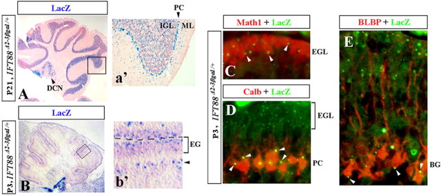
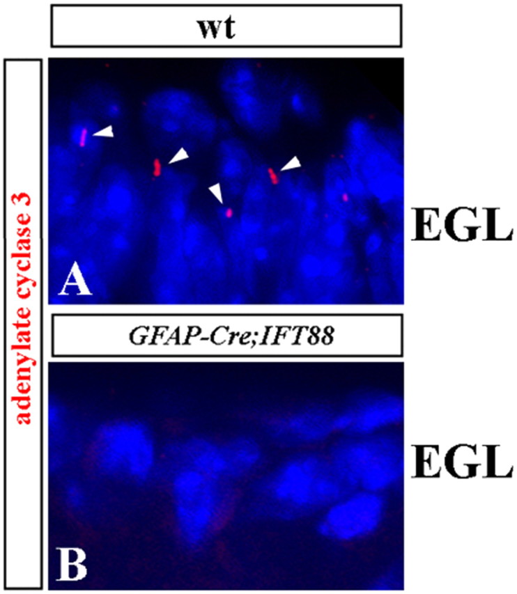
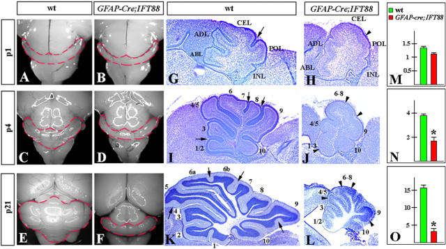
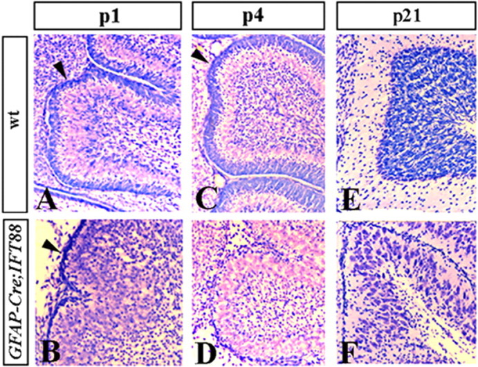
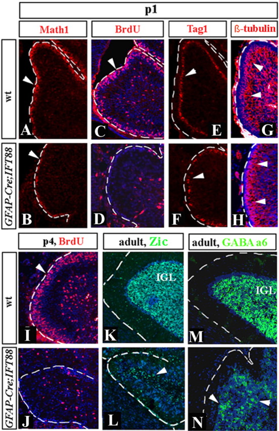
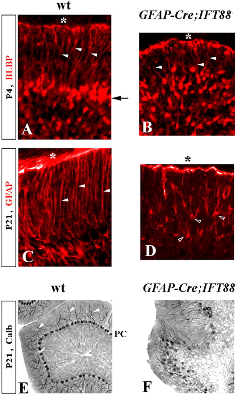
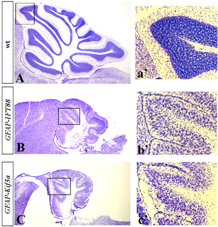
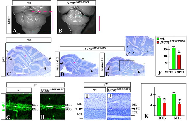
Although human congenital cerebellar malformations are common, their molecular and developmental basis is still poorly understood. Recently, cilia-related gene deficiencies have been implicated in several congenital disorders that exhibit cerebellar abnormalities such as Joubert syndrome, Meckel-Gruber syndrome, Bardet-Biedl syndrome, and Orofaciodigital syndrome. The association of cilia gene mutations with these syndromes suggests that cilia may be important for cerebellar development, but the nature of cilia involvement has not been elucidated. To assess the importance of cilia-related proteins during cerebellar development, we studied the effects of CNS-specific inactivation of two mouse genes whose protein products are critical for cilia formation and maintenance, IFT88, (also known as polaris or Tg737), which encodes intraflagellar transport 88 homolog, and Kif3a, which encodes kinesin family member 3a. We showed that loss of either of these genes caused severe cerebellar hypoplasia and foliation abnormalities, primarily attributable to a failure of expansion of the neonatal granule cell progenitor population. In addition, granule cell progenitor proliferation was sensitive to partial loss of IFT function in a hypomorphic mutant of IFT88 (IFT88(orpk)), an effect that was modified by genetic background. IFT88 and Kif3a were not required for the specification and differentiation of most other cerebellar cell types, including Purkinje cells. Together, our observations constitute the first demonstration that cilia proteins are essential for normal cerebellar development and suggest that granule cell proliferation defects may be central to the cerebellar pathology in human cilia-related disorders.


Figures

Figure 1.
Figure 1.
IFT88 expression in the adult and developing mouse cerebellum. Direct lacZ (A, B) and antibody staining (C–E) of sagittal cerebellar sections of IFT88 Δ2–3βgal/+ mice at the indicated stages. Insets (a′, b′) show higher magnification of boxed regions. Deep cerebellar nuclei (DCN), Purkinje cells (PC), molecular layer (ML), internal granule cell layer (IGL), external granule cell layer (EGL), and Bergmann glia (BG) are indicated. b′, Black arrowhead points to the location of Purkinje cells and Bergmann glia. C–E, White arrowheads point to LacZ+ dots in Math1+ granule cells (C), calbindin+ Purkinje cells (D), and BLBP+ Bergmann glia (E).
Figure 2.
Figure 2.
Depletion of cilia in the EGL of hGFAP–Cre+;IFT88floxed/null mutant mice. Adenylate cyclase type 3 antibody staining (red) identifies some primary cilia (seen as condensed red dots and short lines, labeled by arrowheads) in the P4 wild-type (A; wt) but not hGFAP–Cre+;IFT88floxed/null mutant (B) EGL. Sections were counterstained with 4′,6′-diamidino-2-phenylindole (blue) to visualize nuclei.
Figure 3.
Figure 3.
Size reduction and foliation abnormalities in hGFAP–Cre+;IFT88floxed/null mutant mice. Dorsal whole-mount views (A–F) and sagittal sections of the cerebellar vermis (G–L) of wild-type (wt) (A, C, E, G, I, K) and hGFAP–Cre+;IFT88floxed/null mutant (B, D, F, H, J, L) cerebella at the indicated stages. G, H, Anterobasal (ABL), anterodorsal (ADL), central (CEL), posterior (POL), and inferior (INL) lobes are indicated. I–L, Lobes are indicated by numbers. Fissures underdeveloped in hGFAP–Cre+;IFT88floxed/null mutants are labeled by arrowheads. In normal littermates, the corresponding fissures are indicated by arrows. M–O, Quantification of the cerebellar vermis area (in arbitrary units) in wild-type and hGFAP–Cre+;IFT88floxed/null mutants at P1 (M), P4 (N), and P21 (O). *p < 0.01.
Figure 4.
Figure 4.
EGL is prematurely depleted in hGFAP–Cre+;IFT88floxed/null mutant mice. Sagittal sections of cerebellar vermis of wild-type (A, C, E) and hGFAP–Cre+;IFT88floxed/null mutant (B, D, F) mice at the indicated stages. Sections were stained with cresyl violet. Arrowheads point to EGL (A–D). In hGFAP–Cre+;IFT88floxed/null cerebellum, EGL is thin at P1 and is virtually absent at P4.
Figure 5.
Figure 5.
Granule progenitor proliferation, but not differentiation, is abnormal in hGFAP–Cre+;IFT88floxed/null mutant mice. Sagittal sections of cerebellar vermis of wild-type (wt) (A, C, E, G, I, K, M) and hGFAP–Cre+;IFT88floxed/null mutant (B, D, F, H, J, L, N) mice at the indicated stages. Sections were stained with indicated antibodies. Arrowheads point to Math1+ cells (A, B), BrdU+ cells (C, D, I, J), Tag1+ cells (E, F), β-tubulin III-positive granule cells migrating from the EGL to IGL (G, H), and Zic1+ (K, L) and GABAA α6+ (M, N) granule cells in the adult IGL. hGFAP–Cre+;IFT88floxed/null EGL is BrdU negative at both P1 and P4. Granule cells, however, are clearly present, although in reduced numbers, based on expression of multiple markers of granule cell progenitors and differentiated granule cells.
Figure 6.
Figure 6.
Bergmann glia and Purkinje cell defects in hGFAP–Cre+;IFT88floxed/null mutant mice. Immunostained sections of wild-type (wt) (A, C, E) and hGFAP–Cre+;IFT88floxed/null mutant (B, D, F) cerebella at the indicated stages. A–D, Arrow points to Bergmann glia aligned along Purkinje cells in the wild-type but not in the hGFAP–Cre+;IFT88floxed/null cerebellum. White arrowheads point to radial glia fibers extending to the cerebellar pial surface (asterisk). Black arrowheads point to mutant Bergmann glia with astrocyte-like morphology. E, F, Arrowheads point to wild-type Purkinje cell (PC) dendrites, which are not recognizable in hGFAP–Cre+;IFT88floxed/null cerebellum. calb, Calbindin.
Figure 7.
Figure 7.
hGFAP–Cre+;IFT88floxed/null and hGFAP–Cre+;Kif3afloxed/null mice display similar cerebellar phenotypes. Sagittal sections of the cerebellar vermis of P21 wild-type (A; wt), hGFAP–Cre+;IFT88floxed/null mutant (B), and hGFAP–Cre+;Kif3afloxed/null mutant (C) mice. Insets (a′, b′, c′) show higher magnification of boxed regions.
Figure 8.
Figure 8.
Cerebellar abnormalities detected in IFT88orpk/orpk mice. Dorsal whole-mount views (A, B) and sagittal sections of cerebellar vermis (C–E, G–J) of wild-type (wt) (A, C, G, I) and IFT88orpk/orpk mutant (B, D, E, H, J) cerebella at the indicated stages. A, B, Red bars indicate the extent of wild-type and IFT88orpk/orpk cerebella along the anteroposterior axis. The IFT88orpk/orpk cerebellum is significantly shorter, with hypoplasia of the posterior lobes. C–E, Fissures that are underdeveloped in IFT88orpk/orpk mutants are labeled by arrowheads. In the normal littermate, the corresponding fissures are indicated by arrows. Lobes 4, 5, and 9 are labeled. Pronounced hypoplasia of centrally located posterior lobes, giving a concave appearance to the vermis in IFT88orpk/orpk mutants, is labeled by an asterisk (D). Inset (e′) shows a higher magnification of boxed region. Arrows point to granule cell heterotopia in IFT88orpk/orpk cerebellum. F, Quantification of the cerebellar vermis area (in arbitrary units) in wild-type and IFT88orpk/orpk mutants at P21. *p < 0.01. G, H, BrdU labeling revels reduced proliferation in the EGL of IFT88orpk/orpk mutants. I, J, IGL and molecular layer (ML) are thinner in P21 IFT88orpk/orpk mutants compared with wild-type littermates. PC, Purkinje cells. K, Quantification of the thickness of the IGL and molecular layer (ML) (in arbitrary units) in wild-type and IFT88orpk/orpk mutants at P21. For consistency, all measurements were performed at the dorsal surface of lobe 5 (pink bars, C, D). *p < 0.01.

References

    1. Altman J, Bayer SA. New York: CRC; 1997. The development of the cerebellar system: in relation in its evolution, structure and function.
    1. Ansley SJ, Badano JL, Blacque OE, Hill J, Hoskins BE, Leitch CC, Kim JC, Ross AJ, Eichers ER, Teslovich TM, Mah AK, Johnsen RC, Cavender JC, Lewis RA, Leroux MR, Beales PL, Katsanis N. Basal body dysfunction is a likely cause of pleiotropic Bardet-Biedl syndrome. Nature. 2003;425:628–633. - PubMed
    1. Arts HH, Doherty D, van Beersum SE, Parisi MA, Letteboer SJ, Gorden NT, Peters TA, Marker T, Voesenek K, Kartono A, Ozyurek H, Farin FM, Kroes HY, Wolfrum U, Brunner HG, Cremers FP, Glass IA, Knoers NV, Roepman R. Mutations in the gene encoding the basal body protein RPGRIP1L, a nephrocystin-4 interactor, cause Joubert syndrome. Nat Genet. 2007;39:882–888. - PubMed
    1. Baala L, Romano S, Khaddour R, Saunier S, Smith UM, Audollent S, Ozilou C, Faivre L, Laurent N, Foliguet B, Munnich A, Lyonnet S, Salomon R, Encha-Razavi F, Gubler M-C, Boddaert N, de Lonlay P, Johnson CA, Vekemans M, Antignac C, Attié-Bitach T. The Meckel-Gruber syndrome gene, MKS3, is mutated in Joubert syndrome. Am J Hum Genet. 2007;80:186–194. - PMC - PubMed
    1. Baptista CA, Hatten ME, Blazeski R, Mason CA. Cell-cell interactions influence survival and differentiation of purified Purkinje cells in vitro. Neuron. 1994;12:243–260. - PubMed

Publication types