. 2007 Nov;45(11):3506-13.
doi: 10.1128/JCM.00936-07. Epub 2007 Sep 5.

Highly sensitive and broadly reactive quantitative reverse transcription-PCR assay for high-throughput detection of Rift Valley fever virus

Affiliations

Highly sensitive and broadly reactive quantitative reverse transcription-PCR assay for high-throughput detection of Rift Valley fever virus

Brian H Bird et al. J Clin Microbiol. 2007 Nov.

Abstract

Rift Valley fever (RVF) virus is a mosquito-borne virus associated with large-scale epizootics/epidemics throughout Africa and the Arabian peninsula. Virus infection can result in economically disastrous "abortion storms" and high newborn mortality in livestock. Human infections result in a flu-like illness, with 1 to 2% of patients developing severe complications, including encephalitis or hemorrhagic fever with high fatality rates. There is a critical need for a highly sensitive and specific molecular diagnostic assay capable of detecting the natural genetic spectrum of RVF viruses. We report here the establishment of a pan-RVF virus quantitative real-time reverse transcription-PCR assay with high analytical sensitivity (approximately 5 RNA copies of in vitro-transcribed RNA/reaction or approximately 0.1 PFU of infectious virus/reaction) and efficiency (standard curve slope = -3.66). Based on the alignments of the complete genome sequences of 40 ecologically and biologically diverse virus isolates collected over 56 years (1944 to 2000), the primer and probe annealing sites targeted in this assay are known to be located in highly conserved genomic regions. The performance of this assay relative to serologic assays is illustrated by testing of known RVF case materials obtained during the Saudi Arabia outbreak in 2000. Furthermore, analysis of acute-phase blood samples collected from human patients (25 nonfatal, 8 fatal) during that outbreak revealed that patient viremia at time of presentation at hospital may be a useful prognostic tool in determining patient outcome.


Figures
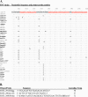
FIG. 1.
FIG. 1.
(A) Alignment of nucleotides 2900 to 3001 of RVF virus L segments from 40 different virus strains. Nucleotide identities throughout the alignment with wild-type strain 1260/78 are indicated by a dot (.) (GenBank accession numbers DQ375395 to DQ375434). The positions of primers RVFL-2912ggfwd and RVFL-2981revAC and fluorogenic probe RVFL-probe-2950 are indicated in underlined red text. The 21 phylogenetically diverse strains tested by Q-RT-PCR amplification are indicated by a boldfaced strain name and an asterisk. All RVF virus strains tested were positively detected by this assay. Note the high nucleotide conservation at the primer-probe annealing sites among these 40 RVF virus strains. (B). Nucleotide sequence of the primer-probe combination of RVFL-2912ggfwd, RVFL-2981revAC, and RVFL-probe-2950 and their respective calculated annealing temperatures using an online calculator (http://www.basic.northwestern.edu/biotools/oligocalc.html).
FIG. 2.
FIG. 2.
Delta Rn (change in fluorescence) versus cycle number. The results of Q-RT-PCR amplification of RVF virus stock strain ZH501 diluted serially 10-fold (10−1 to 10−8) in serum are depicted in the main chart. The inset shows the results of a log transformation of the CT values plotted versus log10 dilutions of extracted RNA of stock ZH501. The resulting equation of the standard curve was y = −3.66x + 44.99. The LOD of this assay with serially diluted stock virus in serum was approximately 0.1 PFU/rxn.
FIG. 3.
FIG. 3.
RVF RNA load among patients with fatal and nonfatal outcomes. Results of Q-RT-PCR amplification of RVF virus-infected patient sera collected during the epidemic/epizootic in Saudi Arabia in 2000. A total of 62 patient specimens are divided into groups according to patient outcome (nonfatal or fatal) and by days after onset of clinical symptoms (days 1 to 4, 5 to 8, and 9 to 14). Significant differences (P < 0.001) between the mean CT value of fatal and nonfatal groups at days 1 to 4 postinfection are indicated by a single asterisk (*) and at days 5 to 8 by a double asterisk (**). At all time points the mean RVF RNA load of fatal cases was significantly higher (P < 0.001) than nonfatal cases and is indicated by a “§” symbol. Note that the mean serum RVF RNA load of both fatal and nonfatal cases decrease as the time after onset increases, presumably due to the development of anti-RVF virus-specific IgM and/or IgG antibody with the subsequent clearance of virus from patient blood. Error bars indicate the mean value ±2 standard errors of the mean. The LOD of the Q-RT-PCR assay is indicated by an arrow and represents a CT value of >40. The scale of the y-axis scale is demarcated every 3.66 CT values, which corresponds to a 1.0 log10 PFU/ml change in virus RNA titer as calculated from titration of the stock RVF virus.
FIG. 4.
FIG. 4.
Results of Q-RT-PCR and RVF virus-specific antigen capture, IgM, and IgG ELISAs of a representative subset of four human patients from whom multiple blood samples were obtained. Patients 2 and 4 represent nonfatal outcomes of RVF. Patients 7 and 23 represent fatal outcomes of RVF. IgM, solid square and solid line; IgG, open square and dashed line; antigen capture, open circle and dashed line; Q-RT-PCR CT values, closed circle and solid line. All serological data are expressed as the adjusted SUMOD of four sample dilutions of 1:4, 1:16, 1:64, and 1:256 for the anti-RVF antigen capture assay and dilutions of 1:100, 1:400, 1:1,600, and 1:6,400 for the anti-RVF specific IgM and IgG assays. The LOD of the Q-RT-PCR assay is indicated by an arrow and represents a CT value of >40. The scale of the right-hand y-axis scale is demarcated every 3.66 U, which corresponds to a 1.0 log10 PFU/ml change in virus RNA titer as calculated from titration of stock RVF virus.

Similar articles

Cited by

References

    1. Anonymous. 2000. Update: outbreak of Rift Valley Fever-Saudi Arabia, August-November 2000. Morb. Mortal. Wkly. Rep. 49:982-985. - PubMed
    1. Bird, B. H., M. L. Khristova, P. E. Rollin, T. G. Ksiazek, and S. T. Nichol. 2007. Complete genome analysis of 33 ecologically and biologically diverse Rift Valley fever virus strains reveals widespread virus movement and low genetic diversity due to recent common ancestry. J. Virol. 81:2805-2816. - PMC - PubMed
    1. Coetzer, J. A. 1977. The pathology of Rift Valley fever. I. Lesions occurring in natural cases in new-born lambs. Onderstepoort J. Vet. Res. 44:205-211. - PubMed
    1. Drosten, C., S. Gottig, S. Schilling, M. Asper, M. Panning, H. Schmitz, and S. Gunther. 2002. Rapid detection and quantification of RNA of Ebola and Marburg viruses, Lassa virus, Crimean-Congo hemorrhagic fever virus, Rift Valley fever virus, dengue virus, and yellow fever virus by real-time reverse transcription-PCR. J. Clin. Microbiol. 40:2323-2330. - PMC - PubMed
    1. Easterday, B. C., M. H. McGavran, J. R. Rooney, and L. C. Murphy. 1962. The pathogenesis of Rift Valley fever in lambs. Am. J. Vet. Res. 23:470-479. - PubMed

Publication types

MeSH terms

LinkOut - more resources