Review
. 2007 Sep;71(3):413-51.
doi: 10.1128/MMBR.00012-07.

Siderophore-based iron acquisition and pathogen control

Affiliations
Review

Siderophore-based iron acquisition and pathogen control

Marcus Miethke et al. Microbiol Mol Biol Rev. 2007 Sep.

Abstract

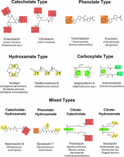
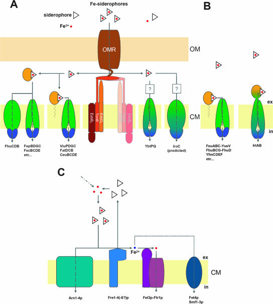
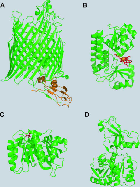
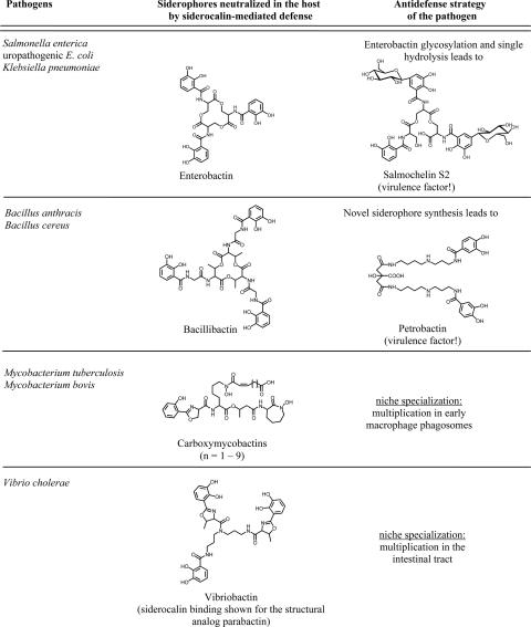
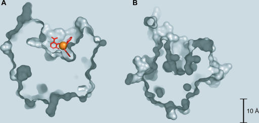
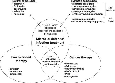
High-affinity iron acquisition is mediated by siderophore-dependent pathways in the majority of pathogenic and nonpathogenic bacteria and fungi. Considerable progress has been made in characterizing and understanding mechanisms of siderophore synthesis, secretion, iron scavenging, and siderophore-delivered iron uptake and its release. The regulation of siderophore pathways reveals multilayer networks at the transcriptional and posttranscriptional levels. Due to the key role of many siderophores during virulence, coevolution led to sophisticated strategies of siderophore neutralization by mammals and (re)utilization by bacterial pathogens. Surprisingly, hosts also developed essential siderophore-based iron delivery and cell conversion pathways, which are of interest for diagnostic and therapeutic studies. In the last decades, natural and synthetic compounds have gained attention as potential therapeutics for iron-dependent treatment of infections and further diseases. Promising results for pathogen inhibition were obtained with various siderophore-antibiotic conjugates acting as "Trojan horse" toxins and siderophore pathway inhibitors. In this article, general aspects of siderophore-mediated iron acquisition, recent findings regarding iron-related pathogen-host interactions, and current strategies for iron-dependent pathogen control will be reviewed. Further concepts including the inhibition of novel siderophore pathway targets are discussed.


Figures

FIG. 1.
FIG. 1.
Representative examples of different siderophores and their natural producers. Moieties involved in iron coordination are highlighted as follows: catecholates are in red, phenolates are in orange, hydroxamates are in pale yellow, α-hydroxy-carboxylates (deriving from citrate units) are in light green, and α-keto-carboxylates (deriving from 2-oxo-glutarate units) are in blue-green.
FIG. 2.
FIG. 2.
Structures of proteins involved in siderophore biosynthesis. (A) The Y. enterocolitica salicylate synthase Irp9 homodimer (PDB accession number 2FN1) with reaction products salicylate and pyruvate (shown in red) and the Mg(II) cofactor (shown in yellow). (B) Homodimer of the E. coli isochorismatase-aryl carrier protein EntB (PDB accession number 2FQ1). The enlarged section is the aryl carrier protein domain with Ser245 (yellow) for cofactor modification and residues involved in minor (bright orange) and major (dark orange) interactions with the nonribosomal peptide synthetase EntF. (C) The E. coli 2,3-dihydro-2,3-DHB dehydrogenase EntA in the homotetrameric state (PDB accession number 2FWM). (D) The lone-standing DHB adenylation domain DhbE of B. subtilis cocrystallized with DHB-AMP (shown in red) (PDB accession number 1MDB). (E) The lone-standing condensation domain VibH of V. cholerae (PDB accession number 1L5A) linking VibB-activated DHB and norspermidine via amide bond formation.
FIG. 3.
FIG. 3.
Alignment (ClustalW, shown in BOXSHADE format) of MFS-type proteins involved in the secretion of siderophores. Sections of predicted TMSs are shown individually for each protein and are indicated with red letters and/or red shading. Nonshaded letters indicate nonsimilar amino acids; gray- or light red-shaded letters indicate similar amino acids; black- and dark red-shaded letters indicate identical amino acids.
FIG. 4.
FIG. 4.
Cellular transport systems for uptake of siderophore-delivered iron in bacteria and fungi. (A) Gram-negative bacteria. Fe-siderophore uptake through the OM is mediated by an OM receptor (OMR), which is energized by the proton motive force transduction system ExbB7-ExbD2-TonB (“TonB complex”). The second TonB complex shown in faint colors suggests a possible 2:1 interaction with the OM receptor (see the text for details). Fe-siderophore uptake through the inner membrane depends on ABC-type transporters with different domain arrangements and localizations (see the text for details): periplasmic binding proteins/domains are shown in bright orange, membrane-spanning proteins/domains are shown in green, and cytoplasmic ATP-binding proteins/domains are shown in dark blue. Domain fusions are indicated by corresponding color transitions. Question marks point to the unknown involvement of periplasmic binding proteins. (B) Gram-positive bacteria. Fe-siderophore uptake through the CM by known ABC-type transporters is shown. (C) S. cerevisiae. Fe-siderophore uptake is either mediated by MFS transporters (Arn proteins) or Fe-siderophores are reduced extracytoplasmatically by membrane-standing metalloreductases (Fre proteins). Released ferrous iron either is then reoxidized and taken up by the multicopper ferroxidase-high-affinity uptake complex (Fet3p-Ftr1p) or may be imported by divalent metal transporters such as Smf proteins (low specificity) or Fet4p (low affinity and specificity).
FIG. 5.
FIG. 5.
Structures of proteins involved in siderophore uptake and iron release. (A) The E. coli OM ferrichrome transporter FhuA in complex with the periplasmic C-terminal TonB domain (with the latter shown in orange) (PDB accession number 2GRX). (B) The periplasmic ferrihydroxamate binding protein FhuD of E. coli cocrystallized with ferricoprogen (coprogen is shown in red, and iron is shown in orange) (PDB accession number 1ESZ). (C) The E. coli enterobactin and diglucosylenterobactin (salmochelin S4) esterase IroE covalently linked to DFP (DFP is shown in red) at the active-site serine (PDB accession number 2GZS). (D) Ferrienterobactin esterase Fes from Shigella flexneri (PDB accession number 2B20).
FIG. 6.
FIG. 6.
Coevolution of siderophore-related defense between the mammalian host and bacterial pathogens.
FIG. 7.
FIG. 7.
Structural comparison of siderocalin (PDB accession number 1L6M) (A) and tear lipocalin (PDB accession number 1XKI) (B) binding pockets. Siderocalin was cocrystallized with Fe-enterobactin (iron is shown in orange, and ligand is shown in red) (the trilactone backbone is partially degraded).
FIG. 8.
FIG. 8.
Representative iron-chelating compounds involved in natural microbial defense and considered as potential therapeutics for treatment of infection, iron overload, and tumorigenesis. PIHs, pyridoxal isonicotinoyl hydrazones.
FIG. 9.
FIG. 9.
Representative inhibitors of aryl-capped siderophore biosynthesis and modification. s.a., see above; obs., observed; norm., normalized; A domain, adenylation domain.
FIG. 10.
FIG. 10.
Models for uptake and iron release inhibition. (A) Crystal structure of the C. jejuni Fe-enterobactin periplasmic binding protein CeuE (PDB accession number 2CHU), which was found in dimerization with (Fe-MECAM)2, a synthetic enterobactin analogue with a benzoyl backbone. (B) Model for (Fe-MECAM)2-dependent dimerization of substrate binding proteins (SBPs), leading to Fe-siderophore uptake inhibition. (C) Model for dimerization of substrate binding proteins by covalently linked siderophore analogues, which may induce dimerization in both iron-charged and nonloaded forms. (D) Model for intracellular inhibition of esterase-dependent iron release by siderophore analogues containing nonhydrolyzable backbones.

References

    1. Abergel, R. J., M. K. Wilson, J. E. Arceneaux, T. M. Hoette, R. K. Strong, B. R. Byers, and K. N. Raymond. 2006. Anthrax pathogen evades the mammalian immune system through stealth siderophore production. Proc. Natl. Acad. Sci. USA 103:18499-18503. - PMC - PubMed
    1. Ackerley, D. F., T. T. Caradoc-Davies, and I. L. Lamont. 2003. Substrate specificity of the nonribosomal peptide synthetase PvdD from Pseudomonas aeruginosa. J. Bacteriol. 185:2848-2855. - PMC - PubMed
    1. Actis, L. A., M. E. Tolmasky, L. M. Crosa, and J. H. Crosa. 1995. Characterization and regulation of the expression of FatB, an iron transport protein encoded by the pJM1 virulence plasmid. Mol. Microbiol. 17:197-204. - PubMed
    1. Adilakshmi, T., P. D. Ayling, and C. Ratledge. 2000. Mutational analysis of a role for salicylic acid in iron metabolism of Mycobacterium smegmatis. J. Bacteriol. 182:264-271. - PMC - PubMed
    1. Adjimani, J. P., and T. Emery. 1987. Iron uptake in Mycelia sterilia EP-76. J. Bacteriol. 169:3664-3668. - PMC - PubMed

Publication types