Review
. 2007 Sep;71(3):477-94.
doi: 10.1128/MMBR.00013-07.

Ribosome biogenesis and the translation process in Escherichia coli

Affiliations
Review

Ribosome biogenesis and the translation process in Escherichia coli

Magdalena Kaczanowska et al. Microbiol Mol Biol Rev. 2007 Sep.

Abstract

Translation, the decoding of mRNA into protein, is the third and final element of the central dogma. The ribosome, a nucleoprotein particle, is responsible and essential for this process. The bacterial ribosome consists of three rRNA molecules and approximately 55 proteins, components that are put together in an intricate and tightly regulated way. When finally matured, the quality of the particle, as well as the amount of active ribosomes, must be checked. The focus of this review is ribosome biogenesis in Escherichia coli and its cross-talk with the ongoing protein synthesis. We discuss how the ribosomal components are produced and how their synthesis is regulated according to growth rate and the nutritional contents of the medium. We also present the many accessory factors important for the correct assembly process, the list of which has grown substantially during the last few years, even though the precise mechanisms and roles of most of the proteins are not understood.


Figures

FIG. 1.
FIG. 1.
Tertiary structures of the 30S (A) and 50S (B) subunits, seen from the interface side. The structures are adapted from the E. coli 3.5-Å crystal structure (187) and were modeled with PyMol (51). Features described in the text are indicated; L1 and L7/L12 are not present in the structure. rRNA is shown as translucent gray spheres, and ribosomal proteins are shown as blue ribbons.
FIG. 2.
FIG. 2.
Schematic drawing of the rrnB operon. (A) Nucleolytic processing of the rrnB primary transcript. The rRNA and tRNA species, promoters P1 and P2, and terminators T1 and T2 are indicated, as well as the processing sites of RNase III (III), RNase G (G), RNase E (E), RNase P (P), RNase T (T), and the unknown RNases (?). (B) Promoter region of the rrnB operon. Locations of FIS- and H-NS-binding sites and the UP, discriminator, and nut sequences are marked. Arrows show the start sites of transcription.
FIG. 3.
FIG. 3.
Tertiary rRNA structures of the 30S and 50S subunits, seen from the interface side. The structures are adapted from the E. coli 3.5-Å crystal structure (187) and were modeled with PyMol (51). The division into domains is adapted from reference . (A) 16S rRNA and its four domains: 5′ (purple), central (gray), 3′ major (red), and 3′ minor (yellow). (B) The 5S subunit (orange) and the six domains of 23S rRNA: domain I (purple-blue), domain II (cyan), domain III (green), domain IV (yellow), domain V (red), and domain VI (violet).
FIG. 4.
FIG. 4.
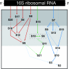
Assembly map of the 30S subunit (a kind gift from G. Culver). The 16S rRNA is represented by a rectangle, and the binding order of the ribosomal proteins is shown. The dark gray area indicates the primary binding proteins, the light gray area indicates the secondary binding proteins, and the white area indicates the tertiary binding proteins. The thick, thin, and dashed arrows show strong, weak, and very weak interactions between the proteins, respectively. Proteins S6 and S18 bind as a complex and are therefore enclosed in a dashed box. Red arrows indicate the assembly of the body, green arrows indicate the platform, and blue arrows indicate the head.
FIG. 5.
FIG. 5.
Assembly map of the 50S subunit. The 23S rRNA is represented by a rectangle with its main fragments and the binding order of the ribosomal proteins and 5S rRNA. The red arrows indicate strong dependence for binding, and the black arrows indicate weaker dependence. The blue line encloses proteins essential for RI*50(1) complex formation, and the green triangle encloses proteins essential for 5S rRNA integration. The horizontal orange line shows the division between the RI50(1) and RI50(2) proteins. (Reproduced from reference with permission of the publisher, Wiley-VCH Verlag GmbH & Co., KGaA.)

References

    1. Adilakshimi, T., P. Ramaswamy, and S. A. Woodson. 2005. Protein-independent folding pathway of the 16S rRNA 5′ domain. J. Mol. Biol. 351:508-519. - PubMed
    1. Agrawal, R. K., R. K. Lata, and J. Frank. 1999. Conformational variability in Escherichia coli 70S ribosome as revealed by 3D cryo-electron tomography. Int. J. Biochem. Cell Biol. 31:243-254. - PubMed
    1. Alix, J. H., and M. F. Guerin. 1993. Mutant DnaK chaperones cause ribosome assembly defects in Escherichia coli. Proc. Natl. Acad. Sci. USA 90:9725-9729. - PMC - PubMed
    1. Alix, J. H., D. Hayes, and K. N. Nierhaus. 1979. Properties of ribosomes and RNA synthesized by Escherichia coli grown in the presence of ethionine. V. Methylation dependence of the assembly of E. coli 50S ribosomal subunits. J. Mol. Biol. 127:375-395. - PubMed
    1. Alix, J. H., and K. N. Nierhaus. 2003. DnaK-facilitated ribosome assembly in Escherichia coli revisited. RNA 9:787-793. - PMC - PubMed

LinkOut - more resources