. 2007 Sep 1;67(17):8032-42.
doi: 10.1158/0008-5472.CAN-07-0083.

Genetic screening reveals an essential role of p27kip1 in restriction of breast cancer progression

Affiliations

Genetic screening reveals an essential role of p27kip1 in restriction of breast cancer progression

Yuhui Yuan et al. Cancer Res. .

Abstract

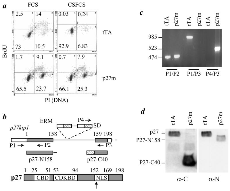
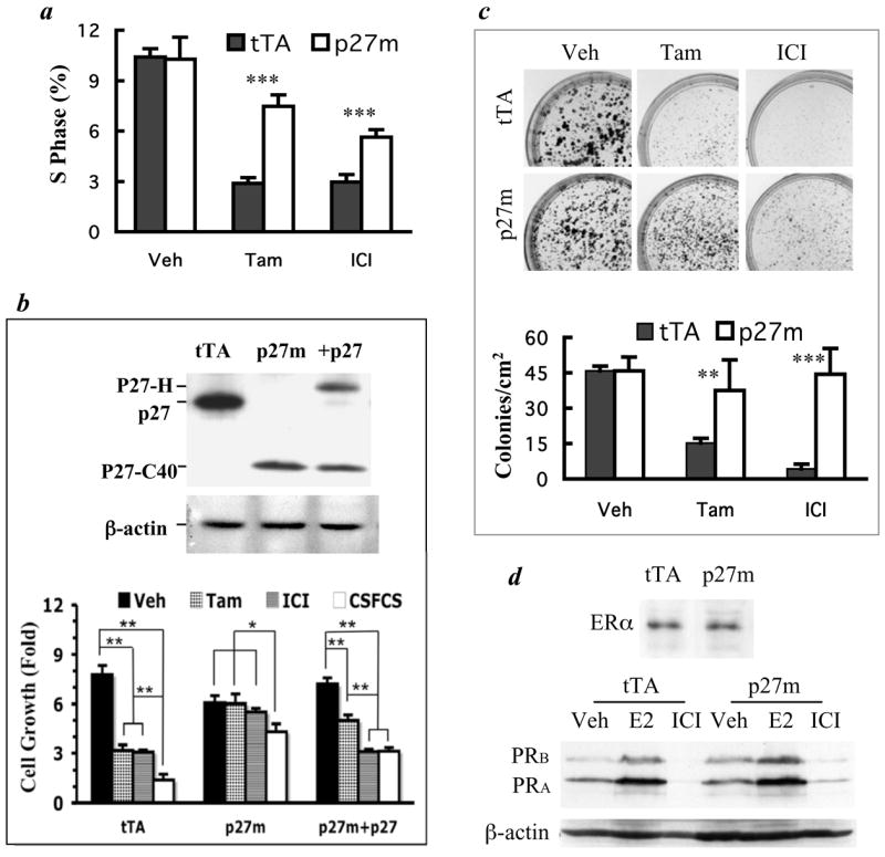
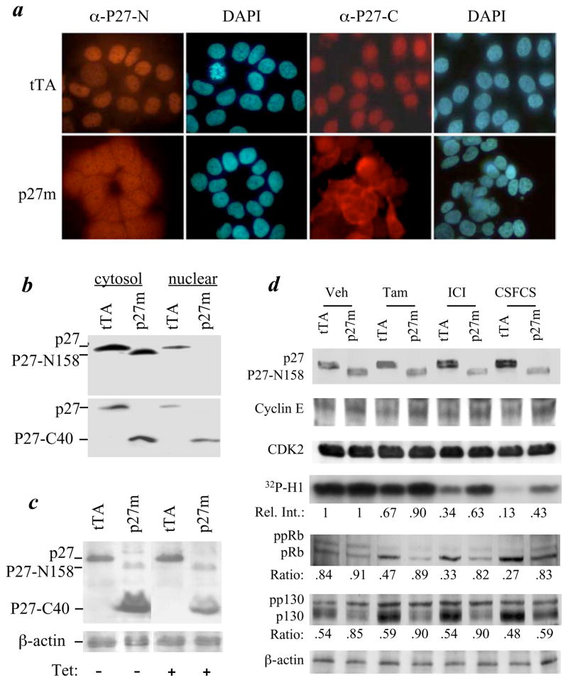
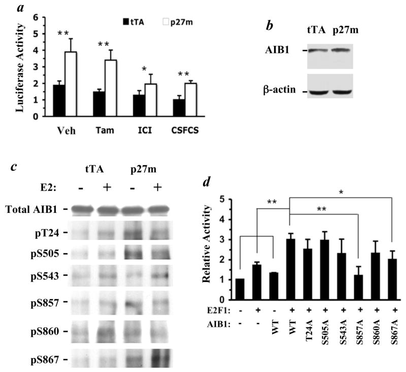
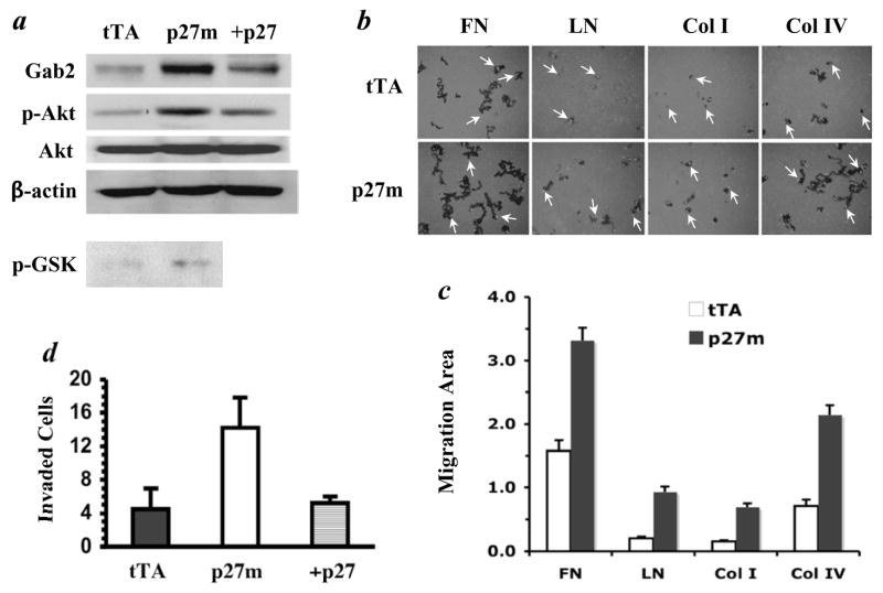
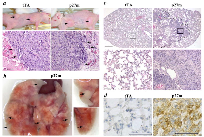
The genetic changes and mechanisms underlying the progression of estrogen-dependent breast cancers to estrogen-independent, antiestrogen-resistant, and metastatic breast cancers are unclear despite being a major problem in endocrine therapy. To identify genes responsible for this progression, we carried out a genetic screening by an enhanced retroviral mutagen (ERM)-mediated random mutagenesis in the estrogen-dependent T47D breast cancer cells. We found that T47D cells contain only one p27kip1 (p27) allele coding for the p27 cyclin-dependent kinase (CDK) inhibitor. An ERM insertion into the p27 locus of T47D cells disrupted the p27 gene and created estrogen-independent and antiestrogen-resistant breast cancer cells that still maintained functional estrogen receptors. Disruption of p27 in T47D cells resulted in several changes, and most of these changes could be rescued by p27 restoration. First, CDK2 activity was increased in the absence of estrogen or in the presence of estrogen antagonists tamoxifen or ICI 182780; second, amplified in breast cancer 1 (AIB1), a cancer overexpressed transcriptional coactivator, was hyperphosphorylated, which made AIB1 a better coactivator for E2F1; and third, growth factor receptor binding protein 2-associated binder 2 (Gab2) and Akt activity were increased following E2F1 overactivation, leading to a significant enhancement of cell migration and invasion. Furthermore, the p27-deficient cells, but not T47D control cells, developed lung metastasis in an ovarian hormone-independent manner when they were i.v. injected into nude mice. In sum, loss of p27 activated AIB1, E2F1, Gab2, and Akt; increased cell migration and invasion; caused antiestrogen insensitivity; and promoted metastasis of breast cancer cells. These findings suggest that p27 plays an essential role in restriction of breast cancer progression.


Figures

Fig. 1
Fig. 1
Disruption of p27 in T47D cells causes loss of G1/S checkpoint and estrogen-independent growth. a. T47DtTA (tTA) and T47DtTA/p27m (p27m) cells were cultured in medium containing BrdU and 10% FCS or 10% CSFCS. BrdU incorporation and DNA content per cell were analyzed by fluorescence labeling and flow cytometry. Cell fractions (%) are indicated. Note the increase in G2/M fraction of T47DtTA/p27m cells cultured in medium with FCS and the unchanged proliferation status of T47DtTA/p27m cells cultured in medium with CSFCS. b. The p27 gene structure and the insertional disruption of p27 by ERM. The first and second exon of p27 and p27 protein are outlined. The ERM insertion site and the locations of 4 primers (P1 – P4) used for PCR are indicated. White box, noncoding regions; gray box, amino acid coding regions; SD, splice donor; stretched box, the ERM tag. CBD, cyclin binding domain; CDKBD, cyclin dependent kinase binding domain; and NLS, nuclear localization signal. c. PCR analysis of the p27 gene. Note the absence of the 985-bp P1/P3 product of the wild type allele and the presence of 523-bp P4/P3 product of the ERM allele in T47DtTA/p27m cells. The 474-bp P1/P2 products were amplified from exon 1 and served as a positive control for the PCR. The ERM insertion was longer than 3 kb and thereby PCR using P1/P3 and Taq DNA polymerase was unable to amplify this long fragment in T47DtTA/p27m cells. d. Immunoblotting analyses using antibodies against p27 C-terminus (α-C) (BD Transduction Lab) and N-terminus (α-N) (Santa Cruz Biotech.). Note the absence of full-length p27 (lane 1 and 4) and the presence of p27-C40 (lane 2) and p27-N158 (lane 4) in T47DtTA/p27m cells.
Fig. 2
Fig. 2
T47DtTA/p27m cells are resistant to antiestrogens although they express functional ERα. a. T47DtTA (tTA) and T47DtTA/p27m (p27m) cells in growth medium were treated with vehicle (Veh), 4-OH-TAM (Tam) or ICI for 4 days. The mean of the S phase fraction was calculated from 4 assays of flow cytometry. ***, p < 0.001, unpaired t-test. b. p27 restoration in T47DtTA/p27m cells rescues their sensitivity to antiestrogens. Immunoblotting assay using the p27 C-terminal antibody detected p27 in T47DtTA cells, the truncated p27-C40 in T47DtTA/p27m and the His-tagged p27 (p27-H) and p27-C40 in T47DtTA/p27m/+p27 (+p27) cells (upper panel). For growth assay (lower panel), T47DtTA, T47DtTA/p27m and T47DtTA/p27m/+p27 cells (105/well, n = 4) were cultured in medium containing 10% CSFCS overnight and then changed to either the medium containing 10% FCS and Veh, Tam or ICI or the medium with 10% CSFCS. Cells were cultured for 4 days before harvested and counted. Data are presented as fold of cell number increase (Mean ± S.D.). *, p < 0.05 and **, p < 0.01 by One-Way ANOVA. c. Photographs of crystal violet-stained colonies formed from T47DtTA and T47DtTA/p27m cells treated with Veh, Tam and ICI (upper panel). Note the significantly larger and more numerous colonies formed from T47DtTA/p27m cells than T47DtTA cells treated with Tam and ICI. The lower panel presents the quantitative data of colony formation assays in the upper panel. For Veh and Tam treated cells, colonies bigger than 1 mm in diameter were counted. For ICI treated cells, colonies bigger than 0.5 mm were counted. Data are presented as mean ± SD colony numbers in eight 1-cm2 areas of culture wells in two repeat experiments. ** and ***, p < 0.01 and p < 0.001, unpaired t-tests. d. Immunoblotting analysis of ERα in T47DtTA and T47DtTA/p27m cells cultured in growth medium and PRB and PRA in T47DtTA and T47DtTA/p27m cells treated with Veh, 17β-estradiol (E2) and ICI in estrogen-free medium. Note that the PRs were increased by estradiol treatment and reduced by ICI treatment in both types of cells. Protein loading amounts were reflected by immunoblotting analysis of β-actin.
Fig. 3
Fig. 3
Subcellular localization and function of p27, p27-N158 and p27-C40 proteins and insensitive inhibition of the CDK2 kinase activity in T47DtTA/p27m cells by 4-OH-TAM (Tam), ICI and CSFCS. a. Immunocytofluorescence labeling of T47DtTA (tTA) and T47DtTA/p27m (p27m) cells with antibodies against p27 N-terminus (α-P27-N) and C-terminus (α-P27-C). Note the cytoplasmic localization of immunoactivity in T47DtTA/p27m cells detected by both antibodies. DAPI staining was used to show cell nuclei. b. Immunoblotting analysis of cytosol and nuclear fractions of T47DtTA and T47DtTA/p27m cells using α-P27-N (upper panel) and α-P27-C (lower panel) antibodies. The subcellular fractions were prepared using the NE-PER Nuclear and Cytoplasmic Extraction Reagents (Pierce Chemical). c. Immunoblotting analysis of T47DtTA and T47DtTA/p27m cells using mixed α-P27-N and α-P27-C antibodies after cells were treated with or without tetracycline (Tet). All samples were assayed in the same blot; the image of the left two lanes and the image of the right two lanes were aligned together after the image of two unused lanes between them was deleted. β-actin was assayed as a loading control. d. Treatment of T47DtTA and T47DtTA/p27m cells with vehicle (Veh), Tam, ICI or CSFCS (estrogen-free medium) and immunoblotting assays of p27, p27-N158, cyclin E, CDK2, 32P-histone H1, ppRB, pRB, pp130, P130 and β-actin control. For CDK2 kinase activity assay, the cyclin E-CDK2 complexes were immunoprecipitated by a cyclin E antibody from equal amounts of cell lysates. CDK2 kinase activity was assayed by using [γ-32P]-ATP and histone H1 as substrates. Relative band intensity (Rel. Int.) of the phosphorylated histone H1 (32P-H1) in each lane is indicated by setting the first lane as 1. For immunoblotting analyses of the phosphorylated Rb and p130, the ratios of hyper-phosphorylated Rb (ppRb) and pp130 to hypo-phosphorylated Rb (pRb) and p130 are indicated for each lane, respectively. Note that the increases in pRb and p130 were observed only in T47DtTA cells but not in T47DtTA/p27m cells after these cells were treated with Tam, ICI and CSFCS.
Fig. 4
Fig. 4
p27 deficiency enhances AIB1 phosphorylation and increases the transcriptional activity of E2F1 and AIB1. a. T47DtTA/p27m (p27m) cells have higher E2F1 activity compared with T47DtTA cells (tTA). E2F1 activity was measured by transfecting cells with an E2F1 luciferase reporter. Transfected cells were cultured in growth medium containing vehicle (Veh), tamoxifen (Tam) and ICI or in medium with 10% CSFCS for two days. Transfection efficiency was normalized to β-galactosidase activity, which was expressed by co-transfecting cells with the pRSV-β-gal vector. Data are presented by setting relative average luciferase activity in CSFCS-treated T47DtTA cells as 1 unit. b. Immunoblotting analysis, showing comparable AIB1 levels in T47DtTA and T47DtTA/p27m cells. Immunoblotting analysis of β-actin serves as a loading control. c. p27 deficiency and 17β-estradiol (E2) enhance AIB1 phosphorylation. T47DtTA and T47DtTA/p27m cells were cultured in the presence or absence of E2 for 1 hour in medium with 10% CSFCS. Total AIB1 was immunoprecipitated with AIB1 antibody and then subjected to immunoblotting analysis with antibodies specific to the phosphorylation sites as indicated. These antibodies were developed and affinity-purified as described (29). Note the total amount of precipitated AIB1 is similar among groups, but the different sites are differentially phosphorylated. d. Mutation of specific phosphorylation sites impairs AIB1 coactivator activity for E2F1-mediated target gene transcription. Cells were transfected with E2F1, E2F1-responsive luciferase reporter and AIB1 or its mutants as indicated. Luciferase activity was assayed as described under Methods. Data are presented as Mean ± S.D. (n = 4) by setting the first bar as 1 unit. *, p < 0.05; **, p < 0.01 by One-Way ANOVA.
Fig. 5
Fig. 5
Inactivation of p27 increases Gab2 protein and Akt activity, cell motility and cell invasive ability. a. p27 deficiency increases Gab2 protein and Akt activity. Gab2 levels in T47DtTA (tTA), T47DtTA/p27m (p27m) and T47DtTA/p27m/+27 (+27) cells were assayed by immunoblotting. Total Akt and phosphorylated active Akt in these cells were detected by respective Akt antibody and phosphorylation site-specific antibody by immunoblotting. Akt activity in T47DtTA and T47DtTA/p27m cells was assayed using GSK-3β as substrates and the phosphorylated GSK-3β (p-GSK-3β) was detected by immunoblotting using a p-GSK-3β-specific antibody. The β-actin assay served as a loading control. b. Measurement of cell migration. T47DtTA and T47DtTA/p27m cells were cultured on plates coated with FN, LN, Col I or Col IV and cell migration was traced by using blue fluorescence beads. Individual cells (arrows) were stained with Rhodamine-Phalloidin. Dark areas indicate cell migration tracks. Cells migrated faster on FN and Col IV. c. Measurement of migration areas. Cell migration tracks of 25 to 30 cells were analyzed by using NIH image software. Data are presented as average (relative migration area) ± standard deviation. The migration areas of T47DtTA/p27m cells on all coated matrices examined are significantly bigger compared with T47DtTA cells (p < 0.01, unpaired t test). d. Cell invasion assay. The invasive capability of T47DtTA, T47DtTA/p27m and T47DtTA/p27m/+27 in the matrigel was measure as describe in Materials and Methods. Data represent the mean of 3 independent experiments.
Fig. 6
Fig. 6
Primary and metastatic tumor formation in ovariectomized nude mice. a. The upper panels show the primary tumors (arrow heads) formed from T47DtTA (tTA) and T47DtTA/p27m (p27m) cells in nude mice with 17β-estradiol pellets. The lower panels show the images of H&E stained tumor sections prepared from T47DtTA and T47DtTA/p27m tumors. Note the different morphologies of the tumor edges indicated by arrowheads in the two types of tumors. The scale bars in the lower panels represent 50 μm. b. Photographs of lungs from different ovariectomized nude mice receiving T47DtTA/p27m cells from their tail veins. Arrowheads indicate visible metastatic lung tumors. c. H&E stained lung sections prepared from ovariectomized nude mice receiving intravenous injection of T47DtTA and T47DtTA/p27m cells. An invasive big lung tumor is outlined in the right upper panel. The lower panels are larger images of the boxed areas in the upper panels. The scale bars for the upper and lower panels represent 500 and 50 μm, respectively. d. Immunohistochemical staining of K8 (brown color) on lung sections prepared from ovariectomized nude mice injected with T47DtTA or T47DtTA/p27m cells as indicated. The scale bars represent 50 μm.

Similar articles

Cited by

References

    1. Doisneau-Sixou SF, Sergio CM, Carroll JS, Hui R, Musgrove EA, Sutherland RL. Estrogen and antiestrogen regulation of cell cycle progression in breast cancer cells. Endocr Relat Cancer. 2003;10:179–86. - PubMed
    1. Foster JS, Fernando RI, Ishida N, Nakayama KI, Wimalasena J. Estrogens down-regulate p27Kip1 in breast cancer cells through Skp2 and through nuclear export mediated by the ERK pathway. J Biol Chem. 2003;278:41355–66. - PubMed
    1. Jordan VC. Third annual William L. McGuire Memorial Lecture “Studies on the estrogen receptor in breast cancer”--20 years as a target for the treatment and prevention of cancer. Breast Cancer Res Treat. 1995;36:267–85. - PubMed
    1. Schiff R, Massarweh S, Shou J, Osborne CK. Breast cancer endocrine resistance: how growth factor signaling and estrogen receptor coregulators modulate response. Clin Cancer Res. 2003;9(Pt 2):447S–54S. - PubMed
    1. Brodie A. Aromatase inhibitor development and hormone therapy: a perspective. Semin Oncol. 2003;30(Suppl 14):12–22. - PubMed

Publication types

MeSH terms