Comparative Study
. 2007 Sep 11;104(37):14825-30.
doi: 10.1073/pnas.0705046104. Epub 2007 Sep 5.

Astrocytes regulate GluR2 expression in motor neurons and their vulnerability to excitotoxicity

Affiliations
Comparative Study

Astrocytes regulate GluR2 expression in motor neurons and their vulnerability to excitotoxicity

Philip Van Damme et al. Proc Natl Acad Sci U S A. .

Abstract

Influx of Ca(2+) ions through alpha-amino-3-hydroxy-5-methylisoxazole-4-propionic acid (AMPA) receptors contributes to neuronal damage in stroke, epilepsy, and neurodegenerative disorders such as ALS. The Ca(2+) permeability of AMPA receptors is largely determined by the glutamate receptor 2 (GluR2) subunit, receptors lacking GluR2 being permeable to Ca(2+) ions. We identified a difference in GluR2 expression in motor neurons from two rat strains, resulting in a difference in vulnerability to AMPA receptor-mediated excitotoxicity both in vitro and in vivo. Astrocytes from the ventral spinal cord were found to mediate this difference in GluR2 expression in motor neurons. The presence of ALS-causing mutant superoxide dismutase 1 in astrocytes abolished their GluR2-regulating capacity and thus affected motor neuron vulnerability to AMPA receptor-mediated excitotoxicity. These results reveal a mechanism through which astrocytes influence neuronal functioning in health and disease.


Conflict of interest statement

The authors declare no conflict of interest.

Figures

Fig. 1.
Fig. 1.
Wistar motor neurons have AMPA receptors with a higher Ca2+ permeability and are more vulnerable to excitotoxicity. (A) Coculture of motor neurons (SMI32; red) grown on a preestablished monolayer of astrocytes (GFAP; green). (Scale bar, 20 μm.) (B) PCa/PNa values of AMPA receptor currents in motor neurons when cultured on astrocytes derived from the same rat strain (W/W, Wistar motor neurons on Wistar astrocytes; H/H, Holtzman motor neurons on Holtzman astrocytes; n = 20–23; *, P = 0.017). (C) Ca2+ transients measured with the low-affinity dye Indo-1FF and induced by application of 300 μM kainate in the presence of 100 μM verapamil in cultured Wistar motor neurons (n = 19) compared with Holtzman motor neurons (n = 18; *, P = 0.014). (D) Vulnerability of cultured Wistar and Holtzman motor neurons to AMPA receptor-mediated excitotoxicity induced by a short (30-min) exposure to 300 μM kainate (n = 12–14; *, P = 0.0001).
Fig. 2.
Fig. 2.
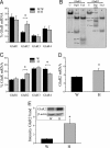
GluR2 mRNA and protein expression in Wistar and Holtzman. (A) Relative GluR mRNA expression in cultured Wistar and Holtzman motor neurons determined by single-cell RT-PCR (n = 14–18; *, P = 0.018). (B) Example of analysis of editing efficiency of GluR2 in a single-cell sample from a Wistar motor neuron. Lane 1, Undigested (U) PCR fragment of PCR specific for GluR1; lane 2, same fragment as in lane 1, digested with BglI (cleaves only GluR1 fragment); lane 3, same fragment as in lane 1, digested with TseI (which cleaves the 190-bp band into two smaller bands when not edited); thus, GluR1 is completely unedited; lane 4, undigested PCR fragment of PCR specific for GluR2; lane 5, same fragment as in lane 4, digested with Bsp 1286I (cleaves only GluR2 fragment); lane 6, same fragment as in 4, digested with TseI (which only cleaves the 190-bp band when not edited). Thus, GluR2 is completely edited. (C) Relative GluR mRNA expression in the ventral spinal cord from Wistar and Holtzman rats determined by RT-PCR (n = 5–7; *, P ≤ 0.01). (D) Real-time PCR for GluR2 normalized to 18S RNA with SYBR green on cDNA prepared from the ventral part of the spinal cord of Wistar (W) and Holtzman (H) rats (n = 8; *, P = 0.04). (E) Western blot of GluR2 in the ventral part of the spinal cord from Wistar and Holtzman rats (n = 14–16; P = 0.002). Equal loading was demonstrated by β-actin staining, and the intensities of bands were normalized to the β-actin signal.
Fig. 3.
Fig. 3.
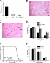
Differences in vulnerability to excitotoxicity between both rat strains in vivo. (A) Motor score after spinal cord ischemia [0 = normal, 6 = complete paraplegia (18); n = 6–8; *, P < 0.05, different from Wistar]. (B and C) H&E staining of spinal cord section after spinal cord ischemia induced by clamping of the aortic arch and left subclavian artery for 14 min from a Wistar rat (B) and a Holtzman rat (C). gm, gray matter; wm, white matter. (Scale bar, 50 μm.) Quantification of number of neurons (divided into three size categories) in the ventral horn of the lumbar spinal cord after spinal cord ischemia, with or without pretreatment with the AMPA receptor antagonist, NBQX (n = 4–8; *, P < 0.05). Survival of Wistar and Holtzman mt SOD1 rats (n = 55–62 per group). Quantification of number of neurons (divided into three size categories) in the ventral horn of the lumbar spinal cord of end-stage Wistar and Holtzman mt SOD1 rats (n = 3; P > 0.3).
Fig. 4.
Fig. 4.
Astrocytes regulate GluR2 expression in motor neurons. (A) Schematic representation of the different motor neuron and astrocyte combinations. W/W, Wistar motor neurons on Wistar astrocytes; H/H, Holtzman motor neurons on Holtzman astrocytes; W/H, Wistar motor neurons on Holtzman astrocytes; H/W, Holtzman motor neurons on Wistar astrocytes. (B) Functional determination of the GluR2 expression in motor neurons when cultured on astrocytes from the same and the other rat strain. The PCa/PNa values of AMPA receptor currents are shown (n = 21–23, W/W and H/W not different; *, P < 0.05, significantly different from W/W and H/W). (C) Kainate-induced AMPA receptor-mediated excitotoxicity in W/W, W/H, H/H, and H/W (n = 4–7, W/W and H/W not different; *, P ≤ 0.005, significantly different from W/W and H/W). (D) Relative GluR mRNA expression determined by single-cell RT-PCR in W/W, W/H, H/H, and H/W (n = 21–26; *, P ≤ 0.03, significantly different from neurons cultured on Wistar astrocytes). (E) Effect of presence of astrocytes (A) or ACM (added directly after seeding, for 48 h) on luciferase activity in cortical neurons (normalized to β-galactosidase activity and background activity of empty vector; n = 5–10; *, P < 0.05). (F) AMPA receptor-mediated excitotoxicity in Wistar motor neurons grown on Holtzman astrocytes from different regions. VSC, ventral spinal cord; DSC, dorsal spinal cord; CER, cerebellum; CTX, cortex. n = 3–7; *, P < 0.025, different from other groups. (G) AMPA receptor-mediated excitotoxicity in W/W in the presence of astrocytic membrane fractions prepared from Wistar or Holtzman astrocytes (n = 3; P = 0.03). (H) AMPA receptor-mediated excitotoxicity in W/W in the presence of ACM from Wistar or Holtzman astrocytes, with or without pretreatment with heat or trypsin (n = 4; *, P < 0.015, significantly different from other groups).
Fig. 5.
Fig. 5.
mt SOD1 in astrocytes abolishes their GluR2-regulating capacity. (A) PCa/PNa values of AMPA receptor currents in W/W, H/H, W/H, and W/H (n = 11–23; *, P < 0.03). (B) AMPA receptor-mediated excitotoxicity in W/W, H/H, W/H, and W/H (n = 5–18; *, P < 0.04). (C) AMPA receptor-mediated excitotoxicity in Wistar motor neurons with or without mt SOD1 grown on different combinations of astrocytes with or without mt SOD1 (n = 4–9; *, P ≤ 0.01, significantly different from all other groups). (D) Relative GluR mRNA expression in W/H and W/HG93A (n = 14–17; *, P < 0.04). (E) Effect of presence of mt SOD1 in Holtzman astrocytes on luciferase activity in cortical neurons seeded on Holtzman astrocytes (normalized to β-galactosidase activity and background activity of empty vector, n = 6–8; *, P = 0.015). (F) Relative GluR mRNA expression in the ventral spinal cord from Wistar and Holtzman rats with or without mt SOD1 determined by RT-PCR (n = 5–7; *, P ≤ 0.01, significantly different from other groups). (G) Real-time PCR for GluR2 normalized to 18S RNA with SYBR green on cDNA prepared from the ventral part of the spinal cord of Wistar and Holtzman rats with or without mt SOD1 (n = 8–10; *, P < 0.04, significantly different from other groups). (H) Western blot of GluR2 in the ventral part of the spinal cord from Wistar and Holtzman rats with or without mt SOD1 (n = 8–11; *, P < 0.05). Equal loading was demonstrated by β-actin staining, and the intensity of bands was normalized to the β-actin signal.

Similar articles

Cited by

References

    1. Lipton SA, Rosenberg PA. N Engl J Med. 1994;330:613–622. - PubMed
    1. Van Den Bosch L, Van Damme P, Bogaert E, Robberecht W. Biochim Biophys Acta. 2006;1762:1068–1082. - PubMed
    1. Hollmann M, Hartley M, Heinemann S. Science. 1991;252:851–853. - PubMed
    1. Sommer B, Kohler M, Sprengel R, Seeburg PH. Cell. 1991;67:11–19. - PubMed
    1. Gorter JA, Petrozzino JJ, Aronica EM, Rosenbaum DM, Opitz T, Bennett MV, Connor JA, Zukin RS. J Neurosci. 1997;17:6179–6188. - PMC - PubMed

Publication types

MeSH terms

LinkOut - more resources