. 2007 Sep 11;104(37):14700-5.
doi: 10.1073/pnas.0702305104. Epub 2007 Sep 5.

R-Spondin1 regulates Wnt signaling by inhibiting internalization of LRP6

Affiliations

R-Spondin1 regulates Wnt signaling by inhibiting internalization of LRP6

Minke E Binnerts et al. Proc Natl Acad Sci U S A. .

Abstract

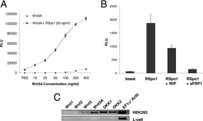
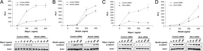
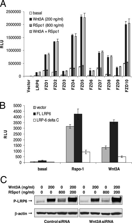
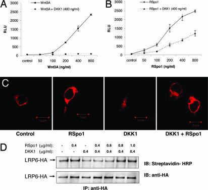
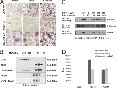
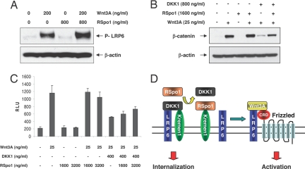
The R-Spondin (RSpo) family of secreted proteins act as potent activators of the Wnt/beta-catenin signaling pathway. We have previously shown that RSpo proteins can induce proliferative effects on the gastrointestinal epithelium in mice. Here we provide a mechanism whereby RSpo1 regulates cellular responsiveness to Wnt ligands by modulating the cell-surface levels of the coreceptor LRP6. We show that RSpo1 activity critically depends on the presence of canonical Wnt ligands and LRP6. Although RSpo1 does not directly activate LRP6, it interferes with DKK1/Kremen-mediated internalization of LRP6 through an interaction with Kremen, resulting in increased LRP6 levels on the cell surface. Our results support a model in which RSpo1 relieves the inhibition DKK1 imposes on the Wnt pathway.


Conflict of interest statement

The authors declare no conflict of interest.

Figures

Fig. 1.
Fig. 1.
RSpo1 activity is linked to canonical Wnt signaling. (A) HEK-293 cells stably expressing a 16TCF luciferase reporter (HEK-293 A6) were treated with recombinant Wnt3A protein in the absence or presence of a low dose of recombinant RSpo1 protein (20 ng/ml). (B) HEK-293 A6 cells were incubated with RSpo1 (200 ng/ml) in the presence or absence of recombinant Wnt pathway inhibitors WIF (1 μg/ml) and sFRP1 (1 μg/ml). (C) RSpo1-responsive HEK-293 cells and nonresponsive mouse L-cells were analyzed for mRNA expression of canonical Wnt ligands and Wnt inhibitors by semiquantitative RT-PCR.
Fig. 2.
Fig. 2.
RSpo1 activity in HEK-293 cells depends critically on an endogenous pool of canonical Wnt ligands. HEK-293 A6 cells were transfected with Wnt3A or control siRNA duplexes (50 nM), treated with increasing amounts of RSpo1 (A) or Wnt3A (B) proteins, and assayed for luciferase reporter activity or cytosolic β-catenin levels. HEK-293 A6 cells were transfected with WLS or control siRNA duplexes (50 nM), treated with increasing amounts of recombinant RSpo1 (C), or Wnt3A (D) protein and analyzed as described above.
Fig. 3.
Fig. 3.
RSpo1 regulates Wnt signaling at the level of the LRP6 receptor. (A) S2 cells were transfected with 16TCF luciferase reporter, TCF4, hLRP6, and hFZD1–10 expression constructs, followed by treatment with Wnt3A (200 ng/ml) or RSpo1 (800 ng/ml) recombinant proteins or a combination of RSpo1 and Wnt3A. (B) HEK-293 A6 cells were transfected with WT LRP6 or LRP6ΔC expression constructs and treated with RSpo1 (200 ng/ml) or Wnt3A (200 ng/ml) proteins, and luciferase reporter activity was determined. (C) HEK-293 cells transfected with control or Wnt3A siRNA duplexes (50 nM) were treated 48 hours after transfection with RSpo1 (800 ng/ml), Wnt3A (200 ng/ml), or with a combination of RSpo1 and Wnt3A proteins. LRP6 phosphorylation was analyzed 4 hours after treatment by using a phospho-Ser-1490-specific anti-LRP6 antibody.
Fig. 4.
Fig. 4.
RSpo1 inhibits DKK1/Kremen1-mediated internalization of LRP6. HEK-293 A6 cells were treated with recombinant DKK1 protein (400 ng/ml) in the presence of increasing amounts of recombinant Wnt3A (A) or RSpo1 (B) proteins, and TCF reporter activity was determined. (C) HEK-293 cells were cotransfected with LRP6-HA and Kremen1 constructs and treated for 10 minutes with recombinant DKK1 (200 ng/ml) in the presence or absence of RSpo1 protein (800 ng/ml). Distribution of immunostained, HA-tagged LRP6 was evaluated by confocal microscopy. (D) HEK-293 cells transfected with LRP6-HA and Kremen1 constructs were treated with recombinant DKK1 in the presence or absence of recombinant RSpo1 for 1 hour. Cell-surface proteins were biotinylated and HA-LRP6 was immunoprecipitated with anti-HA mAb and analyzed with streptavidin-HRP or anti-HA mAb.
Fig. 5.
Fig. 5.
RSpo1 interacts with Kremen. (A) HEK-293T cells transiently transfected with LRP6-HA, Kremen1, or control plasmids were incubated with DKK1-AP (15 nM) conditioned medium or with RSpo1-AP (5 nM) conditioned medium in the presence of heparin (2 μg/ml) and were stained for bound AP activity. HEK-293T cells transfected as described above were incubated with RSpoΔC-AP (180 nM) in the absence of heparin and stained for bound AP activity. (B) Recombinant DKK1 or RSpo1 proteins were mixed with monomeric recombinant LRP6 ECD, and complex formation was analyzed by monitoring coelution of injected proteins on a analytical size-exclusion chromatography column. (C) HEK-293 cells stably expressing LRP6-Flag were transfected with Kremen1-HA and subjected to coimmunoprecipitation with anti-HA antibodies. Immunoprecipitates were analyzed by Western blotting by using the indicated antibodies. (D) HEK-293 A6 reporter cells were transfected with Kremen2 siRNA complexes (2 nM), treated with recombinant RSpo1 (200 ng/ml) or Wnt3A (200 ng/ml) 48 hours after transfection, and analyzed for reporter activity 18 hours after treatment.
Fig. 6.
Fig. 6.
Analysis of RSpo1 signaling in RSpo1 nonresponsive L-cells. (A) Mouse L-cells were treated with recombinant Wnt3A (200 ng/ml), RSpo1 (800 ng/ml), or a combination of RSpo1 and Wnt3A and analyzed for LRP6 phosphorylation 4 hours after treatment, as described above. Mouse L-cells were treated with Wnt3A, DKK1, or RSpo1 proteins as indicated and analyzed for cytosolic β-catenin levels (B) or 16TCF reporter activation (C). (D) Model of RSpo1 function. Wnt/β-catenin signaling is initiated upon binding of a canonical Wnt ligand to Frizzled and association with LRP5/6 receptors. In the absence of RSpo1, Wnt signaling is limited by the amount of LRP6 on the cell surface, which is kept low by DKK1/Kremen1-mediated internalization of LRP6. RSpo1 enhances Wnt signaling by antagonizing DKK1/Kremen-mediated LRP6 turnover, resulting in increased cell surface levels of LRP6.

Similar articles

Cited by

References

    1. Logan CY, Nusse R. Annu Rev Cell Dev Biol. 2004;20:781–810. - PubMed
    1. Moon RT, Kohn AD, De Ferrari GV, Kaykas A. Nat Rev Genet. 2004;5:691–701. - PubMed
    1. Bejsovec A. Cell. 2005;120:11–14. - PubMed
    1. Gordon MD, Nusse R. J Biol Chem. 2006;281:22429–22433. - PubMed
    1. He X, Semenov M, Tamai K, Zeng X. Development (Cambridge, UK) 2004;131:1663–1677. - PubMed

Publication types

MeSH terms