

Disrupted cardiac development but normal hematopoiesis in mice deficient in the second CXCL12/SDF-1 receptor, CXCR7
- PMID: 17804806
- PMCID: PMC1976222
- DOI: 10.1073/pnas.0702229104
Disrupted cardiac development but normal hematopoiesis in mice deficient in the second CXCL12/SDF-1 receptor, CXCR7
Abstract
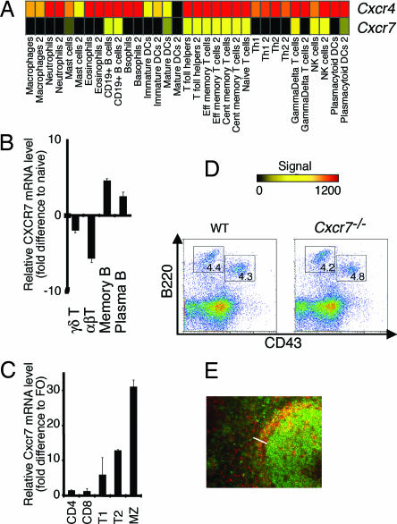
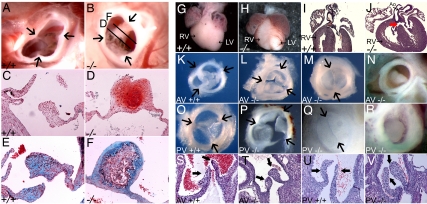
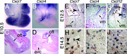
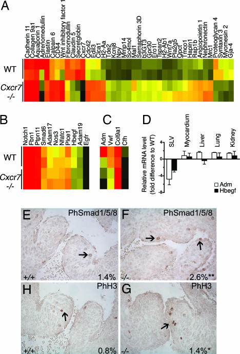
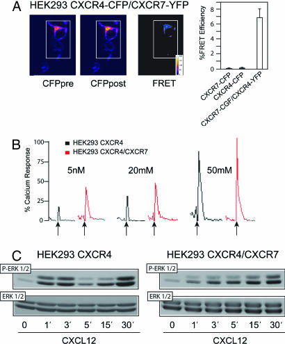
Chemotactic cytokines (chemokines) attract immune cells, although their original evolutionary role may relate more closely with embryonic development. We noted differential expression of the chemokine receptor CXCR7 (RDC-1) on marginal zone B cells, a cell type associated with autoimmune diseases. We generated Cxcr7(-/-) mice but found that CXCR7 deficiency had little effect on B cell composition. However, most Cxcr7(-/-) mice died at birth with ventricular septal defects and semilunar heart valve malformation. Conditional deletion of Cxcr7 in endothelium, using Tie2-Cre transgenic mice, recapitulated this phenotype. Gene profiling of Cxcr7(-/-) heart valve leaflets revealed a defect in the expression of factors essential for valve formation, vessel protection, or endothelial cell growth and survival. We confirmed that the principal chemokine ligand for CXCR7 was CXCL12/SDF-1, which also binds CXCR4. CXCL12 did not induce signaling through CXCR7; however, CXCR7 formed functional heterodimers with CXCR4 and enhanced CXCL12-induced signaling. Our results reveal a specialized role for CXCR7 in endothelial biology and valve development and highlight the distinct developmental role of evolutionary conserved chemokine receptors such as CXCR7 and CXCR4.
Conflict of interest statement
The authors declare no conflict of interest.
Figures
Similar articles
-
The chemokine receptor CXCR7 functions to regulate cardiac valve remodeling.Dev Dyn. 2011 Feb;240(2):384-93. doi: 10.1002/dvdy.22549. Dev Dyn. 2011. PMID: 21246655 Free PMC article.
-
A novel chemokine receptor for SDF-1 and I-TAC involved in cell survival, cell adhesion, and tumor development.J Exp Med. 2006 Sep 4;203(9):2201-13. doi: 10.1084/jem.20052144. Epub 2006 Aug 28. J Exp Med. 2006. PMID: 16940167 Free PMC article.
-
Pattern of CXCR7 Gene Expression in Mouse Brain Under Normal and Inflammatory Conditions.J Neuroimmune Pharmacol. 2016 Mar;11(1):26-35. doi: 10.1007/s11481-015-9616-y. Epub 2015 May 22. J Neuroimmune Pharmacol. 2016. PMID: 25997895 Free PMC article.
-
The role of the CXCL12-CXCR4/CXCR7 axis in the progression and metastasis of bone sarcomas (Review).Int J Mol Med. 2013 Dec;32(6):1239-46. doi: 10.3892/ijmm.2013.1521. Epub 2013 Oct 11. Int J Mol Med. 2013. PMID: 24127013 Review.
-
The peculiarities of the SDF-1/CXCL12 system: in some cells, CXCR4 and CXCR7 sing solos, in others, they sing duets.Cell Tissue Res. 2014 Feb;355(2):239-53. doi: 10.1007/s00441-013-1747-y. Epub 2013 Nov 29. Cell Tissue Res. 2014. PMID: 24292718 Review.
Cited by
-
CXCR4 or CXCR7 antagonists treat endometriosis by reducing bone marrow cell trafficking.J Cell Mol Med. 2020 Feb;24(4):2464-2474. doi: 10.1111/jcmm.14933. Epub 2020 Jan 6. J Cell Mol Med. 2020. PMID: 31904910 Free PMC article.
-
IL-6R/Signal Transducer and Activator of Transcription 3 Signaling in Keratinocytes rather than in T Cells Induces Psoriasis-Like Dermatitis in Mice.J Invest Dermatol. 2022 Apr;142(4):1126-1135.e4. doi: 10.1016/j.jid.2021.09.012. Epub 2021 Oct 7. J Invest Dermatol. 2022. PMID: 34626614 Free PMC article.
-
Transcriptional Changes in Pulmonary Phagocyte Subsets Dictate the Outcome Following Interaction With The Fungal Pathogen Cryptococcus neoformans.Front Immunol. 2021 Sep 28;12:722500. doi: 10.3389/fimmu.2021.722500. eCollection 2021. Front Immunol. 2021. PMID: 34650554 Free PMC article.
-
Decreased ACKR3 (CXCR7) function causes oculomotor synkinesis in mice and humans.Hum Mol Genet. 2019 Sep 15;28(18):3113-3125. doi: 10.1093/hmg/ddz137. Hum Mol Genet. 2019. PMID: 31211835 Free PMC article.
-
Regulation of neutrophil trafficking from the bone marrow.Cell Mol Life Sci. 2012 May;69(9):1415-23. doi: 10.1007/s00018-011-0870-8. Epub 2011 Nov 2. Cell Mol Life Sci. 2012. PMID: 22045556 Free PMC article. Review.
References
-
- Doitsidou M, Reichman-Fried M, Stebler J, Koprunner M, Dorries J, Meyer D, Esguerra CV, Leung T, Raz E. Cell. 2002;111:647–659. - PubMed
-
- Mackay CR. Nat Immunol. 2001;2:95–101. - PubMed
-
- Zou YR, Kottmann AH, Kuroda M, Taniuchi I, Littman DR. Nature. 1998;393:595–599. - PubMed
-
- Nagasawa T, Hirota S, Tachibana K, Takakura N, Nishikawa S, Kitamura Y, Yoshida N, Kikutani H, Kishimoto T. Nature. 1996;382:635–638. - PubMed
Publication types
MeSH terms
Substances
Associated data
- Actions
Grants and funding
LinkOut - more resources
Full Text Sources
Other Literature Sources
Molecular Biology Databases
Research Materials
Miscellaneous
