. 2007 Nov;18(11):4420-37.
doi: 10.1091/mbc.e07-02-0149. Epub 2007 Sep 5.

ARL4D recruits cytohesin-2/ARNO to modulate actin remodeling

Affiliations

ARL4D recruits cytohesin-2/ARNO to modulate actin remodeling

Chun-Chun Li et al. Mol Biol Cell. 2007 Nov.

Abstract

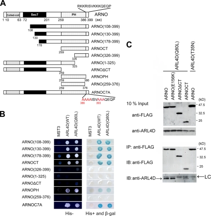
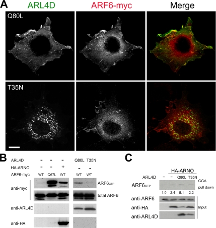
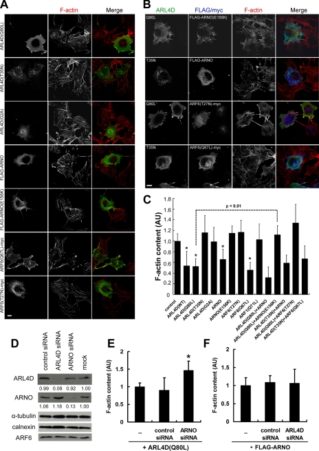
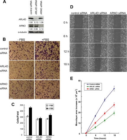
ARL4D is a developmentally regulated member of the ADP-ribosylation factor/ARF-like protein (ARF/ARL) family of Ras-related GTPases. Although the primary structure of ARL4D is very similar to that of other ARF/ARL molecules, its function remains unclear. Cytohesin-2/ARF nucleotide-binding-site opener (ARNO) is a guanine nucleotide-exchange factor (GEF) for ARF, and, at the plasma membrane, it can activate ARF6 to regulate actin reorganization and membrane ruffling. We show here that ARL4D interacts with the C-terminal pleckstrin homology (PH) and polybasic c domains of cytohesin-2/ARNO in a GTP-dependent manner. Localization of ARL4D at the plasma membrane is GTP- and N-terminal myristoylation-dependent. ARL4D(Q80L), a putative active form of ARL4D, induced accumulation of cytohesin-2/ARNO at the plasma membrane. Consistent with a known action of cytohesin-2/ARNO, ARL4D(Q80L) increased GTP-bound ARF6 and induced disassembly of actin stress fibers. Expression of inactive cytohesin-2/ARNO(E156K) or small interfering RNA knockdown of cytohesin-2/ARNO blocked ARL4D-mediated disassembly of actin stress fibers. Similar to the results with cytohesin-2/ARNO or ARF6, reduction of ARL4D suppressed cell migration activity. Furthermore, ARL4D-induced translocation of cytohesin-2/ARNO did not require phosphoinositide 3-kinase activation. Together, these data demonstrate that ARL4D acts as a novel upstream regulator of cytohesin-2/ARNO to promote ARF6 activation and modulate actin remodeling.


Figures

Figure 1.
Figure 1.
ARNO interacts with ARL4D. (A) Schematic representation of ARL4D molecule and mutants. G1–G3 are conserved regions important for binding to phosphate/Mg2+ and the guanine base. ARL4D(Q80L) is a putative GTPase-defective mutant, and it is predicted to exist in the GTP-bound active form. ARL4D(T35N) is a putative GTP-binding-defective mutant, and it is predicted to be in a GDP-bound form. ARL4DΔC lacks 16 a.a. (a.a. 186-201) at the C terminus, which contain the bipartite NLS. ARL4D(G2A) is a N-myristoylation–deficient mutant. (B) ARL4D and ARL4A interacted with ARNO in a yeast two-hybrid system. Yeast reporter strain L40 cotransformed with the Gal4 AD fusion constructs of ARNO and the LexA BD fusion of the indicated constructs were grown on synthetic histidine-containing medium lacking leucine and tryptophan (His+ plate) and assayed for β-galactosidase activity by a filter assay. Colonies from His+ plates were also grown on His-minus selective medium lacking histidine, leucine, and tryptophan (His− plate) for a growth assay. (C) ARL4D coprecipitated with ARNO in vitro. Lysates of 293T cells expressing ARL4D or its mutants were incubated with either GST or GST-ARNO immobilized on glutathione beads. Bead-bound ARL4D was probed using anti-ARL4D antibody. Ten percent of the cell lysate (Input) is shown for each of the experiments. The equal input of GST fusion proteins used in the assay and visualized by Coomassie staining is shown on the bottom. (D) Interaction between ARL4D and ARNO. 293T cells cotransfected with ARL4D constructs and FLAG-ARNO were lysed and immunoprecipitated with anti-FLAG M2 affinity agarose gel. Bound proteins were separated by SDS-PAGE and subjected to immunoblot with anti-FLAG and anti-ARL4D antibodies. Ten percent of the cell lysate (Input) was loaded to show the expression level.
Figure 2.
Figure 2.
The PH domain of ARNO interacts with ARL4D. (A) Schematic representation of ARNO and its deletion mutants. ARNO contains a coiled-coil domain (a.a. 10-63), Sec7 domain (a.a. 72-201), PH domain, and polybasic c domain (a.a.386-399). (B) ARL4D and its activated mutants fused to the DNA BD of LexA were cotransformed with deletion mutants of ARNO fused to the Gal4 AD into yeast strain L40, and the transformants were tested for their ability to grow in the absence of histidine (left) and to express β-galactosidase (right). Mammalian STE20-like protein kinase 3 (MST3) was a negative control. (C) Interaction of ARL4D and ARNO is mediated by the C terminus of ARNO. COS-7 cells transfected with the indicated expression vectors were treated with the reducible cross-linker DSP before cells were lysed. Proteins immunoprecipitated from cell lysates with an anti-FLAG M2 affinity agarose gel were separated by SDS-PAGE and immunoblotted with anti-ARL4D or anti-FLAG antibodies. ARL4D(Q80L) was selectively coimmunoprecipitated with FLAG-ARNO constructs containing the C-terminal PH domain and polybasic c domain. The arrows indicate the immunoglobulin light chain (LC) of antibody.
Figure 3.
Figure 3.
Subcellular localization of ARL4D and its mutants. (A) Localization of overexpressed untagged ARL4D. COS-7 cells grown on coverslips were transiently transfected with plasmids encoding ARL4D or its mutants. Forty-eight hours after transfection, cells were fixed, permeabilized, and processed for immunofluorescence with anti-ARL4D antibody. The stacked images were obtained by using a confocal microscope. (B) Detection of endogenous ARL4D by immunoblotting. Total HeLa cell lysate was separated by SDS-PAGE and probed with anti-ARL4D-B antibody alone, or anti-ARL4D-B competed by 1 μg of immunized ARL4D-B peptide, or nonimmunized ARL4D-N peptide dissolved in DMSO. DMSO was used as a mock control. (C) Subcellular distribution of endogenous ARL4D by fractionation. HeLa cells were homogenized and nuclear (N), PNS, cytosolic (C), and membrane (M) fractions prepared as described in Materials and Methods. Equivalent amounts of proteins were separated by SDS-PAGE and analyzed by Western blot by using specific antibodies against ARL4D-B, HP1α (nuclear marker), α-tubulin (cytosol marker), and Na+/K+ATPase (membrane marker). Asterisk (*) indicates a nonspecific band detected in cytosolic fraction. This band might be a degradation product from an unknown protein (∼40-kDa), which was cross-reacted with the ARL4D antibody in the total cell lysate. (D) Subcellular localization of endogenous ARL4D in HeLa cells. Endogenous ARL4D or plasma membrane calcium pump pan PMCA ATPase was detected by immunofluorescence staining with anti-ARL4D-B or anti-PMCA antibody, respectively. The plasma membrane labeling (arrow) was abolished when the antibody was preincubated with ARL4D-B peptide but not by DMSO (mock control) or ARL4D-N peptide. Bar, 10 μm. Peptide competition assays were performed as described in Materials and Methods.
Figure 4.
Figure 4.
ARL4D induces ARNO redistribution to plasma membrane protrusions and ruffles. COS-7 cells were transfected with FLAG-ARNO alone (A) or cotransfected with FLAG-ARNO and ARL4D mutants (B). Cells were fixed, permeabilized, labeled with anti-FLAG M2 and anti-ARL4D antibody and examined by confocal microscopy. Staining intensities measured according to pixel brightness were evaluated by the fluorescence intensity (F.I.) profile of a typical line scan for ARNO in a representative positively stained cell. (C) Quantification of ARNO localization in ARL4D-coexpressing cells. The ratios of the average of the two fluorescence signals of the plasma membrane (PM) to the average signal of the cytosol (CS) were evaluated. (D) Quantification of translocation of ARNO to plasma membrane protrusions and ruffles in ARL4D-expressing cells. The method for quantifying the ARNO plasma membrane localization is described in Materials and Methods. At least 50 cells were analyzed for each condition. Results are the means ± SD of three independent experiments. *p < 0.001 compared with ARNO alone. Bars, 10 μm.
Figure 5.
Figure 5.
ARL4D promotes activation of ARF6. (A) ARL4D(Q80L) and ARF6 colocalize along plasma membrane ruffles and protrusions. COS-7 cells cotransfected with vectors encoding ARL4D(Q80L) or ARL4D(T35N) together with ARF6-myc were processed for immunofluorescence with ARL4D and myc antibodies and examined by confocal microscopy. Bar, 10 μm. (B) Expression of ARL4D(Q80L) results in increased levels of ARF6-GTP. COS-7 cells transfected with plasmids encoding ARF6-myc and/or the indicated proteins were lysed. ARF6-GTP was precipitated using GST-GGA3 coupled to glutathione beads, and the precipitates were immunoblotted with anti-myc antibody. Samples of cell lysates (2% input) were also immunoblotted with myc, HA, and ARL4D antibodies. (C) Overexpression of HA-ARNO increased endogenous ARF6-GTP in HeLa cells when ARL4D(Q80L) was coexpressed. ARF6-GTP levels were calculated as the ratio of the ARF6-GTP to total ARF6 (5% input), and they are reported relative to that in the absence a HA-ARNO = 1.0.
Figure 6.
Figure 6.
ARL4D(Q80L) induces actin stress fiber disassembly. (A) COS-7 cells were transfected with an expression vector encoding FLAG-ARNO, FLAG-ARNO(E156K), ARL4D(Q80L), ARL4D(T35N), ARL4D(G2A), ARF6(Q67L)-myc, and ARF6(T27N)-myc, respectively. Forth-eight hours after transfection, cells were fixed and stained with phalloidin to visualize F-actin and with anti-FLAG, ARL4D, or myc antibody. (B) ARNO(E156K) suppressed decrease of stress fibers induced by ARL4D(Q80L) expression. COS-7 cells cotransfected with a combination of either ARL4D(Q80L) and FLAG-ARNO(E156K), ARL4D(T35N) and FLAG-ARNO, ARL4D(Q80L) and ARF6(T27N)-myc, or ARL4D(T35N) and ARF6(Q67L)-myc. Cells were fixed and labeled with anti-ARL4D and FLAG M2 or myc 9E10 antibody and with phalloidin to visualize F-actin. (C) Quantification of the average fluorescence intensity of F-actin in cells expressing the indicated proteins is described in Materials and Methods. Data are given as the mean ± SD and expressed as arbitrary units (AU). For each population, at least 50 cells were scored. *p < 0.05, calculated by t test and compared with control cells. (D) Depletion of ARNO or ARL4D in COS-7 cells by siRNA. Extracts were prepared 48 h posttransfection, and immunoblots were performed with antibodies against ARNO, ARL4D, ARF6, α-tubulin, or calnexin. Relative ARL4D or ARNO expression was compared after setting the ratio of ARL4D or ARNO signal to α-tubulin signal in the mock-transfected cells as 1.0. (E and F) COS-7 cells were transfected with ARNO siRNAs together with ARL4D(Q80L) or with ARL4D siRNA and FLAG-ARNO. Forty-eight hours after transfection, cells were fixed and stained with phalloidin to visualize F-actin and with anti-ARL4D or FLAG antibody to detect ARL4D(Q80L) and FLAG-ARNO. Cells transfected with each indicated construct to reduced stress fibers were assayed as described above. Error bars represent the SD of three independent experiments. *p < 0.05, calculated by t test and compared with ARL4D(Q80L) alone transfected cells. Bars, 10 μm.
Figure 7.
Figure 7.
Requirement for ARL4D in cell migration. (A) HeLa cells transfected with ARL4D siRNA, ARNO siRNA, or a control siRNA were lysed 48 h after transfection, and then they were assayed for expression by immunoblotting. (B and C) HeLa cells transfected with the indicated siRNAs were subjected to a Transwell migration assay. Cells were plated in the upper chamber of the filters that had been coated on the underside with fibronectin, and their migrations were assessed in the presence or absence of FBS as indicated. Six hours after plating, cells that had migrated to the underside of the filters were fixed, stained with crystal violet, and counted as described in Materials and Methods. Error bars represent the SD of three independent experiments. Bar, 100 μm. (D) Time-lapse microscopy of wound healing migration of HeLa cells transfected with indicated siRNA. Cells were cultured to confluence and then scratched. Cell migration into wounds was monitored and images were captured at the indicated time after wounding. Bar, 200 μm. (E) Quantification of wound-healing migration assays. Migration was measured by calculating the change in the area between migrating cell sheets using MetaMorph software and five repeats per data point. The increase in cellular monolayer area over time is shown. Results are the means ± SD.
Figure 8.
Figure 8.
ARL4D-induced translocation of ARNO is not dependent on PI3K signaling. (A) Inhibitors of PI3K do not inhibit the plasma membrane localization of ARL4D. COS-7 cells transfected with ARL4D or FLAG-ARNO were serum starved, treated with EGF or wortmannin alone, or pretreated with wortmannin followed by EGF. The cells were then fixed immediately and stained with ARL4D or FLAG antibodies. (B) ARL4D recruitment of ARNO to the plasma membrane is PI3K independent. COS-7 cells cotransfected with ARL4D or FLAG-ARNO were examined as described above. (C) Quantification of ARNO localization in ARL4D-coexpressing cells as described in Materials and Methods. The fluorescence signals of ARNO were examine after EGF or wortmannin treatment from transfected FLAG-ARNO alone (black bar) or cotransfected with ARL4D (white bar) cells. At least 50 cells were analyzed for each condition from two separate experiments and error bars represent SD (D) ARL4D(Q80L) binds to ARNO(R279C) in yeast two-hybrid system. (E) ARL4D(Q80L) recruits ARNO(R279C) to the plasma membrane. COS-7 cells transfected with FLAG-ARNO(R279C) alone or cotransfected with ARL4(Q80L) were probed with antibodies against with ARL4D and FLAG. (F) ARNO(R279C), a PIP3-binding abolished mutant, can be recruited to the plasma membrane by overexpressed ARL4D. COS-7 cells cotransfected with ARL4D or FLAG-ARNO(R279C) were examined as described above. (H) ARL4D did not recruit Akt-PH-GFP to the plasma membrane. COS-7 cells cotransfected with ARL4D and Akt-PH-GFP were examined as above. The membrane distribution of FLAG-ARNO(R279C) (G) and Akt-PH-GFP (I) were defined and quantified as per the methods used in Figure 4. More than 50 transfected cells were counted in each group. Data represent means ± SD of two independent experiments. Bar, 10 μm.
Figure 8.
Figure 8.
ARL4D-induced translocation of ARNO is not dependent on PI3K signaling. (A) Inhibitors of PI3K do not inhibit the plasma membrane localization of ARL4D. COS-7 cells transfected with ARL4D or FLAG-ARNO were serum starved, treated with EGF or wortmannin alone, or pretreated with wortmannin followed by EGF. The cells were then fixed immediately and stained with ARL4D or FLAG antibodies. (B) ARL4D recruitment of ARNO to the plasma membrane is PI3K independent. COS-7 cells cotransfected with ARL4D or FLAG-ARNO were examined as described above. (C) Quantification of ARNO localization in ARL4D-coexpressing cells as described in Materials and Methods. The fluorescence signals of ARNO were examine after EGF or wortmannin treatment from transfected FLAG-ARNO alone (black bar) or cotransfected with ARL4D (white bar) cells. At least 50 cells were analyzed for each condition from two separate experiments and error bars represent SD (D) ARL4D(Q80L) binds to ARNO(R279C) in yeast two-hybrid system. (E) ARL4D(Q80L) recruits ARNO(R279C) to the plasma membrane. COS-7 cells transfected with FLAG-ARNO(R279C) alone or cotransfected with ARL4(Q80L) were probed with antibodies against with ARL4D and FLAG. (F) ARNO(R279C), a PIP3-binding abolished mutant, can be recruited to the plasma membrane by overexpressed ARL4D. COS-7 cells cotransfected with ARL4D or FLAG-ARNO(R279C) were examined as described above. (H) ARL4D did not recruit Akt-PH-GFP to the plasma membrane. COS-7 cells cotransfected with ARL4D and Akt-PH-GFP were examined as above. The membrane distribution of FLAG-ARNO(R279C) (G) and Akt-PH-GFP (I) were defined and quantified as per the methods used in Figure 4. More than 50 transfected cells were counted in each group. Data represent means ± SD of two independent experiments. Bar, 10 μm.
Figure 9.
Figure 9.
Model for the role of ARL4D in recruitment of ARNO and activation of ARF6. We speculate that ARL4D can be activated by an unidentified GEF. Through protein–protein interaction, the active form of ARL4D can recruit ARNO to the plasma membrane where ARNO efficiently activates ARF6. ARF6-GTP induces actin reorganization and membrane ruffling formation. ARL4D recruits ARNO to the plasma membrane through a PH domain and polybasic c domain-mediated interaction. The PI3K pathway is not involved in the regulation of ARL4D-mediated translocation of ARNO.

Similar articles

Cited by

References

    1. Balch W. E., Kahn R. A., Schwaninger R. ADP-ribosylation factor is required for vesicular trafficking between the endoplasmic reticulum and the cis-Golgi compartment. J. Biol. Chem. 1992;267:13053–13061. - PubMed
    1. Balla T. Inositol-lipid binding motifs: signal integrators through protein-lipid and protein-protein interactions. J. Cell Sci. 2005;118:2093–2104. - PubMed
    1. Barrios-Rodiles M., et al. High-throughput mapping of a dynamic signaling network in mammalian cells. Science. 2005;307:1621–1625. - PubMed
    1. Beraud-Dufour S., Robineau S., Chardin P., Paris S., Chabre M., Cherfils J., Antonny B. A glutamic finger in the guanine nucleotide exchange factor ARNO displaces Mg2+ and the beta-phosphate to destabilize GDP on ARF1. EMBO J. 1998;17:3651–3659. - PMC - PubMed
    1. Betz S. F., et al. Solution structure of the cytohesin-1 (B2–1) Sec7 domain and its interaction with the GTPase ADP ribosylation factor 1. Proc. Natl. Acad. Sci. USA. 1998;95:7909–7914. - PMC - PubMed

Publication types

MeSH terms

LinkOut - more resources