

Bilateral peripheral infiltrative keratitis after LASIK
- PMID: 17804925
- PMCID: PMC2629674
- DOI: 10.3341/kjo.2007.21.3.172
Bilateral peripheral infiltrative keratitis after LASIK
Abstract
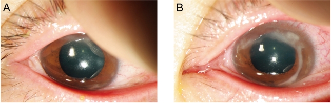
Purpose: To present a case of peripheral infiltrative keratitis mimicking infectious keratitis on the flap margin and limbus, which appeared on the first postoperative day after the laser in situ keratomileusis (LASIK).
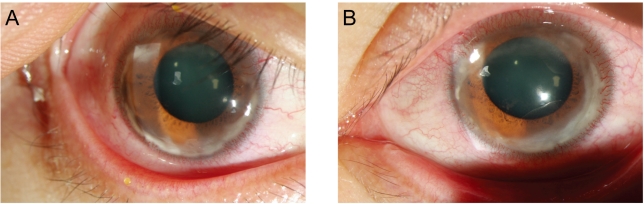
Methods: A 36-year-old woman who underwent uneventful bilateral simultaneous LASIK developed multiple round infiltrate along the flap margin reaching to limbus from the 11 o'clock to 6 o'clock area in both eyes.
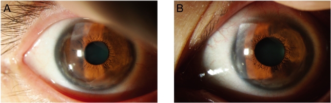
Results: The flap was lifted and irrigation was performed with antibiotics. but infiltration seemed to appear again. The infiltrate was more concentrated at the periphery and was extended to the limbus. Direct smear and culture for bacteria and fungus were negative. Topical prednisolone acetate 1% eye drops was added, infiltrative condition was resolved.
Conclusions: LASIK induced peripheral infiltrative keratitis, in which infectious origin was ruled out, is reported.
Figures
Similar articles
-
Bilateral peripheral infiltrative keratitis after LASIK.J Refract Surg. 2005 Jul-Aug;21(4):402-4. doi: 10.3928/1081-597X-20050701-18. J Refract Surg. 2005. PMID: 16128341
-
Pseudomonas keratitis 4 years after laser in situ keratomileusis.Optom Vis Sci. 2011 Oct;88(10):1252-4. doi: 10.1097/OPX.0b013e318223c0c4. Optom Vis Sci. 2011. PMID: 21666521
-
Idiopathic peripheral necrotizing keratitis after femtosecond laser in situ keratomileusis.J Cataract Refract Surg. 2012 Mar;38(3):544-7. doi: 10.1016/j.jcrs.2011.12.017. Epub 2012 Jan 21. J Cataract Refract Surg. 2012. PMID: 22265184
-
Unilateral Candida parapsilosis interface keratitis after laser in situ keratomileusis: case report and review of the literature.Cornea. 2009 Jan;28(1):105-7. doi: 10.1097/ICO.0b013e318184e69b. Cornea. 2009. PMID: 19092419 Review.
-
Bilateral infectious keratitis after laser in situ keratomileusis: a case report and review of the literature.Ophthalmology. 2001 Jan;108(1):121-5. doi: 10.1016/s0161-6420(00)00435-8. Ophthalmology. 2001. PMID: 11150275 Review.
Cited by
-
Retrospective Analysis of Sterile Corneal Infiltrates in Patients with Keratoconus after Cross-Linking Procedure.J Clin Med. 2022 Jan 25;11(3):585. doi: 10.3390/jcm11030585. J Clin Med. 2022. PMID: 35160037 Free PMC article.
References
-
- Linebarger EJ, Hardten DR, Lindstrom RL. Diffuse lamellar keratitis : Diagnosis and management. J Cataract Refract Surg. 2000;26:1072–1077. - PubMed
-
- Linebarger EJ, Hardten DR, Lindstrom RL. Diffuse lamellar keratitis: identification and management. Int Ophthalmol Clin. 2000;40:77–86. - PubMed
-
- Blustein JN, Hitchins VM, Woo EK. Diffuse lamellar keratitis, endotoxin, and ophthalmic sponges. J Cataract Refract Surg. 2004;30:2027–2028. - PubMed
-
- Lifshitz T, Levy J, Mahler O, Levinger S. Peripheral sterile corneal infiltrates after refractive surgery. J Cataract Refract Surg. 2005;31:1392–1395. - PubMed
-
- Lahner WJ, Hardten DR, Lindstrom RL. Peripheral keratitis following laser in situ keratomileusis. J Refract Surg. 2003;19:671–675. - PubMed
Publication types
MeSH terms
Substances
LinkOut - more resources
Full Text Sources
