. 2007 Oct 3;26(19):4177-88.
doi: 10.1038/sj.emboj.7601844. Epub 2007 Sep 6.

Sequential steps and checkpoints in the early exocytic compartment during secretory IgM biogenesis

Affiliations

Sequential steps and checkpoints in the early exocytic compartment during secretory IgM biogenesis

Tiziana Anelli et al. EMBO J. .

Abstract

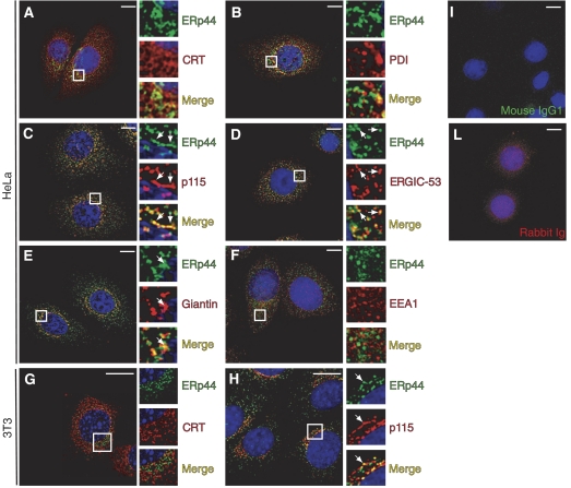
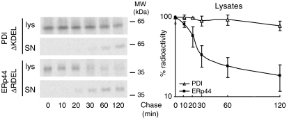
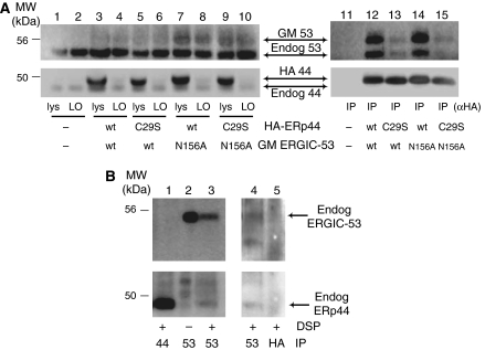
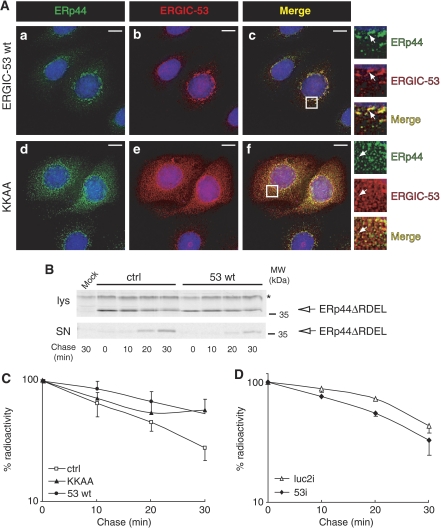
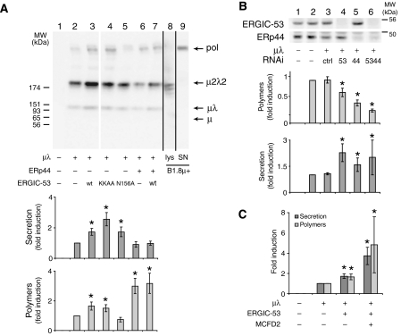
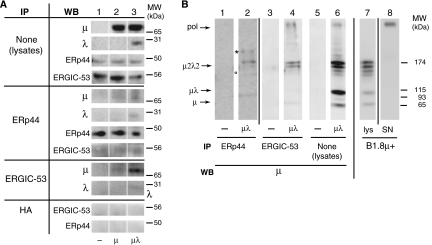
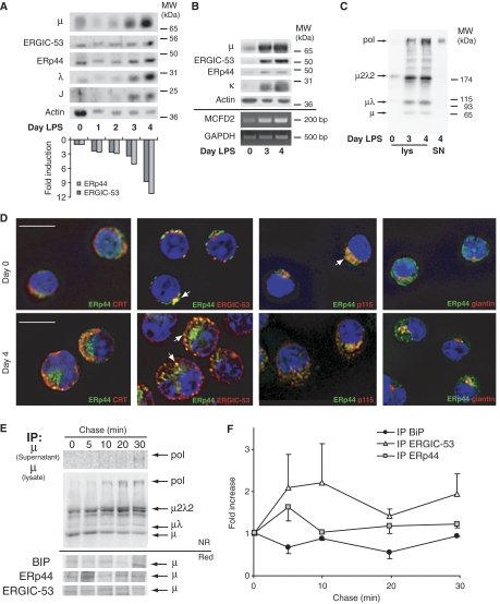
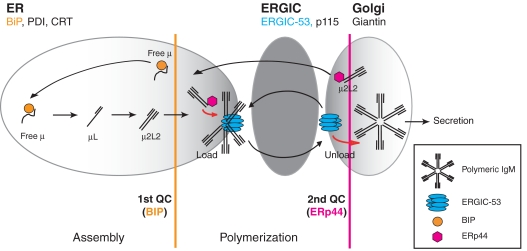
The biogenesis of secretory IgM occurs stepwise under stringent quality control, formation of micro(2)L(2) preceding polymerization. How is efficiency of IgM secretion coupled to fidelity? We show here that ERp44, a soluble protein involved in thiol-mediated retention, interacts with ERGIC-53. Binding to this hexameric lectin contributes to ERp44 localization in the ER-golgi intermediate compartment. ERp44 and ERGIC-53 increase during B-lymphocyte differentiation, concomitantly with the onset of IgM polymerization. Both preferentially bind micro(2)L(2) and higher order intermediates. Their overexpression or silencing in non-lymphoid cells promotes or decreases secretion of IgM polymers, respectively. In IgM-secreting B-lymphoma cells, micro chains interact first with BiP and later with ERp44 and ERGIC-53. Our findings suggest that ERGIC-53 provides a platform that receives micro(2)L(2) subunits from the BiP-dependent checkpoint, assisting polymerization. In this process, ERp44 couples thiol-dependent assembly and quality control.


Figures

Figure 1
Figure 1
Endogenous ERp44 is primarily localized in the ERGIC. HeLa cells were co-stained with antibodies specific for ERp44 and markers of the ER (CRT, PDI), ERGIC (ERGIC-53, p115), Golgi (Giantin) and early endosomes (EEA1), as indicated (AF). (G, H) 3T3 cells co-stained with antibodies specific for ERp44 and CRT or p115, respectively. Images were taken with a fluorescence microscope and analyzed with deconvolution techniques. For each panel, boxes show a higher magnification of the indicated area. Arrows indicate examples of colocalizing structures. Controls of HeLa cells stained with an unrelated mouse IgG1 antibody and a rabbit serum are shown (I, L, respectively). Endogenous ERp44 colocalizes mainly with ERGIC-53 and p115, although some overlapping signal can be observed also with CRT, PDI and giantin. No colocalization at all is observed with the early endosome marker EEA1. Bar=10 μm.
Figure 2
Figure 2
ERp44ΔRDEL is secreted more rapidly than PDIΔKDEL. HeLa cells transiently co-transfected with myc-PDIΔKDEL or HA-ERp44ΔRDEL were pulsed for 10 min with 35S-labeled methionine and cysteine and chased for the indicated times. Culture supernatants (SN) and cell lysates (lys) were IP with anti-myc or anti-HA (to isolate PDI and ERp44, respectively), resolved by reducing SDS–PAGE and subjected to autoradiography. Densitometric quantification of the disappearance of the two proteins from the lysates is shown on the right (level of radioactive signal present at each time chase with respect to time 0; average of two independent experiments quantified in duplicate, ±s.e.m.). Note that at the end of the chase almost all ERp44 is secreted, whereas a considerable amount of PDI is still present intracellularly.
Figure 3
Figure 3
ERp44 interacts with ERGIC-53. (A) After crosslinking with DSP, aliquots (100 μg of protein) of the RIPA lysates from HeLa transfectants expressing various combinations of wt or mutant HA-ERp44 and GM ERGIC-53 as indicated were IP with anti-HA antibodies and resolved by reducing SDS–PAGE (lanes 11–15). Smaller aliquots of the lysates (30 μg), before (lys) and after IP (left overs (LO), lanes 1–10), were loaded to verify the levels of expression and immunoprecipitation efficiency. After transferring to nitrocellulose, blots were decorated with polyclonal anti-ERGIC-53 (upper panels) and anti-ERp44 (lower panels). Both antibodies recognize doublets: the upper band corresponds to the tagged exogenous molecules, and the faster migrating one to the endogenous ones. The diffuse appearance of exogenous GM ERGIC-53 is due to the presence of N-glycans. Note that anti-HA antibodies efficiently precipitate exogenous, but not endogenous ERp44 molecules (lanes 3–10). In cells overexpressing wt ERp44 (lanes 12 and 14), abundant exogenous and endogenous ERGIC-53 co-IP with ERp44. Co-immunoprecipitation is less efficient in cells expressing the inactive ERp44 C29S mutant (lanes 13 and 15). In contrast, mutation in ERGIC-53 active site (N156A) has only minor effects (lane 14). When inactive mutants of both ERp44 and ERGIC-53 (C29S and N156A, respectively) are coexpressed, some ERGIC-53 can still be co-IP with ERp44, suggesting the two molecules can directly, but transiently, interact. ERGIC-53 did not co-immunoprecipitate in non-transfected cells (lane 11). (B) Endogenous ERp44 and ERGIC-53 non-covalently interact. HeLa cells (300 μg), treated with or without DSP as indicated and lysed in RIPA buffer supplemented with 2 mM Ca2+, were IP with anti-ERGIC-53 (lanes 2 and 3), or anti-ERp44 monoclonal antibodies (lane 1), to confirm the identity of the bands co-IP by anti-ERGIC-53, and resolved by reducing SDS–PAGE. The nitrocellulose was sequentially decorated with rabbit anti-ERGIC-53 (upper panel) and anti-ERp44 antibodies (lower panel), as indicated. Endogenous ERp44 can be co-IP with endogenous ERGIC-53, the association being more evident after crosslinking. In a similar, independent experiment (right panel), anti-HA (lane 5) was used in parallel with anti-ERGIC-53 (lane 4), as a further specificity control for the ERp44 co-IP with anti-ERGIC-53. The blot was first probed with anti-ERp44 and then with anti-ERGIC-53. In this experiment, the ERp44-specific monoclonal antibody did not co-immunoprecipitate detectable ERGIC-53, possibly because of its lower efficiency in immunoprecipitation.
Figure 4
Figure 4
ERGIC-53 mediates ERp44 localization in the early secretory pathway. (A) HeLa cells transiently transfected with vectors driving the expression of wt ERGIC-53 (a–c) or the ER-localized mutant ERGIC-53 KKAA (d–f) were co-stained with antibodies specific to ERp44 and ERGIC-53. Images were taken with a fluorescence microscope and analyzed with deconvolution techniques. In each panel, boxes show a higher magnification of the indicated areas. Arrows indicate examples of colocalizing structures. Mutant ERGIC-53 KKAA acts as a localization dominant negative and causes the ER accumulation of endogenous ERGIC-53 molecules as well (Vollenweider et al, 1998 and data not shown); note that endogenous ERp44 is in part delocalized with the mutant lectin, as demonstrated by the diffuse pattern shown in panel d and by the colocalization of the two molecules (f). However, some ERp44 is able to reach the ERGIC region (not shown). Bar=10 μm. (B, C) HeLa cells were transiently transfected with HA-ERp44ΔRDEL alone (ctrl, empty square) or with wt ERGIC-53 (53 wt, filled circles) or the ER-localized KKAA mutant (KKAA, filled triangles). Forty-eight hours after transfection, cells were pulse-labeled and chased for the indicated times as described in legend to Figure 2. Cell lys and SN were IP with anti-HA (to isolate ERp44), resolved by reducing SDS–PAGE and subjected to autoradiography (B). Panel C shows densitometric analyses of lys, performed as in legend to Figure 2 (average of four independent experiments, ±s.e.m.); signals were normalized relative to a stable background (see * in panel B). The overexpression of either wt or KKAA ERGIC-53 inhibits ERp44ΔRDEL secretion. (D) HeLa cells were subjected to RNAi for ERGIC-53 (53i, filled diamonds) or treated with control duplexes (luc2i, empty triangles), and then transiently transfected with HA-ERp44ΔRDEL, as indicated. Seventy-two hours from RNAi (48 h from transfection), cells were pulse-labeled, chased and analyzed as described for Figure 2. In the absence of ERGIC-53, ERp44ΔRDEL is secreted more rapidly (average of three independent experiments, ±s.e.m.). As a control of the efficiency of RNAi, see Figure 5B.
Figure 5
Figure 5
ERp44, ERGIC-53 and MCFD2 modulate IgM polymerization in HeLa cells. (A) ERp44 and ERGIC-53 overexpression promotes IgM polymerization in HeLa cells. HeLa cells transiently transfected with various combinations of vectors driving the expression of μ, λ, ERp44, ERGIC-53 (wt or mutants) as indicated, were washed and cultured for 4 h. Aliquots of the lysates were analyzed by Western blotting to monitor the expression levels of the different transgenes (data not shown). Secreted IgM were concentrated with ConA sepharose, resolved under non-reducing conditions, blotted and decorated with anti-μ antibodies (lanes 1–7). A white line separates lanes 3 and 4, which were juxtaposed from the same gel. Lanes 8 and 9 (coming from other gels run in parallel) show the lysates and supernatants from an IgM-producing murine hybridoma (B1.8 μ+). Arrows point to IgM polymers (pol), μ2λ2 and μλ subunits, and free μ. Lower panels show the quantification of total and polymeric secreted IgM. Data are expressed as fold increase with respect to cells expressing μ and λ alone. Total IgM secretion was calculated as the amount of IgM secreted in 4 h relative to the intracellular pool in each transfectant. The efficiency of polymerization was calculated as the percentage of polymers with respect to the total signal obtained with anti-μ in secreted material. Average of five independent experiments like the one shown in the top panel, ±s.e.m.; *P<0.05 t-test. Note that ERp44 and ERGIC-53 overexpression (both wt and KKAA mutant) increases IgM polymerization. (B) ERp44 and ERGIC-53 silencing inhibits IgM polymerization in HeLa cells. RNAi was performed in HeLa cells with control duplexes (ctrl) or duplexes specific to ERp44 or/and ERGIC-53, as indicated. The day after, cells were transfected with plasmids driving the expression of μ and λ, or with empty vector as a control. Seventy-two hours from RNAi, cells were washed and cultured for 4 h. To check for the efficiency of ERp44 and ERGIC-53 silencing, 15 μg of proteins per lane were loaded on reducing SDS–PAGE, and nitrocellulose was then probed with antibodies against endogenous ERp44 and ERGIC-53, as indicated. After 72 h of silencing, about 15% of ERGIC-53 and 25% of ERp44 were detectable in the lys (lanes 4–6). Secreted IgM were treated as described in panel A and WB data analyzed as described above. The lower panels show quantification of total and polymeric IgM secreted by HeLa cells treated as indicated. Data are expressed as fold increase or decrease with respect to cells expressing μ and λ, and not subjected to silencing. Both ERp44 and ERGIC-53 silencing inhibit IgM polymerization (lanes 4 and 5), their effects being additive (lane 6). Average of three independent experiments, ±s.e.m.; *P<0.05 t-test. (C) MCFD2 synergizes with ERGIC-53 in promoting IgM polymerization and secretion. HeLa cells expressing μ, λ and ERGIC-53 were transiently transfected with or without MCFD2 as indicated and processed as described for panel A. Average of two independent experiments, quantified in duplicate ±s.e.m.; *P<0.05 t-test.
Figure 6
Figure 6
ERp44 and ERGIC-53 preferentially interact with μ2λ2 or higher order assemblies. HeLa cells were transiently transfected with vectors driving the expression of μ chains, alone or in combination with λ, or empty vector as control. (A) Aliquots from lys (300 μg) were IP with anti-ERp44, anti-ERGIC-53 or anti-HA as a control, and resolved under reducing conditions. Aliquots (30 μg) of the lys before IP were analyzed to verify the levels of expression of the relevant proteins (upper four panels). After transfer to nitrocellulose, blots were decorated with the indicated antibodies (WB). Note that more μ can be co-IP with ERp44 and ERGIC-53 when λ chain is also present. (B) The anti-ERp44 (lanes 1 and 2) and anti-ERGIC-53 (lanes 3 and 4) immunoprecipitations obtained from HeLa cells transfected with or without μ+λ as described above, were resolved under non-reducing (NR) conditions and blots decorated with anti-μ antibodies. Aliquots (15 μg) from the corresponding lysates were also analyzed under NR conditions (lanes 5 and 6). All the lanes come from two twin gels run in parallel. Lysates and SN of B1.8μ+ hybridoma cells (lanes 7 and 8) are shown for comparison. The migration of polymers and other IgM assembly intermediates is indicated on the left-hand margin. Lanes are separated by white or black lines, as coming from the same or twin gels, respectively. The asterisk (*) and the circle (°) point to bands containing both μ and ERp44, and likely consisting of ERp44-μ2λ2 and ERp44-μλ mixed disulfides, respectively (see Supplementary Figure S6).
Figure 7
Figure 7
Localization and role of ERp44 and ERGIC-53 in IgM-secreting B cells. (AC) Coordinated expression of ERp44 and ERGIC-53 in differentiating B cells, concomitant with the onset of IgM polymerization. I.29μ+ B-lymphoma cells (A) or primary murine splenocytes (B, C) were stimulated in vitro with LPS to induce plasmacytic differentiation. At the indicated days, aliquots were lysed and resolved electrophoretically under reducing (A, B) or NR (C) conditions. An aliquot of the secreted material at day 4 was loaded under NR conditions as a marker of polymers (SN, last lane on the right, panel C). Densitometric quantifications (A, lower panel) were normalized relative to the actin signal. Aliquots of primary B cells at days 0, 3 and 4 of differentiation were also subjected to RNA extraction, RT and PCR to amplify MCFD2 mRNA. GAPDH was used as a control for normalization. Note that ERGIC-53, ERp44 and MCFD2 are simultaneously upregulated during the last stages of B-cell differentiation, concomitantly with J-chain induction and the onset of IgM polymerization. (D) Murine splenocytes at days 0 and 4 of LPS stimulation were stained with the indicated antibodies; images were taken with a fluorescence microscope and analyzed with deconvolution techniques. Arrows indicate examples of colocalizing structures. Not only in B cells (day 0) but also in PCs (day 4 after LPS), ERp44 is located in the ERGIC compartment, showing intense colocalization with ERGIC-53 and p115. Bar=10 μm. (E, F) Dynamic interactions of nascent μ chains with BiP, ERp44 and ERGIC-53. Ramos cells were pulsed for 5 min with 35S-labeled methionine and cysteine, and chased for the indicated times. Culture SN and cell lys were IP with different antibodies as indicated on the left, and then resolved by SDS–PAGE under NR or reducing (red) conditions (E). After transfer to nitrocellulose, blots were subjected to autoradiography. Filters were then decorated with anti-μ antibodies, to verify the identity of the band co-IP with the different interactors (data not shown). Soon after the 5-min pulse, μ, μλ and μ2λ2 are already detectable in the lys. Polymers appear later, being easily detectable after 10 min of chase in the lys, and after 20–30 min in the SN. Densitometric quantifications of radioactive μ chains co-IP with BiP, ERp44 and ERGIC-53 were performed on reduced blots. Signals were normalized relative to a stable background, and data shown as ratio to the signal obtained at time 0 (average of three independent experiments, ±s.e.m.) (F). The level of labeled μ co-IP with BiP decreases immediately after the pulse and is reduced to one-half within 20 min. On the contrary, ERp44- and ERGIC-53-associated μ chains peak at the first chase points, just before IgM polymerization occurs.
Figure 8
Figure 8
Schematic model of the IgM polymerization machinery. The IgM assembly line is schematized in its sequential arrangement. The distribution of ERp44 in the early secretory pathway (primarily in the ERGIC) is depicted as a gradient of gray. BiP-dependent control ensures that μ chains do not proceed to the polymerization machinery unless assembled with L chains (first QC step). ERGIC-53, a hexameric lectin acting in conjunction with MCFD2 (the latter is not shown) captures μ2L2 subunits, likely aligning them in a planar conformation, suitable for polymerization. ERp44 ensures that unpolymerized subunits are not secreted, retrieving them into the assembly line (second QC step). Because of its ability to bind IgM subunits and ERGIC-53, ERp44 may recruit and locally concentrate μ2L2, to stimulate further the polymerization machinery efficacy.

References

    1. Amitay R, Bar-Nun S, Haimovich J, Rabinovich E, Shachar I (1991) Post-translational regulation of IgM expression in B lymphocytes. Selective nonlysosomal degradation of assembled secretory IgM is temperature-dependent and occurs prior to the trans-Golgi. J Biol Chem 266: 12568–12573 - PubMed
    1. Anelli T, Alessio M, Bachi A, Bergamelli L, Bertoli G, Camerini S, Mezghrani A, Ruffato E, Simmen T, Sitia R (2003) Thiol-mediated protein retention in the endoplasmic reticulum: the role of ERp44. EMBO J 22: 5015–5022 - PMC - PubMed
    1. Anelli T, Alessio M, Mezghrani A, Simmen T, Talamo F, Bachi A, Sitia R (2002) ERp44, a novel endoplasmic reticulum folding assistant of the thioredoxin family. EMBO J 21: 835–844 - PMC - PubMed
    1. Appenzeller-Herzog C, Roche AC, Nufer O, Hauri HP (2004) pH-induced conversion of the transport lectin ERGIC-53 triggers glycoprotein release. J Biol Chem 279: 12943–12950 - PubMed
    1. Blond-Elguindi S, Cwirla SE, Dower WJ, Lipshutz RJ, Sprang SR, Sambrook JF, Gething MJ (1993) Affinity panning of a library of peptides displayed on bacteriophages reveals the binding specificity of BiP. Cell 75: 717–728 - PubMed

Publication types

MeSH terms