. 2007 Nov;9(11):1963-70.
doi: 10.1089/ars.2007.1745.

Preclinical evaluation of targeting the Nrf2 pathway by triterpenoids (CDDO-Im and CDDO-Me) for protection from LPS-induced inflammatory response and reactive oxygen species in human peripheral blood mononuclear cells and neutrophils

Affiliations

Preclinical evaluation of targeting the Nrf2 pathway by triterpenoids (CDDO-Im and CDDO-Me) for protection from LPS-induced inflammatory response and reactive oxygen species in human peripheral blood mononuclear cells and neutrophils

Rajesh K Thimmulappa et al. Antioxid Redox Signal. 2007 Nov.

Abstract

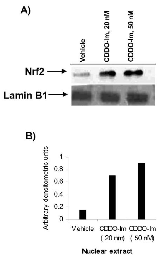
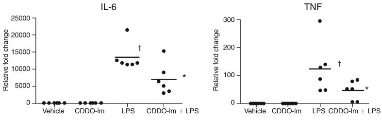
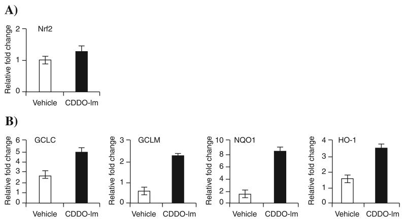
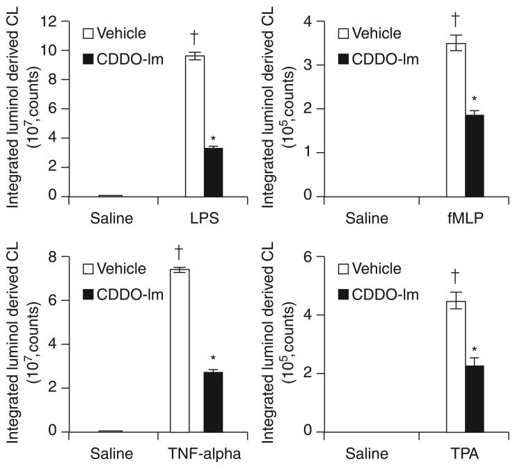
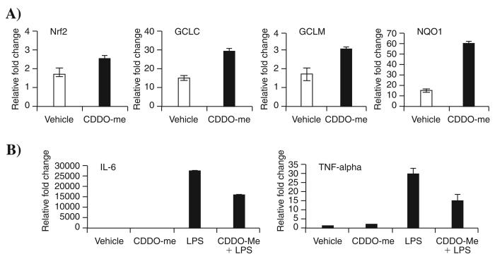
Sepsis is characterized by an inappropriate host immune-inflammatory response and sustained oxidative damage. Nrf2, a bZIP oxidant-responsive transcription factor, regulates a battery of cytoprotective genes including antioxidants and maintains cellular redox homeostasis. Mouse studies have demonstrated a critical role of Nrf2 in improving survival during sepsis. This preclinical ex vivo study using neutrophils and peripheral blood mononuclear cells (PBMCs) as a surrogate cells evaluates the efficacy of CDDO-Im and CDDO-Me [imidazole and methyl ester derivative of 2-cyano-3,12-dioxooleana-1,9(11)-dien-28-oic acid (CDDO)] to activate the Nrf2 pathway and protect from lipopolysaccharide (LPS)-induced inflammatory response in humans. CDDO-Im treatment significantly induced Nrf2-dependent antioxidative genes (HO-1, GCLC, GCLM, and NQO1) in PBMCs isolated from six normal subjects. CDDO-Im increased nuclear accumulation of Nrf2 protein. Pretreatment of PBMC by CDDO-Im significantly attenuated LPS-induced cytokine expression. Similar increases in levels of antioxidant genes and suppression of LPS-induced cytokine expression was observed after CDDO-Me pretreatment. CDDO-Im also greatly inhibited LPS, fMLP, TNF-alpha, and TPA-induced ROS generation in neutrophils. In conclusion, these results demonstrate that activation of the Nrf2-dependent antioxidative pathway by CDDO-Im or CDDO-Me protects against the LPS-induced inflammatory response and suggest that they can be potential therapeutic candidates for intervening sepsis syndrome.


Figures

FIG. 1
FIG. 1. CDDO-Im pretreatment up-regulates expression of Nrf2-dependent antioxidative genes in human PBMCs
(A) mRNA expression of Nrf2 in PBMCs isolated from six normal subjects. (B) mRNA expression of Nrf2-dependent antioxidants NQO1, GCLM, HO-1, and GCLC in PBMCs isolated from six normal subjects. Isolated PBMCs were treated either with vehicle or CDDO-Im (20 nM) for 20 h, and gene-expression analysis was done by real-time PCR. Data are expressed as fold changes relative to the lowest value in the vehicle-treated groups. Horizontal line represents the mean fold change. *Distribution differs from vehicle-treated group (p < 0.05).
FIG. 2
FIG. 2. CDDO-Im induces nuclear accumulation of Nrf2 protein
(A) Immunoblot demonstrating increase levels of Nrf2 protein in nuclear extracts of PBMCs treated with CDDO-Im. PBMCs were treated with either vehicle or CDDO-Im (20 nM or 50 nM) for 6 h. Cells were then harvested and processed for nuclear extraction. Nuclear proteins (20 μg) were resolved on a 10% PAGE, and immunoblotting was performed with an Nrf2 antibody, as described in Methods. The blot was reprobed with anti-Lamin B1 antibody as a marker of nuclear proteins. (B) Quantification of Nrf2 protein. For band densitometry, bands in nuclear extract blot were normalized to Lamin B1.
FIG. 3
FIG. 3. CDDO-Im pretreatment attenuated LPS-induced cytokine expression in PBMCs isolated from normal subjects
CDDO-Im pretreatment suppressed LPS-induced mRNA expression of IL-6 and TNF-α in PBMCs of six subjects measured 4 h after LPS treatment. Ex vivo PBMCs were pretreated with DMSO or CDDO-Im for 20 h followed by LPS (100 ng/ml) stimulation. Horizontal line represents the mean fold change. Differs from vehicle control. *Differs from only LPS-treated group (p < 0.05).
FIG. 4
FIG. 4. Treatment of peripheral blood neutrophils with CDDO-Im upregulated Nrf2–dependent antioxidative genes
(A) mRNA expression of Nrf2 in neutrophils after CDDO-Im. (B) CDDO-Im induced expression of Nrf2–dependent antioxidative genes (GCLC, GCLM, NQO1, and HO-1). Ex vivo blood neutrophils pooled from two subjects were treated with either vehicle or CDDO-Im (20 nM) for 20 h and processed immediately for measurement of antioxidant gene expression. Experiments were performed in triplicate.
FIG. 5
FIG. 5. CDDO-Im treatment attenuated ROS generation in peripheral blood neutrophils induced by LPS, fMLP, TNF-α, and TPA
Ex vivo pooled peripheral blood neutrophils isolated from two subjects were treated with DMSO or CDDO-Im (20 nM) for 20 h. Immediately after this, cells were harvested and stimulated with different inflammatory agents (LPS, 100 ng/ml; fMLP, 10 μM; TNF-α, 10 ng/ml; TPA, 50 nM). Each bar is the mean ± SD (n = 3) of values presenting the integration of the area under curve for 30 min, expressed in counts. *Differs from LPS alone. δ, Differs from saline (p < 0.05).
FIG. 6
FIG. 6. CDDO-Me, a methyl ester derivative of CDDO, significantly upregulated Nrf2–dependent antioxidants and suppressed LPS-induced cytokine expression in PBMCs isolated from a normal subject
(A) mRNA levels of Nrf2 and antioxidative genes (GCLC, GCLM, and NQO1) in PBMCs after CDDO-Me treatment. (B) IL-6 and TNF-α expression in PBMCs pretreated with CDDO-Me after LPS challenge. Ex vivo PBMCs isolated from normal subjects were treated with either vehicle or CDDO-Me (20 nM) for 20 h and processed immediately for measurement of antioxidant gene expression. For assessing LPS-induced inflammation, cells were incubated with LPS for additional 4 h, and expression of IL-6 and TNF-α was assessed by real-time PCR. Experiments were performed in triplicate.

Similar articles

Cited by

References

    1. Abraham E. Nuclear factor-kappaB and its role in sepsis-associated organ failure. J Infect Dis. 2003;187(suppl 2):S364–S369. - PubMed
    1. Angus DC, Linde-Zwirble WT, Lidicker J, Clermont G, Carcillo J, Pinsky MR. Epidemiology of severe sepsis in the United States: analysis of incidence, outcome, and associated costs of care. Crit Care Med. 2001;29:1303–1310. - PubMed
    1. Asehnoune K, Strassheim D, Mitra S, Kim JY, Abraham E. Involvement of reactive oxygen species in toll-like receptor 4-dependent activation of NF-kappaB. J Immunol. 2004;172:2522–2529. - PubMed
    1. Cho HY, Jedlicka AE, Reddy SP, Kensler TW, Yamamoto M, Zhang LY, Kleeberger SR. Role of NRF2 in protection against hyperoxic lung injury in mice. Am J Respir Cell Mol Biol. 2002;26:175–82. - PubMed
    1. Cho HY, Reddy SP, Yamamoto M, Kleeberger SR. The transcription factor NRF2 protects against pulmonary fibrosis. FASEB J. 2004;18:1258–1260. - PubMed

Publication types

MeSH terms