. 2007 Sep;26(6):1479-88.
doi: 10.1111/j.1460-9568.2007.05775.x. Epub 2007 Sep 6.

Presynaptic regulation of dendrodendritic dopamine transmission

Affiliations

Presynaptic regulation of dendrodendritic dopamine transmission

Michael J Beckstead et al. Eur J Neurosci. 2007 Sep.

Abstract

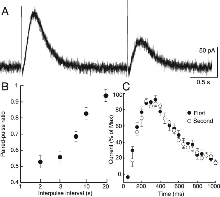
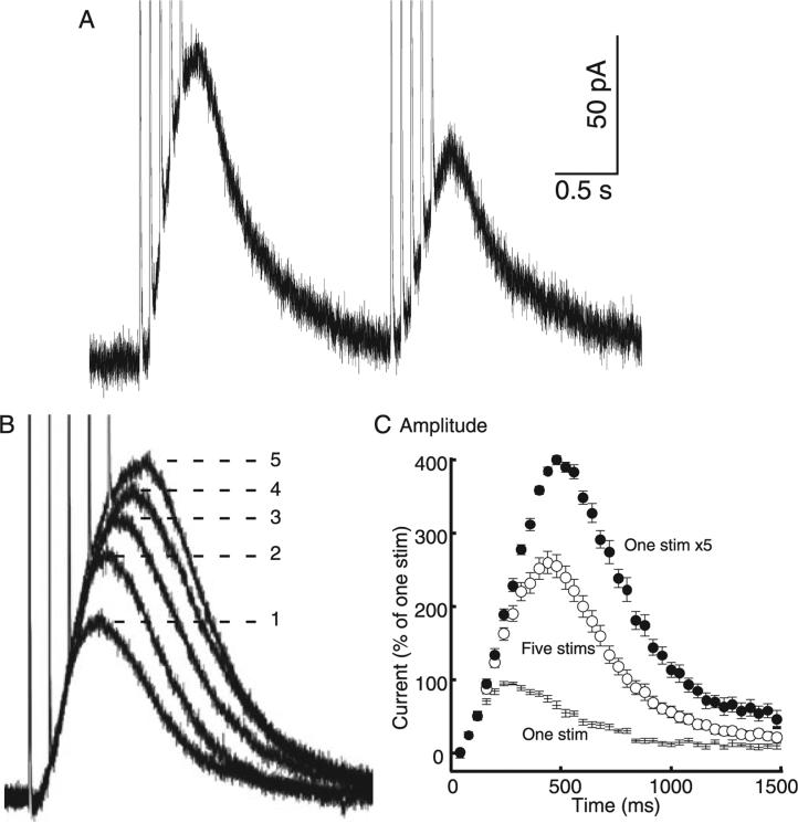
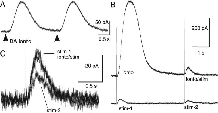
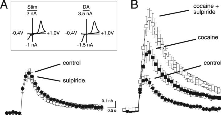
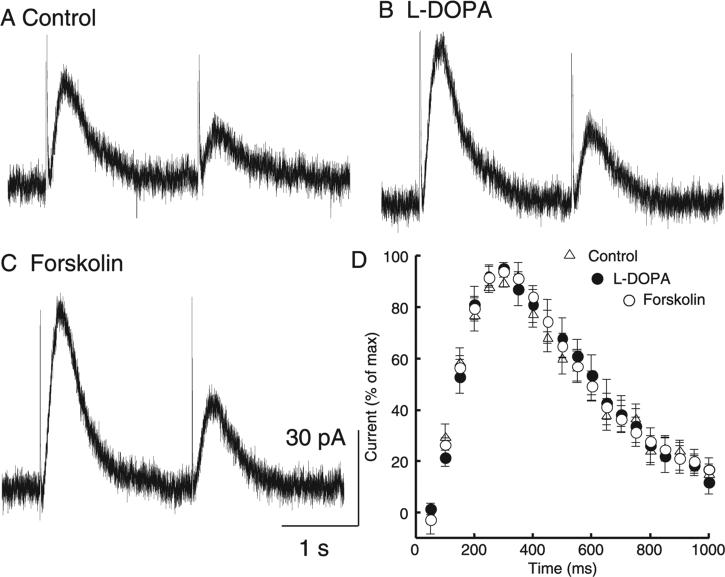
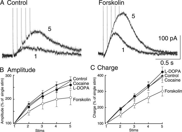
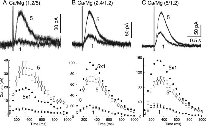
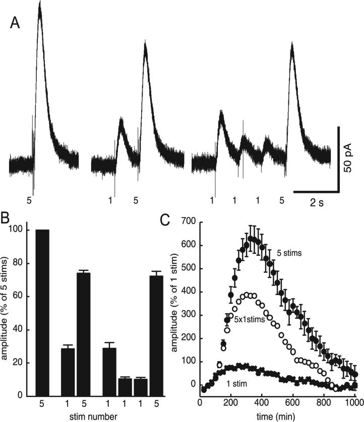
The amount of dopamine release from terminals in the forebrain following an electrical stimulus is variable. This dynamic regulation, both between and within trains of electrical stimuli, has fostered the notion that burst firing of dopamine neurons in vivo may be a determinant of dopamine release in projection areas. In the present study dendritic dopamine release was examined in the substantia nigra and ventral tegmental area in mouse brain slices using whole-cell recording of a dopamine-mediated inhibitory postsynaptic current (IPSC). Paired stimuli produced a depression of the IPSC that was not observed with paired pulses of exogenously applied dopamine. Increasing the number of electrical stimuli from one to five produced an increase in the amplitude the dopamine IPSC but the increase was less than additive, indicating a depression of transmission with each successive stimulus. Analysis with fast-scan cyclic voltammetry demonstrated that presynaptic D2-autoreceptors did not contribute to the depression. Facilitation of the IPSC was observed only after the probability of release was reduced. Thus the regulation of dopamine release in the cell body region was dependent on dopamine neuron impulse activity. Under circumstance where there was initially little activity the probability of dopamine release was high and repetitive activation resulted in depression of further release. With increased activity, the release probability decreased and a burst of activity caused a relative facilitation of dopamine release. This form of regulation would be expected to limit activity within the cell body region.


Figures

Fig. 1
Fig. 1
Paired-pulse depression of dopamine IPSCs. (A) Using a pair of single pulses, the dopamine IPSC consistently exhibited substantial paired-pulse depression. (B) This depression subsided as the interpulse duration was extended from 2 to 20 s (n = 5). (C) Although the first pulse produced a larger amplitude current, there was no difference between the kinetics of the dopamine IPSC subsequent to the first and second pulse when the traces were normalized to their maxima (n = 10).
Fig. 2
Fig. 2
Depression was observed when multiple stimuli were used to mimic bursting. (A) The paired-pulse depression produced by a pair of single pulses was maintained but somewhat blunted by using a pair of stimulus trains. (B) However, as the number of stimuli was increased from one to five (at 12.5 Hz) each successive pulse produced less postsynaptic current. (C) The convolution can be observed by mathematically simulating perfect temporal summation of five single IPSCs. The measured time course of 11 normalized single pulse IPSCs is plotted as ‘One stim’. Perfect additivity of five pulses (at 12.5 Hz) would produce the simulated time course predicted with the closed circles labelled ‘One stim × 5’. The measured IPSC evoked by a train of five stimuli is considerably smaller (‘Five stims’; F1,20 = 23.5, P < 0.0001, n = 11), illustrating the extent of subadditivity in the IPSC produced by a train of five stimuli.
Fig. 3
Fig. 3
Paired-pulse depression was presynaptic. (A) Paired-pulse inhibition was not observed when dopamine (1 m) was applied exogenously through iontophoresis (dark arrows). Furthermore, a large iontophoretic pulse did not depress the dopamine IPSC. (B, bottom trace) Two IPSCs (stim-1 and stim-2) separated by 4 s exhibited paired-pulse depression. (B, top trace) In the same cell a large iontophoretic pulse (ionto) did not depress the dopamine IPSC evoked 4 s later (ionto/stim). (C) IPSC tracings from this experiment are overlaid, showing that paired stimuli produced depression while iontophoresis of dopamine did not.
Fig. 4
Fig. 4
Fast-scan cyclic voltammetry determination of the regulation of dopamine release from the VTA by D2 autoreceptors. (A) Summarized voltammetric current vs. time responses were generated by three pulses at 5 Hz (n = 7) under control conditions and in the presence of sulpiride (200 nm). (Inset) Cyclic voltammagrams recorded at the peak of the stimulated dopamine response and after the exogenous application of dopamine with iontophoresis (100 nA, 50 ms). Peak oxidation and reduction peaks were +637 and –352 mV for the electrically evoked signal and +609 and –352 mV for the exogenous dopamine signal. Voltammagrams were generated by a voltage waveform from –0.4 to +1.0 V at 300 V/s. (B) Summarized voltammetric current vs. time responses were generated by three pulses at 5 Hz (n = 9) under control conditions, in the presence of cocaine (1 μm) and cocaine (1 μm) + sulpiride (200 nm).
Fig. 5
Fig. 5
Increasing the amplitude of the IPSC did not change paired-pulse depression or IPSC kinetics. A pair of single pulses 2 s apart (A) always yielded paired-pulse depression that (B) did not change when vesicular content was increased with L-DOPA (10 μm) or (C) release probability was increased with forskolin (10 μm). These manipulations did yield larger amplitude IPSCs (control, 26.2 ± 3.0 pA; L-DOPA, 36.6 ± 4.7 pA; forskolin, 64.2 ± 14.3 pA; n = 9–11). (D) Normalizing the traces to their maxima revealed that the increase in size was not accompanied by a change in time course of the IPSC (n = 6–11).
Fig. 6
Fig. 6
When multiple stimuli were applied, forskolin increased the relative contribution of the first pulse. (A) Increasing release probability with forskolin (10 μm) produced IPSCs with larger amplitudes whether one or five stimuli were applied. (B) The IPSC peak amplitude and (C) total charge were subsequently plotted as a function of number of stimuli and normalized to the first pulse. Pharmacological manipulations (cocaine, L-DOPA and forskolin) did not change the general observation of a decreasing contribution of each successive pulse (n = 6–11). The difference in the forskolin curve demonstrates an increased relative contribution of the first pulse. This was not observed subsequent to cocaine or L-DOPA application, both of which also increased the amplitude of the IPSC.
Fig. 7
Fig. 7
Facilitation of dopamine release was demonstrated by lowering release probability. Extracellular calcium and magnesium concentrations were altered to manipulate release probability (PR): (A) low PR (1.2 mm Ca, 5 mm Mg), (B) control PR (2.4 mm Ca, 1.2 mm Mg) and (C) high PR (5 mm Ca, 1.2 mm Mg). Dopamine IPSCs were evoked with single stimuli or trains of five rapid (10 ms interval) stimuli. (A, top) When PR was low, one stimulus produced very little current while five stimuli still produced a substantial IPSC. If the current produced by a single stimulus (‘1’) was constant over the next four stimuli, the IPSC would be the amplitude and time course predicted by the closed circles labelled ‘5 × 1’. (A, bottom, ‘5’) This predicted IPSC is smaller than the measured IPSC evoked by five stimuli. (B and C, bottom) When PR was increased the predicted IPSC ‘5 × 1’ was larger than the measured IPSC evoked by five stimuli, suggesting that facilitation of dopamine release occurred only when PR was low (n = 7–9). Total charged passed by one vs. five stimuli: low PR, 8.8 ± 1.6%; normal PR, 27.6 ± 2.3%; high PR, 28.8 ± 2.9%; n = 7–9.
Fig. 8
Fig. 8
Presynaptic depression of release unmasked facilitation. (A) Dopamine IPSCs were measured subsequent to three patterns of discharge activity: a train of five stimuli alone (5), a train preceded by one prepulse (1, 5) and a train preceded by three prepulses (1, 1, 1, 5). A single prepulse was sufficient to depress release and the amplitude of the IPSC. (B) Summarized data suggest that, subsequent to a prepulse, the IPSC produced by a single pulse was much more depressed than the IPSC produced by a train. (C) As this IPSC (‘5 stims’) is more than the arithmetic summation of five single-pulse IPSCs (‘5 × 1 stim’), presynaptic depression may be in effect unmasking the facilitation of release.

References

    1. Beckstead MJ, Grandy DK, Wickman K, Williams JT. Vesicular dopamine release elicits an inhibitory postsynaptic current in midbrain dopamine neurons. Neuron. 2004;42:939–946. - PubMed
    1. Beckstead MJ, Williams JT. Long-term depression of a dopamine IPSC. J. Neurosci. 2007;27:2074–2080. - PMC - PubMed
    1. Benoit-Marand M, Borrelli E, Gonon F. Inhibition of dopamine release via presynaptic D2 receptors: time course and functional characteristics in vivo. J. Neurosci. 2001;21:9134–9141. - PMC - PubMed
    1. Bigornia L, Allen CN, Jan CR, Lyon RA, Titeler M, Schneider AS. D2 dopamine receptors modulate calcium channel currents and catecholamine secretion in bovine adrenal chromaffin cells. J. Pharmacol. Exp. Ther. 1990;252:586–592. - PubMed
    1. Bjorklund A, Lindvall O. Dopamine in dendrites of substantia nigra neurons: suggestions for a role in dendritic terminals. Brain Res. 1975;83:531–537. - PubMed

Publication types