. 2007 Sep 6:8:75.
doi: 10.1186/1471-2199-8-75.

The promoter regions of the Myb-regulated Adora2B and Mcm4 genes co-localize with origins of DNA replication

Affiliations

The promoter regions of the Myb-regulated Adora2B and Mcm4 genes co-localize with origins of DNA replication

Holger Gundelach et al. BMC Mol Biol. .

Abstract

Background: The retroviral oncogene v-myb encodes a transcription factor (v-Myb) which is responsible for the transformation of myelomonocytic cells by avian myeloblastosis virus (AMV). v-Myb is thought to exert its biological effects by deregulating the expression of specific target genes. We have recently demonstrated that the chicken Gas41 gene, whose promoter co-localizes with an origin of DNA replication, is a bona fide Myb target gene. Because of this finding we have asked whether other Myb-regulated genes are also associated with DNA replication origins.

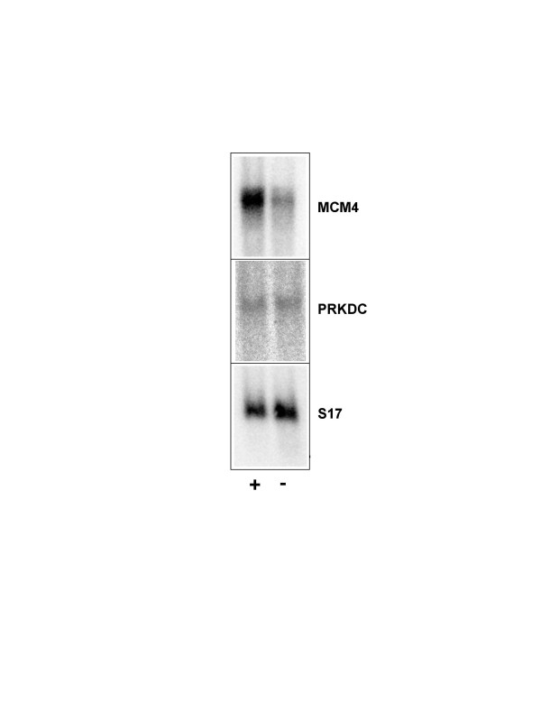
Results: We show that the promoter region of the chicken adenosine receptor 2B gene (Adora2B), a known Myb-target gene, acts as a DNA replication origin. Furthermore, we have examined known replication origins for the presence of Myb binding sites. We found that the intergenic region between the genes for the minichromosome maintenance 4 protein (Mcm4) and the catalytic subunit of DNA-dependent protein kinase (Prkdc), whose human counterpart has been identified as a replication origin, contains a number of Myb binding sites. Our data show that this region also acts as an origin of replication in chicken cells. Interestingly, we found that the chicken Mcm4 gene is also Myb-regulated.

Conclusion: Our work identifies the chicken Mcm4 gene as a novel Myb target gene and presents evidence for the co-localization of two novel origins of DNA replication with Myb-regulated genes. Our work raises the possibility that a fraction of Myb target gene promoters is associated with DNA replication origins.


Figures

Figure 1
Figure 1
Nucleotide sequence of the Mcm4-Prkdc intergenic region. The translational start codons of the Mcm4 and Prkdc genes are marked by arrowheads. Potential Myb binding sites are marked by boxes.
Figure 2
Figure 2
v-Myb binds to the Mcm4 promoter region in vitro. A. Electrophoretic mobility shift experiments were performed using bacterially expressed full-length v-Myb protein and radiolabeled oligonucleotides corresponding to Myb binding sites MBS-1, MBS-4 and MBS-6. Binding reactions were performed without Myb protein (lanes 1) or with increasing amounts of v-Myb (lanes 2 to 4). Complexes of full-length v-Myb and the oligonucleotides are marked by an arrowhead. The band at the bottom corresponds to unbound oligonucleotides. B. Radiolabeled oligonucleotide containing the Myb binding site A of the mim-1 promoter were incubated with bacterially expressed full-length v-Myb and excess of unlabeled competitor oligonucleotides, as indicated at the top. Competitors were used at 40-fold or 80-fold molar excess of unlabeled over labeled oligonucleotide, respectively. Only the upper part of the gel containing the complex of v-Myb and the radioactive oligonucleotide is shown.
Figure 3
Figure 3
Chromatin immunoprecipitation of the Mcm4 promoter region. Chromatin fragments prepared from BM2 cells were subjected to immunoprecipitation using two different Myb-specific rabbit antisera raised against the DNA-binding domain of v-Myb (anti Myb a and b). Control immunoprecipitations were performed with two different batches of normal rabbit serum (NRS a and b) or anti-Flag antibodies (anti Flag). DNA isolated from the immunoprecipitates was analyzed by quantitative real-time PCR using primers specific for the Mcm4 promoter region (-0.55 kb, left panel) or for a region approximately 13 kb upstream of the Mcm4 gene (-13 kb, right panel). The columns indicate the amount of PCR product obtained relative to the anti-Flag control reaction.
Figure 4
Figure 4
The chicken Mcm4 gene is regulated by v-Myb. Polyadenylated RNA from 10.4 cells grown for 24 hours in the presence (+ lanes) or absence (- lanes) of 2 μM β-estradiol was analyzed by Northern blotting using probes specific for Mcm4, Prkdc or S17 mRNA.
Figure 5
Figure 5
Replication origin activity of the Mcm4-Prkdc intergenic region in chicken cells. A. Schematic illustration of the Mcm4-Prkdc intergenic region. The black boxes and the arrows denote the transcriptional start sites of both genes. The positions of primer pairs used in the PCR analysis are shown below. The scale at the top is in kilobases. B. Nascent strand abundance assay. For quantification, real-time PCR was performed using the primer pairs indicated at the bottom. The columns show the average abundance of the corresponding sequences in nascent strand preparations of BM2 (black columns) or HD11 (white columns) cells relative to the sequences amplified with the -0.5 kb primer pair. Error bars mark the standard deviations. C. Nascent strand abundance analysis of the Mcm4 promoter region using wild-type (black columns) or c-myb deficient (white columns) DT40 cells.
Figure 6
Figure 6
Replication origin activity of the promoter region of the chicken adenosine receptor 2B gene. A. Schematic illustration of the chicken A2B receptor gene. The black boxes show the two exons of the gene. The transcriptional start site is marked by an arrow. The position of primer pairs used in the PCR analysis is shown below. The scale at the top is in kilobases. B. Nascent strand abundance assay of the adenosine receptor 2B promoter region using the PCR primer pairs indicated at the bottom. The columns show the average abundance of the corresponding sequences in nascent strand preparations of BM2 (black columns) or HD11 (white columns) cells relative to the sequences amplified with the -3.9 kb primer pair. Error bars mark the standard deviations. C. Nascent strand abundance analysis of the promoter region using wild-type (black columns) or c-myb deficient (white columns) DT40 cells.

References

    1. Lipsick JS, Wang DM. Transformation by v-Myb. Oncogene. 1999;18:3047–3055. doi: 10.1038/sj.onc.1202745. - DOI - PubMed
    1. Oh I-H, Reddy EP. The myb gene family in cell growth, differentiation and apoptosis. Oncogene. 1999;18:3017–3033. doi: 10.1038/sj.onc.1202839. - DOI - PubMed
    1. Weston K. Myb proteins in life, death and differentiation. Curr Opin Genet Dev. 1998;8:76–81. doi: 10.1016/S0959-437X(98)80065-8. - DOI - PubMed
    1. Biedenkapp H, Borgmeyer U, Sippel AE, Klempnauer K-H. Viral myb oncogene encodes a sequence-specific DNA-binding activity. Nature. 1988;355:835–837. doi: 10.1038/335835a0. - DOI - PubMed
    1. Ness SA, Marknell A, Graf T. The v-myb oncogene product binds to and activates the promyelocyte-specific mim-1 gene. Cell. 1989;59:1115–1125. doi: 10.1016/0092-8674(89)90767-8. - DOI - PubMed

Publication types