. 2007 Oct 15;310(2):291-303.
doi: 10.1016/j.ydbio.2007.07.041. Epub 2007 Aug 9.

Cartilage link protein 1 (Crtl1), an extracellular matrix component playing an important role in heart development

Affiliations

Cartilage link protein 1 (Crtl1), an extracellular matrix component playing an important role in heart development

Elaine E Wirrig et al. Dev Biol. .

Abstract

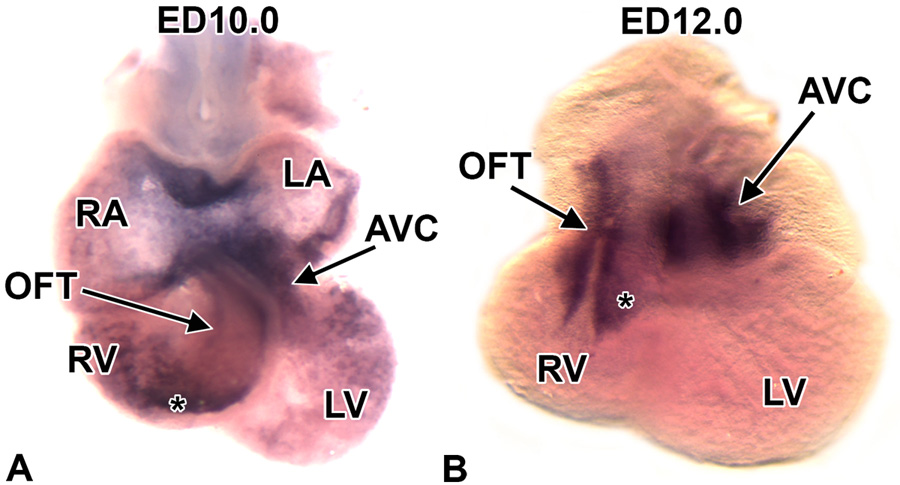
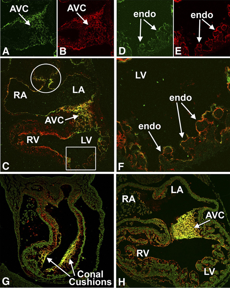
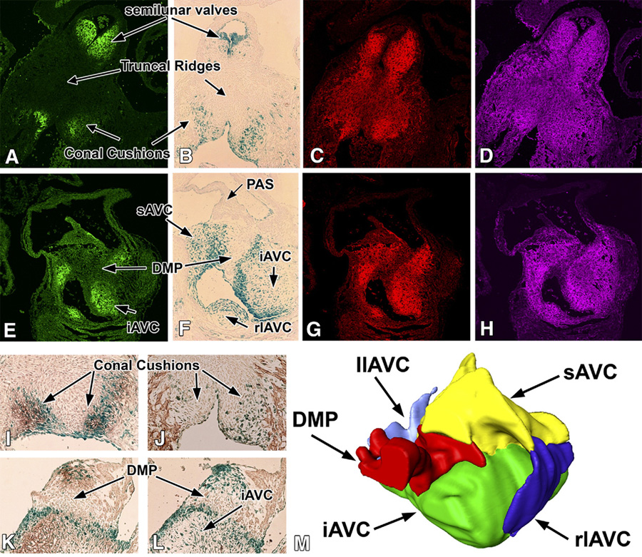
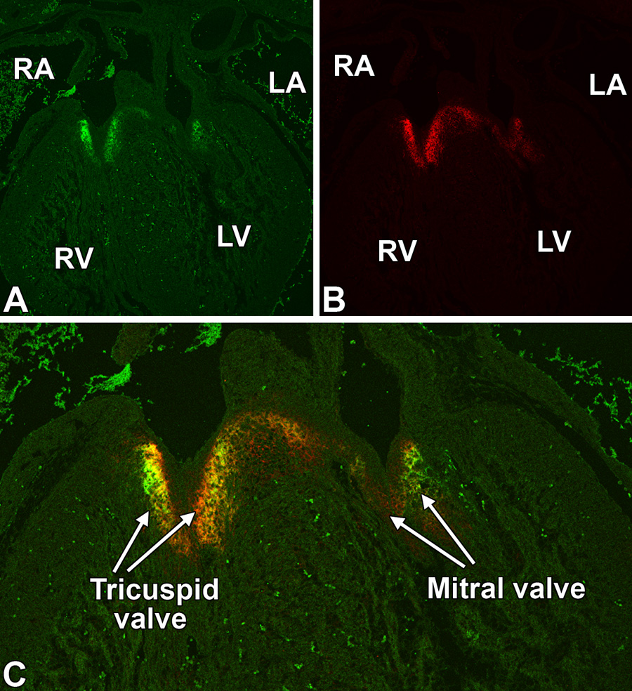
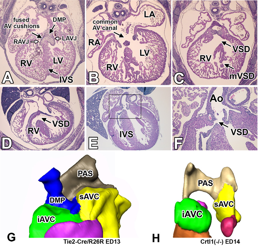
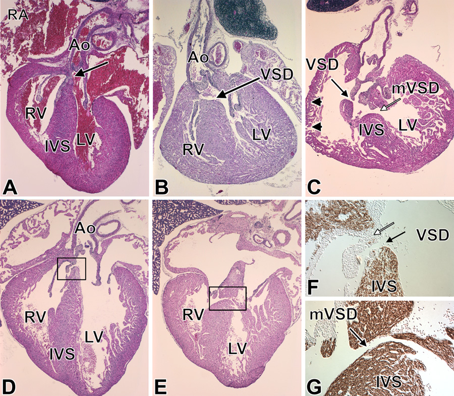
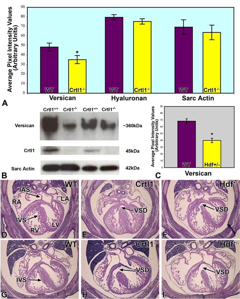
To expand our insight into cardiac development, a comparative DNA microarray analysis was performed using tissues from the atrioventricular junction (AVJ) and ventricular chambers of mouse hearts at embryonic day (ED) 10.5-11.0. This comparison revealed differential expression of approximately 200 genes, including cartilage link protein 1 (Crtl1). Crtl1 stabilizes the interaction between hyaluronan (HA) and versican, two extracellular matrix components essential for cardiac development. Immunohistochemical studies showed that, initially, Crtl1, versican, and HA are co-expressed in the endocardial lining of the heart, and in the endocardially derived mesenchyme of the AVJ and outflow tract (OFT). At later stages, this co-expression becomes restricted to discrete populations of endocardially derived mesenchyme. Histological analysis of the Crtl1-deficient mouse revealed a spectrum of cardiac malformations, including AV septal and myocardial defects, while expression studies showed a significant reduction in versican levels. Subsequent analysis of the hdf mouse, which carries an insertional mutation in the versican gene (CSPG2), demonstrated that haploinsufficient versican mice display septal defects resembling those seen in Crtl1(-/-) embryos, suggesting that reduced versican expression may contribute to a subset of the cardiac abnormalities observed in the Crtl1(-/-) mouse. Combined, these findings establish an important role for Crtl1 in heart development.


Conflict of interest statement

Conflict of Interest None

Figures

Figure 1
Figure 1. Expression of Crtl1 mRNA in the developing mouse heart
Panels A and B show Crtl1 mRNA expression by whole mount in situ hybridization in the developing mouse heart at ED10.0 (panel A) and ED12.0 (panel B). At ED10.0, Crtl1 mRNA is abundantly expressed in the AVJ and conal region of the OFT (*). Relatively high expression is also seen in the atrial roof. Lower levels of expression are seen in the lining off the atria and ventricles. A ED12.0 (panel B) Crtl1 mRNA expression becomes more restricted, with high expression in the AVJ and OFT (*). At this stage, Crtl1 mRNA expression is no longer detected in the atria and ventricles. LA=left atrium, RA=right atrium, LV=left ventricle, RV=right ventricle, OFT=outflow tract, AVJ=atrioventricular junction, (*)=OFT conal cushions.
Figure 2
Figure 2. Expression of Crtl1 and versican at ED9.5 and ED10.5
This figure shows immunofluorescently-stained sections co-labeled for Crtl1 (green) and versican (red), (co-exprssion shown in yellow), at ED9.5 (panels A–F) and ED10.5 (G and H). At ED9.5, Crtl1 is expressed in the extracellular matrix surrounding the endocardial and endocardial-derived cells of the AV cushion mesenchyme (panels A and C) and in the lining of the atria (panel C) and ventricles (panels C, D, F). Crtl1 is also expressed in the reflections of the dorsal mesocardium (white circle, panel C). Versican (panels B and E) shows a high degree of co-expression (yellow) with Crtl1 (panels C and F). Panels D–F are higher magnifications of the boxed region in C, showing Crtl1 and versican expression in the extracellular matrix surrounding the endocardial lining of the ventricular trabeculae. At ED10.5, Crtl1 and versican are highly expressed in the sub-endocardial extracellular matrix of the conal cushions in the proximal OFT (panel G). In addition, versican is expressed in the sub-endocardial mesenchyme of the truncal OFT ridges, regions where little Crtl1 expression is detected (panel G). In the AV cushions, Crtl1 and versican also show a high degree of co-expression (panel H). RA=right atrium, LA=left atrium, RV=right ventricle, LV=left ventricle, AVC=atrioventricular cushions, endo=endocardium.
Figure 3
Figure 3. Overlapping expression of Crtl1, versican, and hyaluronan in the embryonic mouse heart at ED13.0
This figure shows the expression of Crtl1, versican and HA in the respective mesenchymal cell populations in the developing heart at ED13.0. Panels A–D and panels I and J show sections of the OFT. Panels E–H and panels K and L show sections of the AV mesenchymal structures. Panels A and E show Crtl1 expression (green), panels C and G show versican staining (red), and panels D and H show hyaluronan staining (purple). Furthermore, panels B, F, I, J, K and L are sections from Tie2-cre/ROSA26 embryos in which endocardially-derived cells are expressing lacZ (blue). The sections in panels I and K are co-stained immunohistochemically for Crtl1 (brown), while the section in J is stained for sarcomeric actin. Combined, panels A, B, I, and J demonstrate that Crtl1 expression is largely confined to the endocardially-derived mesenchyme in the conal cushions and developing semilunar valves. The (proximal) truncal ridges (arrows in A and B) do not express Crtl1. Panels C and D show that in the conal cushions and semilunar valves, the expression of Crtl1, versican and HA overlap. Additionally, versican and HA are also expressed in those parts of the truncal ridges where Crtl1 is absent. Panels E, F, K, and L show that in the AV mesenchymal structures, Crtl1 expression is also restricted to the mesenchymal structures that are of endocardial origin. Thus, Crtl1 is expressed in the inferior, superior, and lateral AV cushions, but not in the dorsal mesenchymal protrusion, which forms a non-endocardially derived mesenchymal wedge in between the inferior and superior cushions (M). Panels G and H show that the DMP expresses lower levels of versican and HA than surrounding mesenchyme. AVC=atrioventricular cushions, sAVC=superior AVC, iAVC=inferior AVC, llAVC=left lateral AVC, rlAVC=right lateral AVC, PAS=primary atrial septum, DMP=dorsal mesenchymal protrusion.
Figure 4
Figure 4. Overlapping expression of Crtl1 and versican at ED14.5
This figure shows a co-labeling for Crtl1 (panel A, green) and versican (panel B, red) expression in a transverse section of a ED14.5 heart. The merged image (panel C) shows the overlapping expression of Crtl1 and versican (yellow) in the leaflets of the tricuspid and mitral AV valves. Note the higher expression of both proteins in the tricuspid valve leaflets when compared to the leaflets of the mitral valve. RA=right atrium, LA=left atrium, RV=right ventricle, LV=left ventricle,
Figure 5
Figure 5. Cardiac phenotype of Crtl1-deficient embryos
This figure shows hematoxylin-eosin stained transverse sections of wildtype (panel A) and Crtl1-deficient embryos (panels B–F). The section in panel A shows a wildtype mouse at ED13.5 with a well-formed interventricular septum, separating the right and left AV junction. Panels B and C show sections of a severely malformed Crtl1−/− specimen at ED14.0 with common AV canal (B), hypoplastic AV cushion tissues (B), absence of the DMP (B), thin compact myocardium in the RV wall (B,C), and a very disorganized interventricular septum (B,C). Panel D shows a Crtl1−/− specimen at ED16.0 with a large mesenchymal VSD. Panels E and F are sections from an ED17.0 Crtl1−/− specimen showing a small mesenchymal sub-aortic VSD. Panel F is a higher magnification of the boxed region in panel E. Panel G shows a three-dimensional AMIRA reconstruction of the AV mesenchymal complex of a wild type mouse at ED13, demonstrating the position of the DMP within this complex. Panel H is a 3D reconstruction of the ED14.0 Crtl1−/− mouse, shown in panels B and C, demonstrating the severely underdeveloped AV cushion tissues and absent DMP, combined contributing to the common AV canal phenotype. RA=right atrium, LA=left atrium, RV=right ventricle, LV=left ventricle, RAVJ=right atrioventricular junction, LAVJ=left atrioventricular junction, IVS=interventricular septum, VSD=ventricular septal defect, mVSD= muscular VSD, Ao=aorta, PAS=primary atrial septum, DMP=dorsal mesenchymal protrusion, AVC=atrioventricular cushions, iAVC=inferior AVC, sAVC=superior AVC.
Figure 6
Figure 6. Cardiac phenotype of the neonatal Crtl1-deficient mouse
Panel A shows a wild type neonatal heart with a well-formed IVS and AV cushion-derived fibrous tissue (arrow in A). Panel B is an example of a neonatal Crtl1−/− mouse with a sub-aortic VSD affecting both muscular and mesenchymal components of the AV septum. Panel C shows a neonatal Crtl1−/− mouse with severe cardiac malformations including a highly abnormal IVS, with a large muscular VSD (white arrow), a smaller mesenchymal VSD (black arrow), and an abnormally thin right ventricular wall (black arrowheads). Panels D–G are from another severely malformed Crtl1−/− specimen that exhibits both a mesenchymal VSD (D, F) and a muscular VSD (E,G). The sections F and G are stained with sarcomeric actin to delineate myocardial from fibrous structures. Panel F is a sister section of D showing a higher magnification of the VSD (boxed in D). The black arrow demarcates the mesenchymal VSD, the white arrow points to the fibrous, mesenchymal-derived tissues of the tricuspid valve leaflet bordering the superior margin of the defect. Panels E and G show a muscular VSD (black arrow in panel G). Panel G is a sister section of E showing a higher magnification of the boxed region in E. The actin staining demarcates the muscular upper and lower boundaries of the muscular defect. RA=right atrium, RV=right ventricle, LV=left ventricle, Ao=aorta, IVS=interventricular septum, VSD=ventricular septal defect, mVSD=muscular VSD.
Figure 7
Figure 7. Thin myocardium syndrome is associated with reduced myocardial proliferation in the Crtl1-deficient mouse
Panels A and B are hematoxylin-eosin stained sections of wild-type (A) and Crtl1−/− (B) specimens at ED13.0. The compact myocardium of the Crtl1−/− mouse has approximately half of the number of cell layers as compared to the compact myocardium in the wild type mouse (Panel B; thickness denoted by double-headed white arrows). PCNA labeling of wild type (C, E) and Crtl1−/− (D, F) ED13.0 specimens shows that the level of proliferation, expressed as PCNA-positive nuclei over total nuclei, is significantly reduced (t stat=4.34, p=0.02) in the ventricular wall of the Crtl1−/− specimens (white arrows D, F) when compared to wild type controls (C, E). However proliferation does not appear to be greatly affected in the interventricular septum (C, D, white star) or in the trabeculae (black arrows). LV=left ventricle, (*)= interventricular septum.
Figure 8
Figure 8. Reduced levels of versican are implicated in the etiology of the cardiac malformations in the Crtl1-deficient and heterozygous hdf mice
Panel A graphically depicts the levels of versican, hyaluronan, and sarcomeric actin expression, as measured by quantitative immunofluorescence, in the AV cushions of Crtl1−/− mouse embryos at ED13.0. While the levels of versican are significantly reduced (t=2.74; df=8; p=0.012), hyaluronan expression in the AV cushions, and sarcomeric actin expression in the adjacent myocardium, are not significantly changed. Panel B shows Western Blot results, using total hearts of wildtype and Crtl1−/− littermate embryos at ED13.0, confirming the decreased levels of versican in Crtl1−/− mice. Quantification of the western blots indicated a reduction of versican in the Crtl1−/− specimens by approximately 45% (t= 2.66, df= 4, p= 0.02). Crtl1 controls show the absence of Crtl1 in the Crtl1−/− mice, while the sarcomeric actin staining is an independent control of protein loading. Panel C is a graph depicting versican levels in ED13.5 hdf+/− embryos, as measured by quantitative immunofluorescence. The graph demonstrates that versican is significantly reduced, when compared to wild type littermates (p = 0.0005). Panels D–I are hematoxylin-eosin stained sections of wildtype (D and G), Crtl1−/− (E and H), and Hdf+/− (F and I) mice, respectively (D–F: stage ED14.0–14.5; G–I: stage ED15.5–16.0). Panels E, H, F, and I show that Crtl1−/− and Hdf+/− embryos are characterized by very similar ventricular septal defects (black arrows). RA=right atrium, LA=left atrium, RV=right ventricle, LV=left ventricle, IAS=interatrial septum, IVS=interventricular septum, VSD=ventricular septal defect.

Similar articles

Cited by

References

    1. Binette F, Cravens J, Kahoussi B, Haudenschild DR, Goetinck PF. Link protein is ubiquitously expressed in non-cartilaginous tissues where it enhances and stabilizes the interaction of proteoglycans with hyaluronic acid. J Biol Chem. 1994;269:19116–19122. - PubMed
    1. Bushdid PB, Osinska H, Waclaw RR, Molkentin JD, Yutzey KE. NFATc3 and NFATc4 are required for cardiac development and mitochondrial function. Circ Res. 2003;92:1305–1313. - PubMed
    1. Camenisch TD, Spicer AP, Brehm-Gibson T, Biesterfeldt J, Augustine ML, Calabro A, Jr, Kubalak S, Klewer SE, McDonald JA. Disruption of hyaluronan synthase-2 abrogates normal cardiac morphogenesis and hyaluronan-mediated transformation of epithelium to mesenchyme. J Clin Invest. 2000;106:349–360. - PMC - PubMed
    1. Campione M, Ros MA, Icardo JM, Piedra E, Christoffels VM, Schweickert A, Blum M, Franco D, Moorman AF. Pitx2 expression defines a left cardiac lineage of cells: evidence for atrial and ventricular molecular isomerism in the iv/iv mice. Dev Biol. 2001;231:252–264. - PubMed
    1. Caterson B, Baker JR, Christner JE, Lee Y, Lentz M. Monoclonal antibodies as probes for determining the microheterogeneity of the link proteins of cartilage proteoglycan. J Biol Chem. 1985;260:11348–11356. - PubMed

Publication types