. 2008 Feb;45(4):1035-43.
doi: 10.1016/j.molimm.2007.07.029. Epub 2007 Sep 5.

The unfolded protein response of B-lymphocytes: PERK-independent development of antibody-secreting cells

Affiliations

The unfolded protein response of B-lymphocytes: PERK-independent development of antibody-secreting cells

Jennifer N Gass et al. Mol Immunol. 2008 Feb.

Abstract

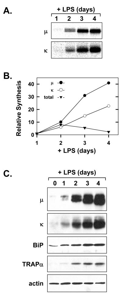
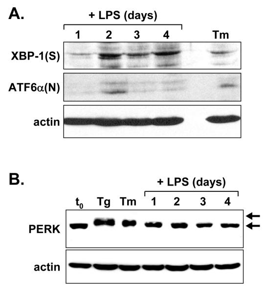
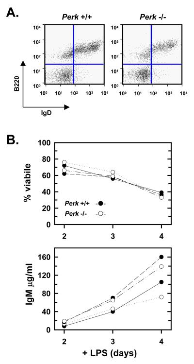
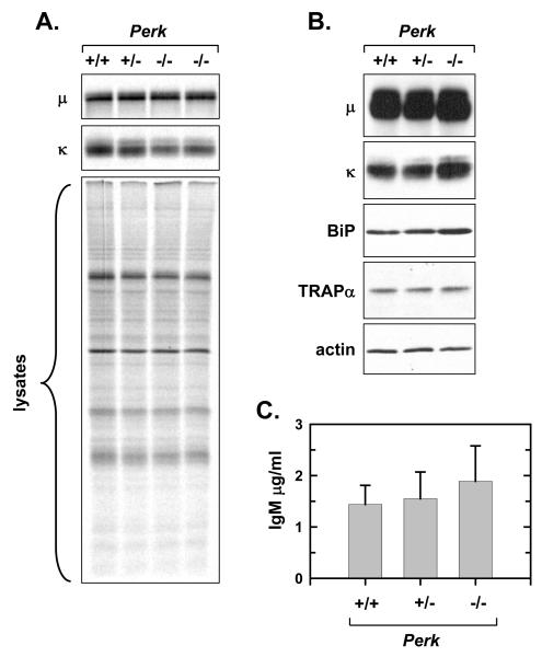
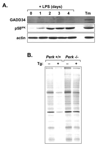
When B-lymphocytes differentiate into plasma cells, immunoglobulin (Ig) heavy and light chain synthesis escalates and the entire secretory apparatus expands to support high-rate antibody secretion. These same events occur when murine B-cells are stimulated with lipopolysaccharide (LPS), providing an in vitro model in which to investigate the differentiation process. The unfolded protein response (UPR), a multi-pathway signaling response emanating from the endoplasmic reticulum (ER) membrane, allows cells to adapt to increasing demands on the protein folding capacity of the ER. As such, the UPR plays a pivotal role in the differentiation of antibody-secreting cells. Three specific stress sensors, IRE1, PERK/PEK and ATF6, are central to the recognition of ER stress and induction of the UPR. IRE1 triggers splicing of Xbp-1 mRNA, yielding a transcriptional activator of the UPR termed XBP-1(S), and activation of the IRE1/XBP-1 pathway has been reported to be required for expansion of the ER and antibody secretion. Here, we provide evidence that PERK is not activated in LPS-stimulated splenic B-cells, whereas XBP-1(S) and the UPR transcriptional activator ATF6 are both induced. We further demonstrate that Perk-/- B-cells develop and are fully competent for induction of Ig synthesis and antibody secretion when stimulated with LPS. These data provide clear evidence for differential activation and utilization of distinct UPR components as activated B-lymphocytes increase Ig synthesis and differentiate into specialized secretory cells.


Figures

Figure 1
Figure 1
Induction of Ig synthesis in LPS-stimulated splenic B-cells. Splenic B-cells were cultured in the presence of LPS. A, At the indicated intervals of LPS stimulation, 2×10⁶ viable cells were metabolically labeled with 75μCi/ml [35S]methionine and cysteine for 10 min. Ig μ and κ chains were immunoprecipitated from the cell lysates and resolved by SDS-PAGE under reducing conditions. Signals from the labeled Ig chains were captured by phosphorimaging. B, Synthesis of Ig μ and κ chains at each interval of LPS stimulation was assessed by quantification of the signals shown in A. Total protein synthesis was assessed by TCA precipitation of cell lysates (2.5×10⁵ cells at each interval) from the experiment in A followed by scintillation spectroscopy. The relative level of synthesis is plotted for μ(●), κ(○), and total protein (▼) at each interval as compared to day 1 (set at 1). C, Cell lysates were prepared at the indicated intervals and equal cell equivalents were assessed by immunoblotting for Ig μ and κ chains, the soluble ER chaperone BiP, the ER translocon component TRAPα, and actin as a loading control.
Figure 2
Figure 2
Immunoblot analysis of UPR components in LPS-stimulated splenic B-cells. Splenic B-cells were cultured in the presence of LPS for the indicated intervals. At day 3, a portion of the cells was treated either for 5 h with 1 μg/ml tunicamycin (Tm) or 1 h with 0.4 μM thapsigargin (Tg) to provide positive controls for UPR activation. Cell lysates were prepared and equal cell equivalents were assessed by immunoblotting for A, XBP-1(S) and ATF6α(N) and B, PERK. Phosphorylated and nonphosphorylated forms of PERK are denoted by upper and lower arrows, respectively. Actin was assessed as a loading control.
Figure 3
Figure 3
Development and LPS response of PERK-deficient B-cells. Splenocytes and splenic B-cells were prepared from age- and sex-matched Perk+/+ and Perk-/- mice. A, Splenocytes were prepared, stained with FITC-conjugated anti-B220 and biotin-conjugated anti-IgD plus streptavidin-APC, and assessed by flow cytometry. Splenocytes scoring as B220+IgD+ mature B-cells appear in the upper right quadrants. Five mice for each genotype were analyzed and representative data are shown. B, Splenic B-cells from two separate Perk+/+ mice (●) and two separate Perk-/- mice (○) were cultured in the presence of LPS. At the indicated intervals, cell viability was assessed by trypan blue dye exclusion (upper panel) and the amounts of IgM present in the culture supernatants were determined by ELISA (lower panel).
Figure 4
Figure 4
Ig synthesis and secretion by LPS-stimulated PERK-deficient B-cells. Splenic B-cells were prepared from age- and sex-matched Perk+/+, Perk+/- and Perk-/- mice and cultured in the presence of LPS for 3 days. A, 2×10⁶ viable cells were metabolically labeled with 75μCi/ml [35S]methionine and cysteine for 10 min. Ig μ and κ chains were immunoprecipitated from the cell lysates and resolved by SDS-PAGE under reducing conditions along with an equivalent amount of cell lysate (2×10⁵ cells). Signals from the labeled Ig chains (upper and middle panels) and from labeled total proteins (lower panel) were captured and quantified by phosphorimaging. As compared to +/+ (set at 1), the mean ± SD for the μ signal: 1.04 ± 0.19 for +/-, n = 3; 0.90 ± 0.28 for -/-, n = 6; κ signal: 0.98 ± 0.14 for +/-, n = 3; 0.68 +/- 0.07 for -/-, n = 6. As compared to +/+ (set at 1), the mean ± SD for total protein synthesis was 0.94 +/- 0.16 for +/-, n = 3 and 0.89 +/- 0.08 for -/-, n = 4. B, Cell lysates were prepared and equal cell equivalents were assessed by immunoblotting for Ig μ and κ chains, the soluble ER chaperone BiP, the ER translocon component TRAPα, and actin as a loading control. C, Cells were harvested, washed and replated at 5×10⁵ c/ml for 4 h. The amount of IgM secreted was determined by ELISA and plotted as mean ±SD (+/+ and +/-, n = 3; -/-, n = 4; p > 0.1).
Figure 5
Figure 5
Induction of PERK regulators and responsiveness of the PERK pathway in LPS-stimulated B-cells. A, Splenic B-cells were cultured in the presence of LPS for the indicated intervals. At day 3, a portion of the cells was treated for 5 h with 1 μg/ml tunicamycin (Tm) to provide a positive control for UPR activation. Cell lysates were prepared and equal cell equivalents were assessed by immunoblotting for GADD34, p58IPK and actin as a loading control. B, Splenic B-cells were prepared from age- and sex-matched Perk+/+ and Perk-/- mice and cultured in the presence of LPS for 3 days. Cells were then treated for 1 h with vehicle alone or 1 μM thapsigargin (Tg) and metabolically labeled with 100μCi/ml [35S]methionine and cysteine for 10 min. Equivalent amounts of cell lysate (2×10⁵ cells) were resolved by SDS-PAGE under reducing conditions and signals from labeled proteins were captured by phosphorimaging. Thapsigargin treatment resulted in a 40% decrease in protein synthesis in +/+ cells and a 14% decrease in -/- cells.

Similar articles

Cited by

References

    1. Andersson J, Sjoberg O, Moller G. Induction of immunoglobulin and antibody synthesis in vitro by lipopolysaccharides. Eur. J. Immunol. 1972;2:349–353. - PubMed
    1. Bertolotti A, Zhang Y, Hendershot LM, Harding HP, Ron D. Dynamic interaction of BiP and ER stress transducers in the unfolded-protein response. Nat. Cell Biol. 2000;2:326–332. - PubMed
    1. Brewer JW, Hendershot LM. Building an antibody factory: a job for the unfolded protein response. Nat. Immunol. 2005;6:23–29. - PubMed
    1. Calame KL, Lin K-I, Tunyaplin C. Regulatory mechanisms that determine the development and function of plasma cells. Annu. Rev. Immunol. 2003;21:205–230. - PubMed
    1. Calfon M, Zeng H, Urano F, Till JH, Hubbard SR, Harding HP, Clark SG, Ron D. IRE1 couples endoplasmic reticulum load to secretory capacity by processing the XBP-1 mRNA. Nature. 2002;415:92–96. - PubMed

Publication types

MeSH terms