

Definition of brainstem afferents to gonadotropin-releasing hormone neurons in the mouse using conditional viral tract tracing
- PMID: 17823269
- PMCID: PMC6101187
- DOI: 10.1210/en.2007-0854
Definition of brainstem afferents to gonadotropin-releasing hormone neurons in the mouse using conditional viral tract tracing
Abstract
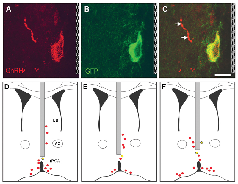
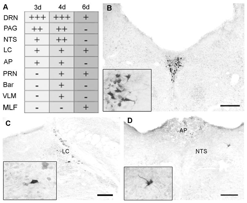
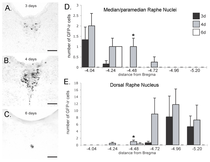
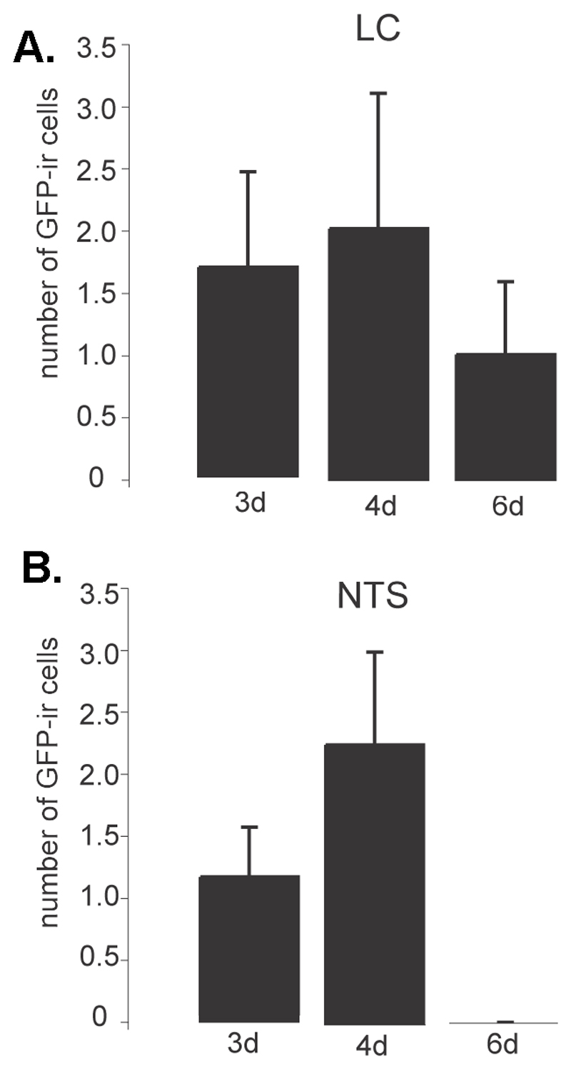
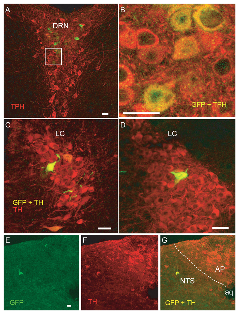
Brainstem monoamines have long been considered to play a role in regulating the activity of GnRH neurons, although their neuroanatomical relationship with these cells has remained unclear. Using a Cre-dependent pseudorabies virus (Ba2001) technique that permits retrograde tracing selectively from GnRH neurons in the mouse, we have examined the organization of brainstem inputs to rostral preoptic area (rPOA) GnRH neurons. Two days after injection of Ba2001 into the rPOA of adult female GnRH-Cre transgenic mice, five to nine GnRH neurons located immediately adjacent to the injection site were found to express green fluorescent protein (GFP), the marker of virus infection, with no GFP expression anywhere else in the brain. In mice killed 24 h later (3 d after injection), GFP-expressing cells were identified (in order of density) in the raphe nuclei, periaqueductal grey, locus coeruleus, nucleus tractus solitarius, and area postrema. This time course is compatible with these neurons representing primary afferent inputs to the GnRH neurons. Four and 6 d after Ba2001 injection, GFP-expressing cells were found in additional brain regions. Dual-label immunofluorescence experiments in 3-d postinjection mice demonstrated that 100% of GFP-expressing neurons in the raphe were positive for tryptophan hydroxylase, whereas 100% and approximately 50% of GFP neurons in the locus coeruleus and nucleus tractus solitarius, respectively, expressed tyrosine hydroxylase. These observations demonstrate that rPOA GnRH neurons receive direct projections from brainstem A2 and A6 noradrenergic neurons and that, surprisingly, the largest afferent input from the brainstem originates from raphe serotonin neurons in the mouse.
Figures
Similar articles
-
Inputs to serotonergic neurons revealed by conditional viral transneuronal tracing.J Comp Neurol. 2009 May 10;514(2):145-60. doi: 10.1002/cne.22003. J Comp Neurol. 2009. PMID: 19274668 Free PMC article.
-
Identification and characterization of estrogen receptor alpha-containing neurons projecting to the vicinity of the gonadotropin-releasing hormone perikarya in the rostral preoptic area of the rat.J Comp Neurol. 1999 Aug 23;411(2):346-58. doi: 10.1002/(sici)1096-9861(19990823)411:2<346::aid-cne13>3.0.co;2-s. J Comp Neurol. 1999. PMID: 10404258
-
Neuronal projections to the medial preoptic area of the sheep, with special reference to monoaminergic afferents: immunohistochemical and retrograde tract tracing studies.J Comp Neurol. 1993 Apr 8;330(2):195-220. doi: 10.1002/cne.903300205. J Comp Neurol. 1993. PMID: 8491868
-
Oestrogen receptors in the brainstem of the female sheep: relationship to noradrenergic cells and cells projecting to the medial preoptic area.J Neuroendocrinol. 1999 Oct;11(10):745-55. doi: 10.1046/j.1365-2826.1999.00370.x. J Neuroendocrinol. 1999. PMID: 10520123
-
Evidence that projections from the bed nucleus of the stria terminalis and from the lateral and medial regions of the preoptic area provide input to gonadotropin releasing hormone (GNRH) neurons in the female sheep brain.Neuroscience. 2005;132(2):421-36. doi: 10.1016/j.neuroscience.2004.12.042. Neuroscience. 2005. PMID: 15802194
Cited by
-
Microdissection of neural networks by conditional reporter expression from a Brainbow herpesvirus.Proc Natl Acad Sci U S A. 2011 Feb 22;108(8):3377-82. doi: 10.1073/pnas.1015033108. Epub 2011 Feb 3. Proc Natl Acad Sci U S A. 2011. PMID: 21292985 Free PMC article.
-
Distinct brainstem and forebrain circuits receiving tracheal sensory neuron inputs revealed using a novel conditional anterograde transsynaptic viral tracing system.J Neurosci. 2015 May 6;35(18):7041-55. doi: 10.1523/JNEUROSCI.5128-14.2015. J Neurosci. 2015. PMID: 25948256 Free PMC article.
-
Regulation of the gonadotropin-releasing hormone neuron during stress.J Neuroendocrinol. 2022 May;34(5):e13098. doi: 10.1111/jne.13098. Epub 2022 Feb 6. J Neuroendocrinol. 2022. PMID: 35128742 Free PMC article. Review.
-
Transneuronal Circuit Analysis with Pseudorabies Viruses.Curr Protoc. 2023 Jul;3(7):e841. doi: 10.1002/cpz1.841. Curr Protoc. 2023. PMID: 37486157 Free PMC article. Review.
-
The alpha-herpesviruses: molecular pathfinders in nervous system circuits.Trends Mol Med. 2008 Mar;14(3):134-40. doi: 10.1016/j.molmed.2007.12.008. Epub 2008 Feb 14. Trends Mol Med. 2008. PMID: 18280208 Free PMC article. Review.
References
-
- Sawyer CH. The seventh Stevenson Lecture. Brain amines and pituitary gonadotrophin secretion. Canadian journal of physiology and pharmacology. 1979;57:667–680. - PubMed
-
- Sawyer CH, Hilliard J, Kanematsu S, Scaramuzzi R, Blake CA. Effects of intraventricular infusions of norepinephrine and dopamine on LH release and ovulation in the rabbit. Neuroendocrinology. 1974;15:328–337. - PubMed
-
- Barraclough CA, Wise PM. The role of catecholamines in the regulation of pituitary luteinizing hormone and follicle-stimulating hormone secretion. Endocr Rev. 1982;3:91–119. - PubMed
-
- Herbison AE. Physiology of the gonadotropin-releasing hormone neuronal network. In: Neill JD, editor. Knobil and Neil's Physiology of Reproduction. Third Edition. New York: Raven Press; 2006. pp. 1415–1482.
-
- Spies HG, Pau KY, Yang SP. Coital and estrogen signals: a contrast in the preovulatory neuroendocrine networks of rabbits and rhesus monkeys. Biology of reproduction. 1997;56:310–319. - PubMed
