Comparative Study
. 2007 Oct;171(4):1168-79.
doi: 10.2353/ajpath.2007.070528. Epub 2007 Sep 6.

Oxidative stress, gene expression, and protein changes induced in the human placenta during labor

Affiliations
Comparative Study

Oxidative stress, gene expression, and protein changes induced in the human placenta during labor

Tereza Cindrova-Davies et al. Am J Pathol. 2007 Oct.

Abstract

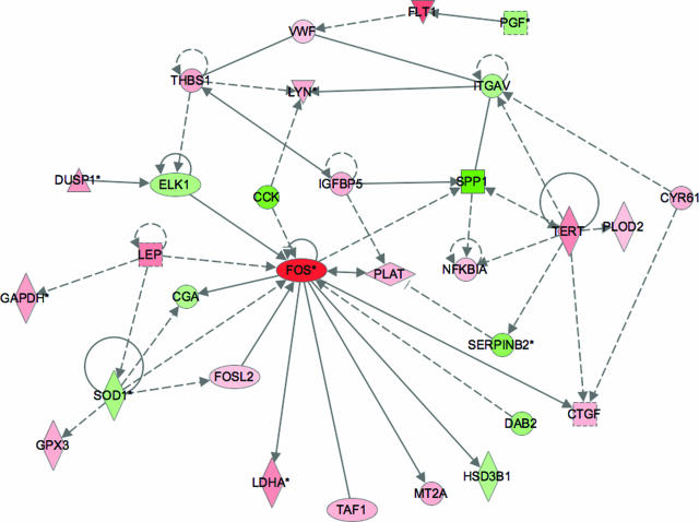
Malperfusion of the placenta has been implicated as a cause of oxidative stress in complications of human pregnancy, leading to release of proinflammatory cytokines and anti-angiogenic factors into the maternal circulation. Uterine contractions during labor are known to be associated with intermittent utero-placental perfusion. We therefore tested whether oxidative stress, proinflammatory cytokines, and angiogenic regulators were increased in placentas subjected to short (<5 hours) and long (>15 hours) labor compared with nonlabored controls delivered by cesarean section. In addition, broader changes in gene transcripts were assessed by microarray analysis. Oxidative stress, activation of the nuclear factor-kappaB pathway, tumor necrosis factor-alpha and interleukin 1beta all increased in placental tissues after labor. Stabilization of hypoxia-inducible factor-1alpha and increased vascular endothelial growth factor soluble receptor-1 were also observed. By contrast, tissue levels of placenta growth factor decreased. Apoptosis was also activated in labored placentas. The magnitude of these changes related to the duration of labor. After labor, 55 gene transcripts were up-regulated and 35 down-regulated, and many of these changes were reflected at the protein level. In conclusion, labor is a powerful inducer of placental oxidative stress, inflammatory cytokines, and angiogenic regulators. Our findings are consistent with intermittent perfusion being the initiating cause. Placentas subjected to labor do not reflect the normal in vivo state at the molecular level.


Figures

Figure 1
Figure 1
Labor induces the expression of oxidative stress markers. Lysates from placentas delivered by CS or vaginally with short labor (<5 hours) or long labor (>15 hours) were immunoblotted with antibodies against Hsp27, Hsp90, and HNE. Immunoblotting with an anti-β-actin antibody served to normalize gel loading. Normalized results (±SE) are plotted, expressing cesarean controls as 100%. Different letters indicate groups that are significantly different using the PLSD test with P < 0.05. Bars indicate groupings of labor samples for comparison with cesarean samples using unpaired Student’s t-test. *P < 0.05.
Figure 2
Figure 2
The effect of labor on antioxidant enzyme expression. Lysates from placentas delivered by CS or vaginally with short labor (<5 hours) or long labor (>15 hours) were immunoblotted with antibodies against catalase, glutathione peroxidase (GPX1), Cu/Zn-SOD, and Mn-SOD. Immunoblotting with an anti-β-actin antibody served to normalize gel loading. Normalized results (±SE) are plotted, expressing cesarean controls as 100%. Different letters indicate groups that are significantly different using the PLSD test with P < 0.05. Bars indicate groupings of labor samples for comparison with cesarean samples using unpaired Student’s t-test. *P < 0.05.
Figure 3
Figure 3
Effect of labor on the expression of inflammatory cytokines, leptin, and COX-2. Lysates from placentas delivered by CS or vaginally with short labor (<5 hours) or long labor (>15 hours) were immunoblotted and analyzed with anti-TNF-α, anti-IL-1β, anti-leptin, anti-COX-2, anti-phospho-IκB, and anti-IκB antibodies. All blots were reprobed with anti-β-actin to normalize gel loading, and normalized results (±SE) are plotted, expressing cesarean controls as 100%. Different letters indicate groups that are significantly different using the PLSD test with P < 0.05.
Figure 4
Figure 4
Effect of labor on the expression of angiogenic factors. Lysates from placentas delivered by CS or vaginally with short labor (<5 hours) or long labor (>15 hours) were immunoblotted and analyzed with anti-HIF-1α, anti-sVEGF-R1, anti-VEGF-A, anti-PlGF, and anti-PAI-2 antibodies. All blots were reprobed with anti-β-actin to normalize gel loading, and normalized results (±SE) are plotted, expressing cesarean controls as 100%. Different letters indicate groups that are significantly different using the PLSD test with P < 0.05.
Figure 5
Figure 5
Labor induces apoptosis. Lysates from placentas delivered by CS or vaginally with short labor (<5 hours) or long labor (>15 hours) were immunoblotted and analyzed with anti-cleaved caspase-3, anti-cleaved caspase-9, and anti-cleaved PARP antibodies. All blots were reprobed with anti-β-actin to normalize gel loading and normalized results (±SE) are plotted, expressing cesarean controls as 100%. Different letters indicate groups that are significantly different using the PLSD test with P < 0.05.
Figure 6
Figure 6
Immunostaining for Hsp27 (A), CuZn-SOD (B), COX-2 (C), TNF-α (D), sVEGF-R1 (E), PlGF (F), PAI-2 (G), and M30 (H) localized these markers principally to the syncytiotrophoblast.
Figure 7
Figure 7
Ingenuity pathway analysis showing regulatory relationships among differentially expressed transcripts. Nodes marked in red indicate transcripts that are increased in prolonged labor relative to CS and those in green, the transcripts that are decreased. Transcripts marked with an asterisk were represented several times in the list of differentially expressed genes, each incidence being derived from a different probe on the microarray. The color intensity reflects the magnitude of the difference. The shape of the symbol denotes the type of protein encoded. Analysis was performed using software from Ingenuity.

Similar articles

Cited by

References

    1. Hubel CA. Oxidative stress in the pathogenesis of preeclampsia. Proc Soc Exp Biol Med. 1999;222:222–235. - PubMed
    1. Myatt L, Cui X. Oxidative stress in the placenta. Histochem Cell Biol. 2004;122:369–382. - PubMed
    1. Burton GJ, Jauniaux E. Placental oxidative stress: from miscarriage to preeclampsia. J Soc Gynecol Invest. 2004;11:342–352. - PubMed
    1. Brosens IA. Morphological changes in the utero-placental bed in pregnancy hypertension. Clin Obstet Gynaecol. 1977;4:573–593. - PubMed
    1. Kim YM, Bujold E, Chaiworapongsa T, Gomez R, Yoon BH, Thaler HT, Rotmensch S, Romero R. Failure of physiologic transformation of the spiral arteries in patients with preterm labor and intact membranes. Am J Obstet Gynecol. 2003;189:1063–1069. - PubMed

Publication types