. 2007 Nov;171(5):1563-75.
doi: 10.2353/ajpath.2007.070147. Epub 2007 Sep 6.

Targeting inflammatory demyelinating lesions to sites of Wallerian degeneration

Affiliations

Targeting inflammatory demyelinating lesions to sites of Wallerian degeneration

Ikuo Tsunoda et al. Am J Pathol. 2007 Nov.

Abstract

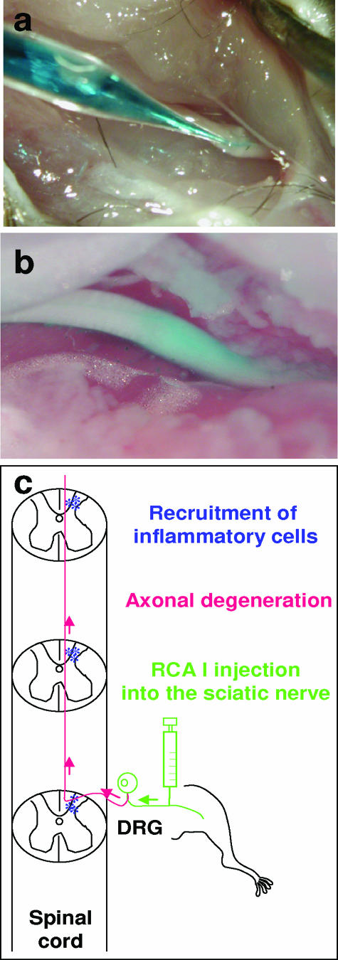
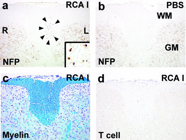
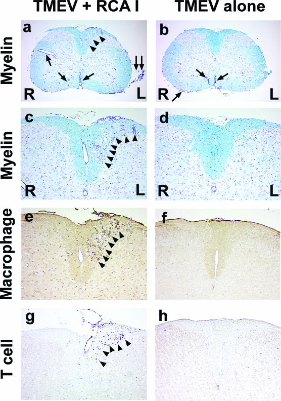
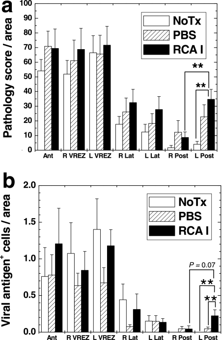
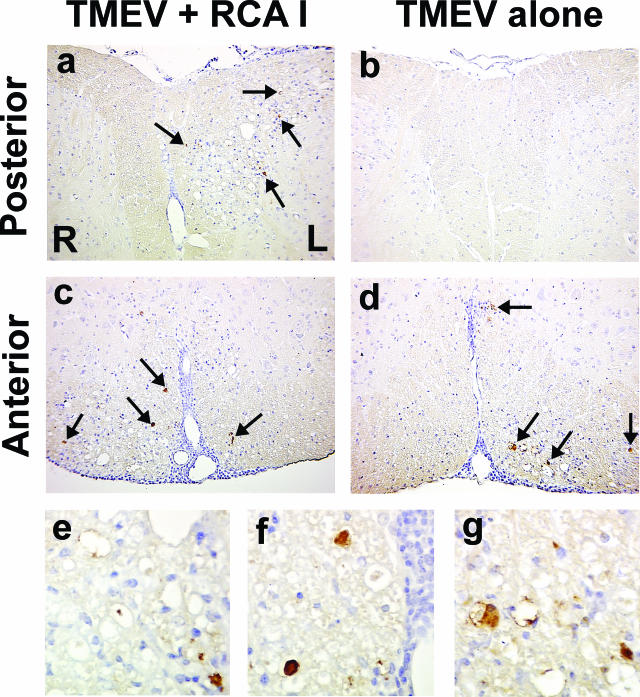
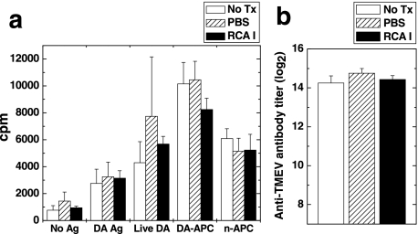
In Theiler's murine encephalomyelitis virus (TMEV) infection, an animal model for multiple sclerosis (MS), axonal injury precedes inflammatory demyelinating lesions, and the distribution of axonal damage present during the early phase of infection corresponds to regions where subsequent demyelination occurs during the chronic phase. We hypothesized that axonal damage recruits inflammatory cells to sites of Wallerian degeneration, leading to demyelination. Three weeks after TMEV infection, axonal degeneration was induced in the posterior funiculus of mice by injecting the toxic lectin Ricinus communis agglutinin (RCA) I into the sciatic nerve. Neuropathology was examined 1 week after lectin injection. Control mice, infected with TMEV but receiving no RCA I, had inflammatory demyelinating lesions in the anterior/lateral funiculi. Other control mice that received RCA I alone did not develop inflammatory lesions. In contrast, RCA I injection into TMEV-infected mice induced lesions in the posterior funiculus in addition to the anterior/lateral funiculi. We found no differences in lymphoproliferative responses or antibody titers against TMEV among the groups. This suggests that axonal degeneration contributes to the recruitment of inflammatory cells into the central nervous system by altering the local microenvironment. In this scenario, lesions develop from the axon (inside) to the myelin (outside) (Inside-Out model).


Figures

Figure 1
Figure 1
a: RCA I was injected into the sciatic nerve using a microinjector. b: The RCA I solution was labeled with Fast Green FCF to ensure the intraneural injection. c: RCA I injected into the left sciatic nerve is transported axonally, leading to death of dorsal root ganglion (DRG) cells and axonal degeneration of the left half of the posterior funiculus, ipsilateral to the injection site. We hypothesized that this axonal degeneration would recruit inflammatory cells into the posterior funiculus.
Figure 2
Figure 2
Spinal cord pathology of mice injected with RCA I. RCA I (a, c, and d) or phosphate-buffered saline (PBS) (b) was injected into the left sciatic nerve of mice. Mice were euthanized 1 week after injection, and neuropathology was examined in the posterior funiculus. a and b: Immunohistochemistry against nonphosphorylated NFP. a: In mice injected with RCA I, the ipsilateral [left (L)] fasciculus gracilis contained many degenerating axons (arrowhead, inset), whereas the contralateral [right (R)] side was devoid of any axonal changes. b: NFP immunoreactivity was detected only in the gray matter (GM) but not in the white matter (WM) of PBS-injected mice. Mice receiving RCA I had no demyelination (c) or T-cell infiltration (d) in the posterior funiculus by Luxol fast blue stain or immunohistochemistry against CD3, respectively. Magnifications: ×120 (a–d); ×962 (a, inset).
Figure 3
Figure 3
Spinal cord pathology of TMEV-infected mice with (a, c, e, and g) or without (b, d, f, and h) RCA I injection. a and c: TMEV-infected mice receiving RCA I injection in the left sciatic nerve had lesions not only in the anterior and lateral funiculi (arrow) but also in the left half (L) of the posterior funiculus (arrowhead). The left side of the spinal cord was labeled by India ink (double arrows). Inflammatory demyelination was seen in the left half of the posterior funiculus, the ipsilateral side of the RCA I injection, but not in the right half (R). b and d: TMEV infection alone induced inflammatory demyelinating lesions in the anterior funiculus and the VREZ (arrow), but not in the posterior funiculus. In the posterior funiculus, macrophage (e and f) and T-cell (g and h) infiltration was seen in RCA I-treated mice (e and g), but not in control mice injected with TMEV alone (f and h). a–d: Luxol fast blue stain. e and f: Lectin histochemistry. g and h: Immunohistochemistry against CD3. Magnifications: ×26 (a and b); ×61 (c–h).
Figure 4
Figure 4
Overall pathology scores (a) and viral antigen-positive cells (b) in the spinal cord of TMEV-infected mice receiving no treatment (no Tx, open column), PBS injection (PBS, hatched column), or RCA I injection (RCA I, closed column). PBS or RCA I was injected into the left sciatic nerve. Higher overall pathology scores and viral antigen-positive cells were detected in the anterior (Ant) funiculus and the VREZ than in the lateral (Lat) and posterior (Post) funiculi in all groups. a: Mean pathology score in the left (L) posterior funiculus for the RCA I group was significantly higher than in the right (R) side in the RCA I group (**P < 0.01, t-test) and the left side of the no treatment group (**P < 0.01, analysis of variance). b: In the posterior funiculus, a significant increase in numbers of viral antigen-positive cells was seen in the left side in mice receiving RCA I, compared with the no treatment and the PBS-injected groups (**P < 0.01), whereas there was no statistical difference between right and left sides in mice injected with RCA I (P = 0.07). Values are mean numbers of viral antigen-positive cells per mouse + SEM for seven to eight mice. Results are representative of two independent experiments.
Figure 5
Figure 5
Immunohistochemical detection of viral antigen-positive cells in the spinal cord of TMEV-infected mice. Spinal cord pathology was examined 4 weeks after TMEV infection with RCA I injection in the left sciatic nerve (RCA I; a, c, e, and f) or without RCA I (TMEV alone; b, d, and g). a: In the posterior funiculus of the RCA I-injected group, viral antigen-positive cells were detected only in the left half (L) (higher magnification shown in e), ipsilateral to injection site, but not in the right half (R). b: Viral antigen was not detected in the posterior funiculus of mice injected with TMEV alone. c and d: In the anterior funiculus, similar numbers of viral antigen-positive cells were seen in the RCA I-treated group (c) and the control group (d) [higher magnification shown in the RCA group (f) and in the control group (g)]. In all groups of mice, viral antigen was seen in macrophages/glial cells but not in neurons. Magnifications: ×112 (a–d); ×424 (e–g).
Figure 6
Figure 6
Correlation of virus persistence with inflammation or demyelination in the spinal cord of infected mice. We compared numbers of virus antigen-positive cells with pathology scores of perivascular cuffing (a and c) or demyelination (b and d) of infected mice, 1 week after RCA I or PBS injection. a and b: In RCA I-injected mice, there was no significant correlation of virus persistence with inflammation (a) or demyelination (b) in the left posterior funiculus, ipsilateral to RCA I injection site. c and d: In the left VREZ of PBS-injected mice, virus antigen-positive cells correlated with perivascular cuffing (c) but not with demyelination (d).
Figure 7
Figure 7
Cellular (a) and humeral (b) immune responses against TMEV. Three weeks after TMEV infection, groups of mice were untreated (open column, no Tx), sham-injected (hatched column, PBS), or injected with RCA I (closed column, RCA I) into the sciatic nerve. One week after treatment, spleen MNCs and sera were collected. a: Lymphoproliferative responses of MNCs that were incubated without antigen (no Ag) or with purified UV-irradiated antigen from Daniels strain of TMEV (DA Ag), live DA virus (live DA), antigen-presenting cells infected with DA virus (DA-APCs), or uninfected syngeneic antigen-presenting cells (n-APCs). All MNCs from the three groups had lymphoproliferative responses against not only DA virus but also n-APC (autoproliferation). No differences were found between the groups. The results are means + SEM of three experiments. b: Anti-TMEV antibody measured by enzyme-linked immunosorbent assay. No significant difference in anti-TMEV antibody titer was detected between the groups (P > 0.05). Values are mean anti-TMEV antibody titers + SEM for seven or eight mice per group. Results are representative of three independent experiments.

Similar articles

Cited by

References

    1. Legido A, Tenembaum SN, Katsetos CD, Menkes JH. Autoimmune and postinfectious diseases. Menkes JH, Sarnat HB, Maria BL, editors. Philadelphia: Lippincott Williams & Wilkins,; Child Neurology. (ed 7) 2006:pp 557–657.
    1. Silber E, Sharief MK. Axonal degeneration in the pathogenesis of multiple sclerosis. J Neurol Sci. 1999;170:11–18. - PubMed
    1. Trapp BD, Peterson J, Ransohoff RM, Rudick R, Mörk S, Bö L. Axonal transection in the lesions of multiple sclerosis. N Engl J Med. 1998;338:278–285. - PubMed
    1. Ferguson B, Matyszak MK, Esiri MM, Perry VH. Axonal damage in acute multiple sclerosis lesions. Brain. 1997;120:393–399. - PubMed
    1. Kuhlmann T, Lingfeld G, Bitsch A, Schuchardt J, Brück W. Acute axonal damage in multiple sclerosis is most extensive in early disease stages and decreases over time. Brain. 2002;125:2202–2212. - PubMed

Publication types

MeSH terms