Review
. 2007 Nov;171(5):1407-18.
doi: 10.2353/ajpath.2007.070251. Epub 2007 Sep 6.

The emerging role of valve interstitial cell phenotypes in regulating heart valve pathobiology

Affiliations
Review

The emerging role of valve interstitial cell phenotypes in regulating heart valve pathobiology

Amber C Liu et al. Am J Pathol. 2007 Nov.

Abstract

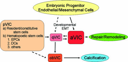
The study of the cellular and molecular pathogenesis of heart valve disease is an emerging area of research made possible by the availability of cultures of valve interstitial cells (VICs) and valve endothelial cells (VECs) and by the design and use of in vitro and in vivo experimental systems that model elements of valve biological and pathobiological activity. VICs are the most common cells in the valve and are distinct from other mesenchymal cell types in other organs. We present a conceptual approach to the investigation of VICs by focusing on VIC phenotype-function relationships. Our review suggests that there are five identifiable phenotypes of VICs that define the current understanding of their cellular and molecular functions. These include embryonic progenitor endothelial/mesenchymal cells, quiescent VICs (qVICs), activated VICs (aVICs), progenitor VICs (pVICs), and osteoblastic VICs (obVICs). Although these may exhibit plasticity and may convert from one form to another, compartmentalizing VIC function into distinct phenotypes is useful in bringing clarity to our understanding of VIC pathobiology. We present a conceptual model that is useful in the design and interpretation of studies on the function of an important phenotype in disease, the activated VIC. We hope this review will inspire members of the investigative pathology community to consider valve pathobiology as an exciting new frontier exploring pathogenesis and discovering new therapeutic targets in cardiovascular diseases.


Figures

Figure 1
Figure 1
The current literature describes numerous VIC functions that can be conveniently organized into five phenotypes: embryonic progenitor endothelial/mesenchymal cells, quiescent VICs (qVICs), activated VICs (aVICs), stem cell-derived progenitor VICs (pVICs), and osteoblastic VICs (obVICs). These represent specific sets of VIC functions in normal valve physiology and pathophysiology. Embryonic progenitor endothelial/mesenchymal cells undergo endothelial-mesenchymal transformation in fetal development to give rise to aVICs and/or qVICs resident in the normal heart valve. The VICs undergoing the transformation possess features of aVICs, including migration, proliferation, and matrix synthesis. When the heart valve is subjected to an insult, be it abnormal hemodynamic/mechanical stress or pathological injury, qVICs become activated, giving rise to aVICs that participate in repair and remodeling of the valve. pVICs including bone marrow-derived cells, circulating cells, and resident valvular progenitor cells are another source of aVICs in the adult. The relationship between bone marrow, circulating and resident pVICs is unknown. In conditions promoting valve calcification, such as in the presence of osteogenic and chondrogenic factors, qVICs can undergo osteoblastic differentiation into obVICs. It is possible that obVICs are derived from pVICs. obVICs actively participate in the valve calcification process. Compartmentalizing VIC function into distinct phenotypes also recognizes the transient behavior of VIC phenotypes as noted (hatched arrows depict possible transitions for which there is currently no solid evidence).
Figure 2
Figure 2
Phase contrast photomicrographs of VICs in monolayer culture at moderate (A), confluent (B), and superconfluent (C) densities. Note the elongated morphology in A and the overlapping growth pattern in B as indicated by arrows. Scale bar = 20 μm. Original magnifications, ×200.
Figure 3
Figure 3
The normal adult heart valve is well adapted to its physiological environment, able to withstand the unique hemodynamic/mechanical stresses under normal conditions. Under conditions of pathological injury or abnormal hemodynamic/mechanical stresses, VICs become activated through activation of VECs and by inflammation and associated cytokine and chemokine signals. Macrophages will also be activated. aVICs increase matrix synthesis, up-regulate expression of matrix-remodeling enzymes, migrate, proliferate and undergo apoptosis, and undergo osteoblast transformation. These processes are regulated by a variety of factors, several secreted by the aVIC. If the aVICs continue to promote these cellular processes, angiogenesis, chronic inflammation, fibrosis, and calcification result, leading to progressive clinical valve disease.
Figure 4
Figure 4
Immunofluorescent photomicrographs of single porcine mitral valve aVICs in culture showing a migrating aVIC. aVICs express prominent amounts of α-SMA (A, green), a widely accepted marker for VIC activation, as well as TGF-β (B, blue), an important factor that regulates this activation as well as several aVIC functions, and is secreted by aVICs in turn. Scale bar = 10 μm.

References

    1. Lester W, Rosenthal A, Granton B, Gotlieb AI. Porcine mitral valve interstitial cells in culture. Lab Invest. 1988;59:710–719. - PubMed
    1. Lester WM, Gotlieb AI. In vitro repair of the wounded porcine mitral valve. Circ Res. 1988;62:833–845. - PubMed
    1. Mulholland DL, Gotlieb AI. Cell biology of valvular interstitial cells. Can J Cardiol. 1996;12:231–236. - PubMed
    1. Butany J, Gotlieb AI. Native valvular heart disease. McManus BM, editor. Philadelphia: Current Medicine Inc.; Atlas of Cardiovascular Pathology for the Clinician. (ed 2) 2001:pp 201–215.
    1. Durbin A, Gotlieb AI. Advances towards understanding heart valve response to injury. Cardiovasc Pathol. 2002;11:69–77. - PubMed

Publication types

MeSH terms

Substances