. 2007 Oct;171(4):1153-67.
doi: 10.2353/ajpath.2007.061164. Epub 2007 Sep 6.

Dissecting the impact of chemotherapy on the human hair follicle: a pragmatic in vitro assay for studying the pathogenesis and potential management of hair follicle dystrophy

Affiliations

Dissecting the impact of chemotherapy on the human hair follicle: a pragmatic in vitro assay for studying the pathogenesis and potential management of hair follicle dystrophy

Eniko Bodó et al. Am J Pathol. 2007 Oct.

Abstract

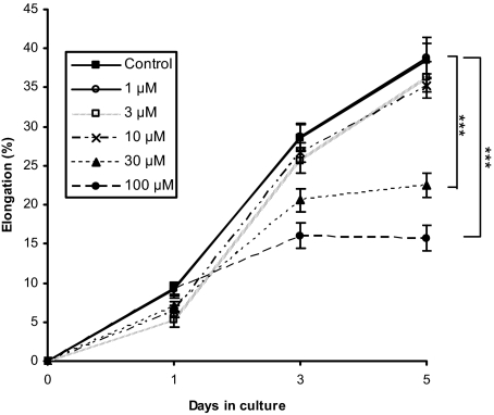
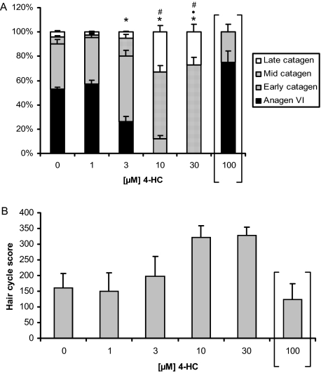
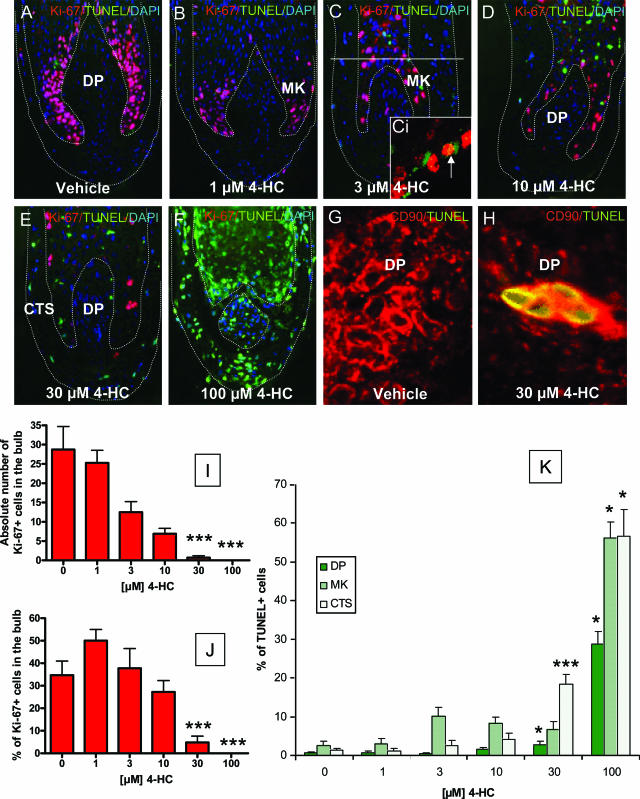
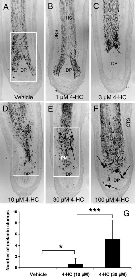
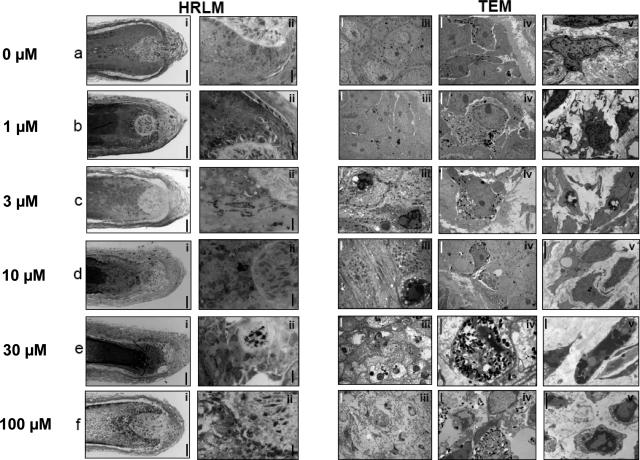
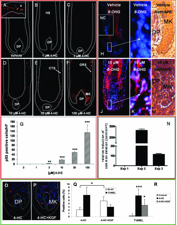
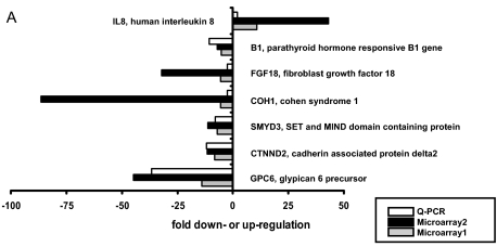
Chemotherapy-induced alopecia represents one of the major unresolved problems of clinical oncology. The underlying molecular pathogenesis in humans is virtually unknown because of the lack of adequate research models. Therefore, we have explored whether microdissected, organ-cultured, human scalp hair follicles (HFs) in anagen VI can be exploited for dissecting and manipulating the impact of chemotherapy on human HFs. Here, we show that these organ-cultured HFs respond to a key cyclophosphamide metabolite, 4-hydroperoxycyclophosphamide (4-HC), in a manner that resembles chemotherapy-induced HF dystrophy as it occurs in vivo: namely, 4-HC induced melanin clumping and melanin incontinence, down-regulated keratinocyte proliferation, massively up-regulated apoptosis of hair matrix keratinocytes, prematurely induced catagen, and up-regulated p53. In addition, 4-HC induced DNA oxidation and the mitochondrial DNA common deletion. The organ culture system facilitated the identification of new molecular targets for chemotherapy-induced HF damage by microarray technology (eg, interleukin-8, fibroblast growth factor-18, and glypican 6). It was also used to explore candidate chemotherapy protectants, for which we used the cytoprotective cytokine keratinocyte growth factor as exemplary pilot agent. Thus, this novel system serves as a powerful yet pragmatic tool for dissecting and manipulating the impact of chemotherapy on the human HF.


Figures

Figure 1
Figure 1
4-HC inhibits hair shaft elongation at high concentrations. Isolated HFs were treated with vehicle/4-HC (1 to 100 μmol/L) for 5 days in supplemented Williams’ E medium. Hair shaft length (distance between the base of the bulb and the cut end of the hair fiber) was measured every 2nd day. Hair shaft elongation (percent) of the treated groups was calculated and compared with those of the control. Elongation data are expressed as mean ± SEM, significance was calculated by Mann-Whitney U-test, ***P < 0.001. Data represents pooled results of three independent experiments.
Figure 2
Figure 2
4-HC induces premature catagen-like transformations. HFs were treated with vehicle/4-HC (1 to 100 μmol/L) and morphological changes were recorded on H&E-stained cryosections according to well-defined criteria. A: Quantitative histomorphometry shows the percentage of HFs in distinct HF stages (anagen, early catagen, mid-catagen, late catagen at day 5). B: Hair cycle score was calculated as follows: each HF was ascribed an arbitrary value (anagen, 100; early catagen, 200; mid-catagen, 300; and late catagen, 400). Values were added within a group and divided by the number of staged follicles. All data represent the results of three independent experiments (10 to 18 follicles/per experiment) and are expressed as mean ± SEM. Statistical analysis was performed using the Mann-Whitney U-test, *P < 0.05 in anagen, P < 0.05 in early catagen, #P < 0.05 in mid-catagen. [ ] HFs arrest in a highly dystrophic pseudo-anagen VI stage.
Figure 3
Figure 3
4-HC inhibits matrix keratinocyte proliferation and stimulates apoptosis in several HF compartments. A–F: HFs were double-labeled with Ki-67 (red)/TUNEL (green) and counterstained with DAPI (blue). Ki-67-positive cells were counted in the matrix keratinocytes below the distal end of the DP (C) and are expressed as relative number (I) or percentage of all (DAPI +) cells (J). K: TUNEL-positive cells were analyzed in two additional compartments: DP and CTS. MK, matrix keratinocytes; Ci, NKI-beteb (melanocyte marker)/Ki-67 double staining. Arrow denotes double-stained melanocyte. Note the proliferative HF pigmentation unit melanocytes in the precortical matrix of 4-HC (3 μmol/L)-treated HFs, as previously reported by Sharov and colleagues for CYP-treated mice in vitro. G and H: Fibroblast marker (red)/TUNEL (green) double staining. Data are expressed as mean ± SEM. Statistical analysis was performed using the Mann-Whitney U-test, *P < 0.05, ***P < 0.001. Original magnifications: ×200 (A–F); ×400 (G and H).
Figure 4
Figure 4
4-CH induces dystrophic melanin production and transfer. A–F: Cultured HFs were embedded and processed for histology. Melanin granules were detected. G: Masson-Fontana+ conglomerates that were larger than keratinocyte nuclei were counted in the defined region (indicated in white). CTS, connective tissue sheath; ORS, outer root sheath; HS, hair shaft; PM, precortical matrix. *P < 0.05, ***P = 0.000. Original magnifications, ×100.
Figure 5
Figure 5
HRLM (i and ii) and TEM (iii–v) show severe ultrastructural changes in diverse cell types of human HF after 4-HC administration. a: No exposure to 4-HC. HFs exhibited normal anagen VI morphology and ultrastructure including dispersion of DP cells in copious extracellular matrix (i and v) and an intact HF pigmentary unit (ii and iv). Keratinocyte differentiation appeared normal (iv), although some thickening of the basal lamina separating the DP and the matrix was apparent (iv). b: Exposure to 1 μmol/L 4-HC. HFs exhibited anagen morphology and ultrastructure with some loss of matrix volume (i). DP cells were dispersed in copious extracellular matrix (i and v) and the HF pigmentary unit appeared intact (ii and iv). Keratinocyte differentiation appeared normal (iv) and proliferation among matrix keratinocytes was evident (ii). c: Exposure to 3 μmol/L 4-HC. HFs exhibited early catagen-like morphology and ultrastructure including some loss of matrix and DP volume (i and v). DP cells contained large vacuoles and phagolysosomal inclusions (v). The HF pigmentary unit displayed some perturbation including pigment accumulation, which suggested a reduction in melanin transfer efficiency (ii). Scattered degenerating keratinocytes were found in the HF precortex. d: Exposure to 10 μmol/L 4-HC. HFs exhibited advanced catagen-like morphology and ultrastructure including a substantial loss of matrix and follicular papilla volume (i and v). DP cells were distributed in a clumped manner and contained large vacuoles and phagolysosomal inclusions, some containing melanin (v). The HF pigmentary unit contained some intact and functioning melanocytes, whereas other melanocytes exhibited degenerative change (ii). Scattered degenerating keratinocytes were detected in the HF precortex. e: Exposure to 30 μmol/L 4-HC. HFs exhibited a dystrophic catagen-like morphology and ultrastructure including a substantial loss of hair bulb epithelial volume and DP volume (i and v). DP cell number seemed to be reduced, and some cells appeared necrotic and were observed to contain numerous large inclusions (v), some of which contained melanin and lipid-like inclusions. The HF pigmentary unit was severely disputed with little evidence of functioning melanocytes remaining. Degenerating melanocytes were also detected high in the precortex, after detachment from the hair bulb (iv). Keratinocytes of the precortex and cortex displayed evidence of widespread necrosis (iii). f: Exposure to 100 μmol/L 4-HC. HFs exhibited a complete cytotoxic stasis characterized by a pseudo-anagen morphology derived from completed necrotic change in all cellular compartments.
Figure 6
Figure 6
4-HC increases the number of the p53-expressing cells, common DNA deletion, and DNA oxidation in a dose-dependent manner in HF organ culture. A–G: KGF slightly, but significantly, diminishes 4-HC-induced inhibition of hair matrix keratinocyte proliferation and inhibits 4-HC-induced HF apoptosis: HFs were treated with 4-HC (1 to 100 μmol/L), and p53 protein was detected on HF cryosections by tyramide signal amplification method. p53-positive nuclei/HFs were counted. Data are expressed as mean ± SEM, statistical analysis was performed using the Mann-Whitney U-test. **P < 0.01, ***P < 0.001. e, epidermis; CTS, connective tissue sheath; ORS, outer root sheath; HS, hair shaft. H–N: Vehicle/4-HC-treated HFs were stained for 8-OHdG and Ref1/APE1 (specific indicators of oxidative DNA damage). NC, negative control; MK, matrix keratinocytes. N: HFs were incubated with vehicle/30 μmol/L 4-HC, and homogenized and genomic DNA was isolated for detection of the common deletion by real-time PCR. Relative induction of mitochondrial DNA deletion were compared with the vehicle (=1). O and P: Double labeling of proliferating (red) and apoptotic (green) cells by Ki-67/TUNEL immunohistochemistry of vehicle (O) and KGF (P) pretreated HFs exposed to 4-HC (30 μmol/L). The percentage of Ki-67- and TUNEL-positive cells is shown (Q and R) as mean ± SEM. *P < 0.05. MK, matrix keratinocytes. R: Human scalp HFs were pretreated overnight with 20 ng/ml KGF before administration of 10 μmol/L 4-HC (under continued stimulation with KGF), and they were cultured for 48 more hours. The percentage of TUNEL+ cells was counted (mean ± SEM, *P < 0.05, ***P < 0.001). Original magnifications: ×100 (A–H, J, and N–R); ×400 (I and K); and ×200 (L and M).
Figure 7
Figure 7
4-HC alters the gene expression program of human HF. Gene expression analysis of HFs from two different individuals (Microarray1 and Microarray2) was performed using Human Whole Genome Oligo Microarray (44K). HFs (20 per group) were treated with vehicle/4-HC (30 μmol/L) for 48 hours. Candidate genes were selected according to the following criteria: equidirectional expression changes in both individuals, P value <0.0001, more than fivefold change. For a confirmation Q-PCR analysis (Q-PCR) of the above-selected genes was performed. Expression changes are displayed as fold down- or up-regulations.

Similar articles

Cited by

References

    1. Hesketh PJ, Batchelor D, Golant M, Lyman GH, Rhodes N, Yardley D. Chemotherapy-induced alopecia: psychosocial impact and therapeutic approaches. Support Care Cancer. 2004;12:543–549. - PubMed
    1. Fraiser LH, Kanekal S, Kehrer JP. Cyclophosphamide toxicity. Characterising and avoiding the problem. Drugs. 1991;42:781–795. - PubMed
    1. Wang J, Lu Z, Au JL. Protection against chemotherapy-induced alopecia. Pharm Res. 2006;23:2505–2514. - PubMed
    1. Merk HF. Drugs affecting hair growth. Lyne AG, Short BF, editors. Sydney: Augus and Robertson,; Biology of the skin and hair growth. 1965:pp 601–609.
    1. Crounse GR, van Scott EJ. Changes in scalp hair roots as a measure of toxicity from cancer chemotherapeutic drugs. J Invest Dermatol. 1960;35:83–90. - PubMed

Publication types

MeSH terms