. 2007 Oct;171(4):1395-404.
doi: 10.2353/ajpath.2007.070190. Epub 2007 Sep 6.

Disordered purinergic signaling inhibits pathological angiogenesis in cd39/Entpd1-null mice

Affiliations

Disordered purinergic signaling inhibits pathological angiogenesis in cd39/Entpd1-null mice

Shaun W Jackson et al. Am J Pathol. 2007 Oct.

Abstract

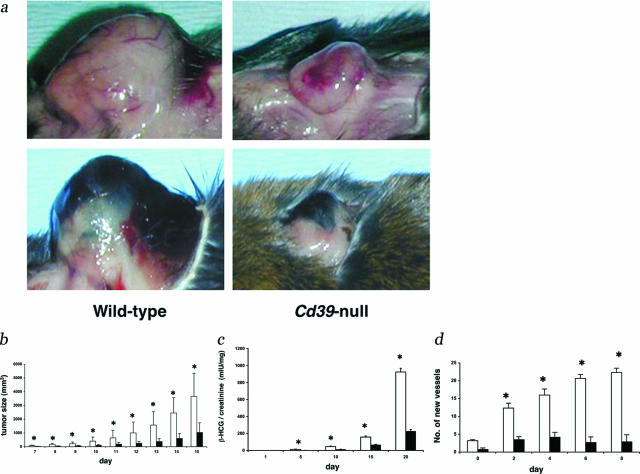
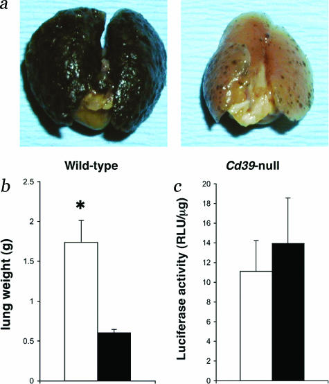
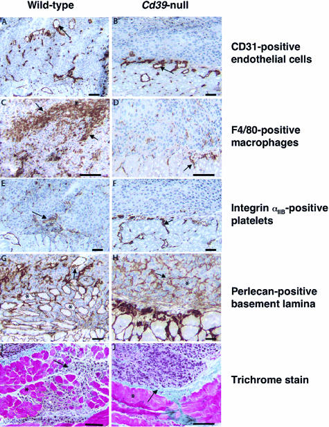
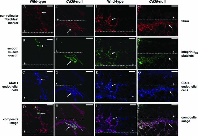
CD39/ecto-nucleoside triphosphate diphosphohydrolase-type-1 (ENTPD1) is the dominant vascular ecto-nucleotidase that catalyzes the phosphohydrolysis of extracellular nucleotides in the blood and extracellular space. This ecto-enzymatic process modulates endothelial cell, leukocyte, and platelet purinergic receptor-mediated responses to extracellular nucleotides in the setting of thrombosis and vascular inflammation. We show here that deletion of Cd39/Entpd1 results in abrogation of angiogenesis, causing decreased growth of implanted tumors and inhibiting development of pulmonary metastases. Qualitative abnormalities of Cd39-null endothelial cell adhesion and integrin dysfunction were demonstrated in vitro. These changes were associated with decreased activation of focal adhesion kinase and extracellular signaling-regulated kinase-1 and -2 in endothelial cells. Our data indicate novel links between CD39/ENTPD1, extracellular nucleotide-mediated signaling, and vascular endothelial cell integrin function that impact on angiogenesis and tumor growth.


Figures

Figure 1
Figure 1
Deletion of CD39 is associated with decreased tumor growth. a: Representative examples of tumor growth in wild-type (left) and Cd39-null (right) mice. Upper tumor is LLC, and lower is B16-F10 melanoma. b: Measurement of murine B16-F10 and LLC (not shown, comparable data) subcutaneous tumor growth in wild-type (□) and Cd39-null (▪) mice. Bars show mean tumor volume in mm3 ± SD. *P < 0.05. c: β-HCG excretion after subcutaneous inoculation with B16-CG tumors. Wild type (□), Cd39-null (▪). Bars show urinary β-HCG/creatinine (mIU/mg) ± SD. *P < 0.05. d: Subperitoneal B16-F10 tumor examined for 8 days after implantation in wild-type and Cd39-null mice. Vessel counts in subperitoneal B16-F10 tumors in wild-type (□) and Cd39-null (▪) mice. Bars show mean number of vessels ± SEM. *P < 0.05.
Figure 2
Figure 2
Decreased growth of inoculated tumor metastases, in Cd39-null mice. a: Representative examples of wild-type (right) and Cd39-null (left) mouse lungs at 15 days after tumor cell injection intravenously. b: Wild-type (□) and Cd39-null (▪) lung weights 15 days after intravenous tumor injection. a and b: Mice were injected with 1.5 × 10⁵ B16-F10 cells per 25 g of body weight into the inferior vena cava and lungs were examined at 15 days. Bars represent mean weight ± SEM. *P < 0.006. c: Retention of cell-associated luciferase activity in wild-type (□) and Cd39-null (▪) mice 1 hour after injection of 5 × 10⁵ luciferase-expressing B16-F10 cells (lucB16/F10). Bars represent relative light units (RLUs) per μg of lung ± SD.
Figure 3
Figure 3
Immunohistochemistry of B16-F10 tumors. Tumors were implanted and grown in the subcutaneous space in wild-type (A, C, E, G, and I) and Cd39-null (B, D, F, H, and J) mice. A and B: CD31-positive endothelial cells can be seen infiltrating tumor (arrow) in wild-type (A), but not Cd39-null animals (B). Endothelial cells are confined to the interface between tumor and surrounding tissue in Cd39-null mice (arrow). C and D: There was marked infiltration of F4/80-positive macrophages (arrows) in tumors grown in wild-type animals (C), in particular in the necrotic central zone (*). In contrast, macrophages were mostly absent within tumors grown in Cd39-null mice (D). E and F: Platelets (integrin αIIb-positive) were associated with vessels within the tumor mass in wild-type mice (E) but were confined to the tumor interface in Cd39-null animals (F). G and H: In tumors grown in wild-type mice (G), perlecan staining showed basal lamina associated with tumor vessels (arrow) but not tumor cell nests. In contrast, in tumors grown in Cd39-null mice (H), which were primarily devoid of vasculature, basal lamina divided individual tumor cell nests into gland-like structures (*). I and J: Trichrome staining showed tumors cells (arrow) in wild-type mice (I) invading deep into the underlying muscle layer (*). In contrast, in tumors grown in Cd39-null mice (J), tumor cell invasion was absent, and the tumor mass and muscle layer (*) was clearly separated. Scale bars = 20 μm.
Figure 4
Figure 4
Triple immunofluorescent staining patterns of B16-F10 tumors. B16-F10 tumors were grown in the subcutaneous space in wild-type (A–D, I–L) and Cd39-null (E–H, M–P) mice and stained with antibodies recognizing: pericytes and fibroblasts (pan-reticular fibroblast marker) (A and E); pericytes and smooth muscle cells (smooth muscle α-actin) (B and F); fibrin (I and M); platelets (integrin αIIb) (J and N); and endothelial cells (CD31) (C, G, K, and O) and their composites (D, H, L, and P). The dotted white line delineates the interface between the tumor mass and adjacent normal tissue. The arrow indicates the direction of the tumor. A–D: In tumors grown in wild-type mice the endothelial lining (blue) of vessels were only partially covered by mature smooth muscle α-actin-expressing pericytes (green) and interspersed in a network of interstitial fibroblasts (red). Note that the interstitial fibroblasts do not express smooth muscle α-actin. E–H: In Cd39-null mice, the endothelial lining (blue) of vessels in normal tissue adjacent to the tumor were to a large extent covered by smooth muscle α-actin-expressing pericytes (green) and surrounded by interstitial fibroblasts (red). Similar structures were absent within the actual tumor mass. I–L: In tumors in wild-type mice, a provisional matrix consisting of fibrin (red) and platelets (green) were associated with a large portion of CD31-positive (blue) tumor vessels (arrow). M–P: In Cd39-null mice fibrin (red) and platelets (green) were concentrated to a compacted rim of CD31 (blue)-positive vessels (arrow) at the interface between the tumor and normal adjacent tissue. Note nonspecific fluorescent staining in the central necrotic zone. Scale bars = 20 μm.
Figure 5
Figure 5
Cd39 deletion and associated integrin αvβ3 dysfunction. a: Phosphorylation of ERK1/2 (Thr202/Tyr204) in wild-type (□) and Cd39-null (▪) endothelial cells at baseline and after stimulation with VEGF (10 ng/ml) for 5 minutes. Bar chart represents densitometry results (means ± SD) of phosphorylated ERK1/2 corrected for total ERK1/2. *P < 0.05. b: Wild-type (□) and Cd39-null (▪) endothelial adhesion to collagen, fibronectin, and vitronectin. Results expressed as a percentage of wild-type adhesion to collagen ± SEM. *P < 0.05. c: Wild-type (□) and Cd39-null (▪) endothelial cell adhesion to vitronectin after a 6-hour pretreatment with soluble apyrase (5 U/ml). Results expressed as mean percentage of wild-type adhesion to vitronectin ± SEM. *P < 0.05. d: The αv-integrin subunit expression in wild-type and Cd39-null endothelial cells was determined by immunoprecipitation and Western blot analysis. Cells were treated with UTP (100 μmol/L) for 0, 2, 4, and 6 hours. No differences were evident (not shown). Bar chart represents wild-type (□) and Cd39-null (▪) endothelial cell adhesion to vitronectin after treatment with MnCl2 (500 μmol/L). Results expressed as a percentage of wild-type adhesion to vitronectin ± SEM. *P < 0.05.
Figure 6
Figure 6
Deletion of Cd39 is associated with defective αvβ3-mediated intracellular signaling. a and c: Graphs represent phosphorylation of FAK (Tyr 397) (a) and ERK1/2 (Thr202/Tyr204) (c) in wild-type (□) and Cd39-null (▪) endothelial cells at 0, 15, and 30 minutes after plating onto vitronectin. b and d: Phosphorylation of FAK (b) and ERK1/2 (d) in wild-type (□) and Cd39-null (▪) endothelial cells pretreated for 6 hours with soluble Apyrase (5 U/ml). Bar charts represent densitometry results (means ± SEM) of phosphorylated FAK or ERK1/2 divided by total FAK or ERK1/2, respectively. *P < 0.05. Representative blots are shown below graphs.

References

    1. Kaczmarek E, Koziak K, Sevigny J, Siegel JB, Anrather J, Beaudoin AR, Bach FH, Robson SC. Identification and characterization of CD39/vascular ATP diphosphohydrolase. J Biol Chem. 1996;271:33116–33122. - PubMed
    1. Fredholm BB, Abbracchio MP, Burnstock G, Daly JW, Harden TK, Jacobson KA, Leff P, Williams M. Nomenclature and classification of purinoreceptors. Pharmacol Rev. 1994;46:143–152. - PMC - PubMed
    1. Enjyoji K, Sevigny J, Lin Y, Frenette PS, Christie PD, Esch JS, Imai M, Edelberg JM, Rayburn H, Lech M, Beeler DL, Csizmadia E, Wagner DD, Robson SC, Rosenberg RD. Targeted disruption of Cd39/ATP diphosphohydrolase results in disordered hemostasis and thromboregulation. Nat Med. 1999;5:1010–1017. - PubMed
    1. Satterwhite CM, Farrelly AM, Bradley ME. Chemotactic, mitogenic, and angiogenic actions of UTP on vascular endothelial cells. Am J Physiol. 1999;45:H1091–H1097. - PubMed
    1. Goepfert C, Sundberg C, Sevigny J, Enjyoji K, Hoshi T, Csizmadia E, Robson SC. Disordered cellular migration and angiogenesis in Cd39-null mice. Circulation. 2001;104:3109–3115. - PubMed

Publication types

MeSH terms