. 2007 Dec;21(12):2941-55.
doi: 10.1210/me.2007-0211. Epub 2007 Sep 6.

RelB, a new partner of aryl hydrocarbon receptor-mediated transcription

Affiliations

RelB, a new partner of aryl hydrocarbon receptor-mediated transcription

Christoph F A Vogel et al. Mol Endocrinol. 2007 Dec.

Abstract

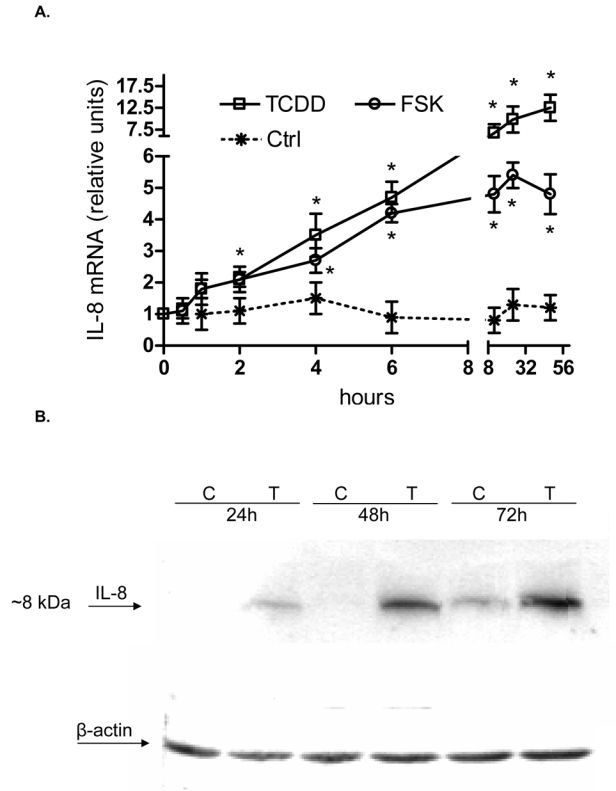
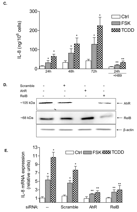
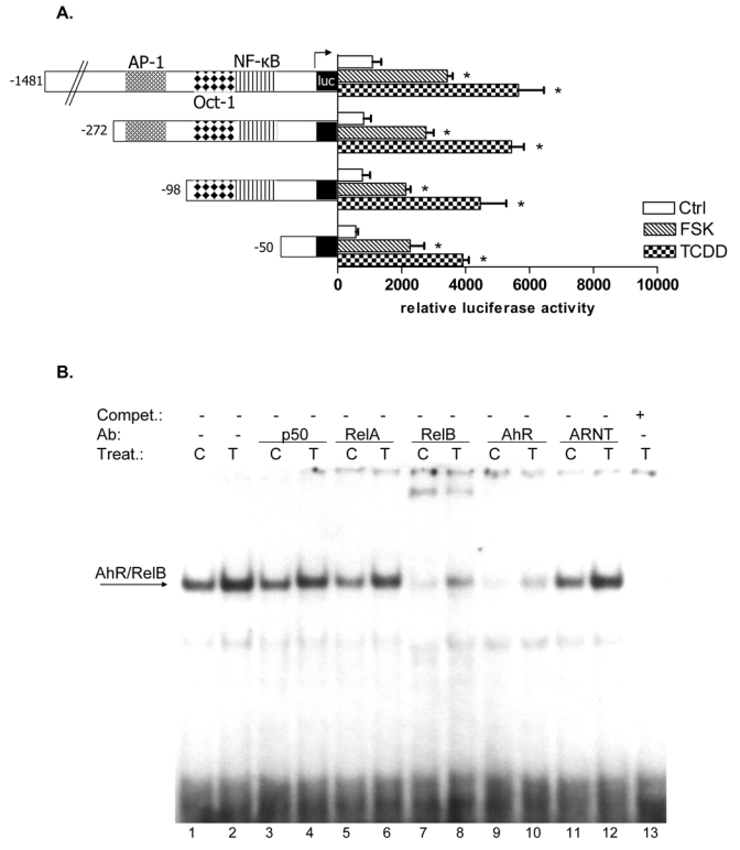
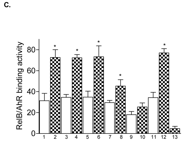
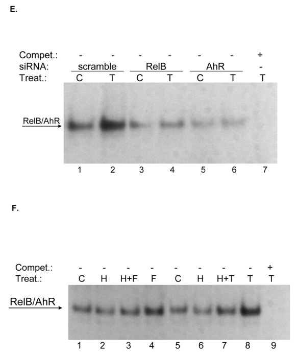
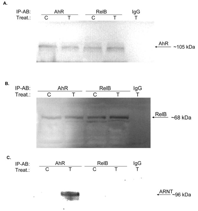
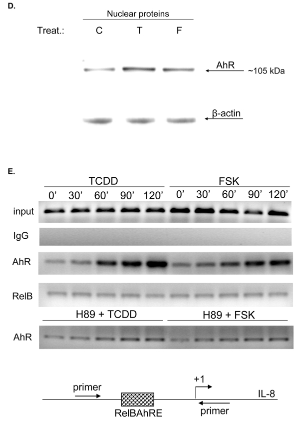
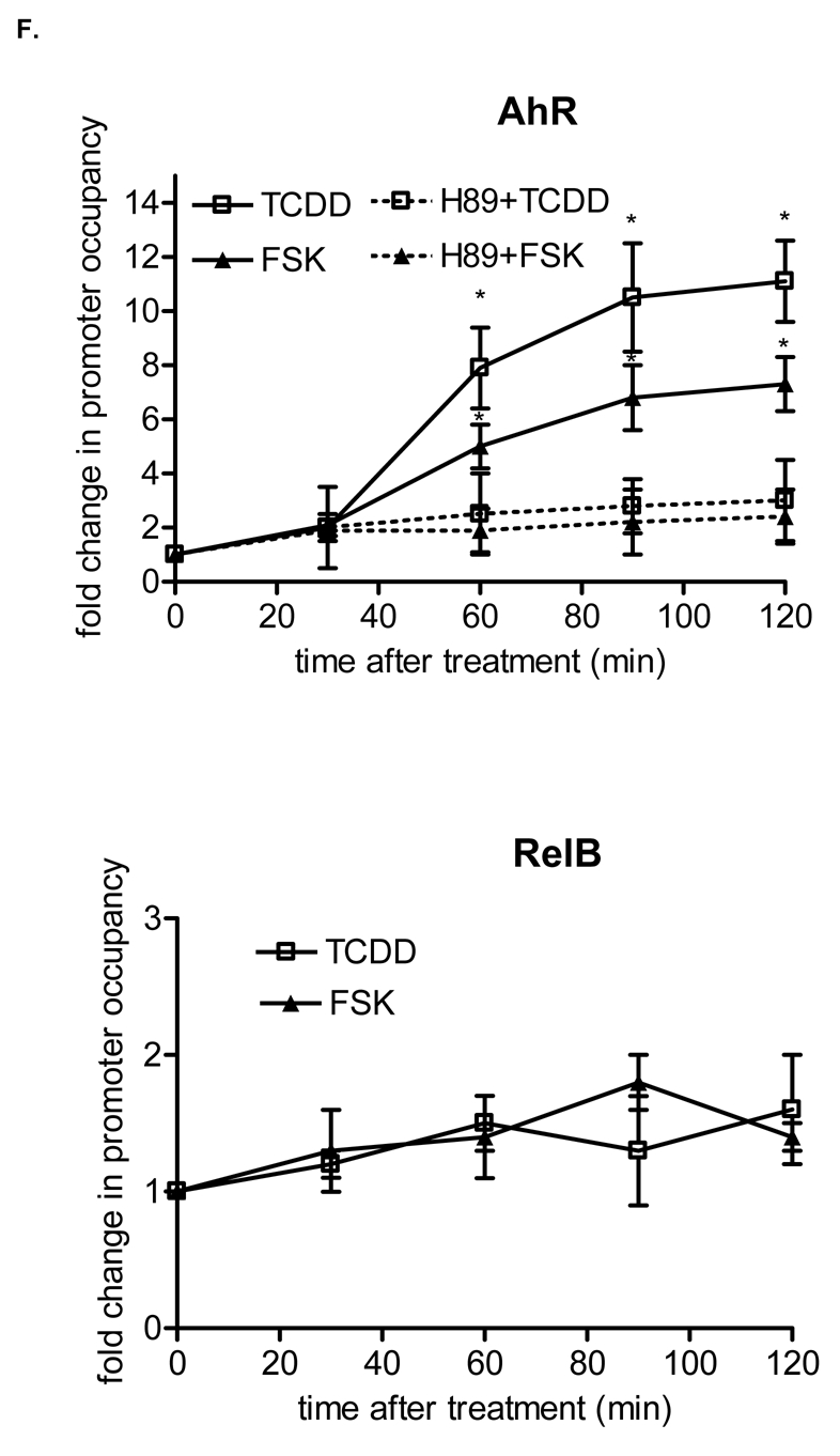
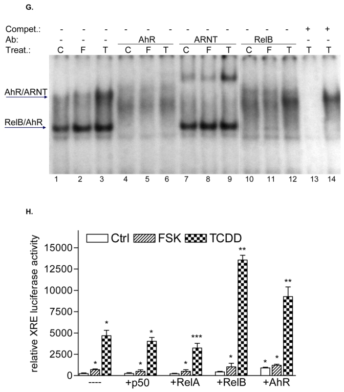
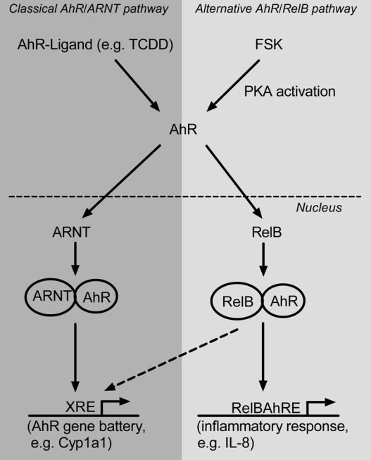
The nuclear factor-kappaB (NF-kappaB) transcription factor family has a crucial role in rapid responses to stress and pathogens. We show that the NF-kappaB subunit RelB is functionally associated with the aryl hydrocarbon receptor (AhR) and mediates transcription of chemokines such as IL-8 via activation of AhR and protein kinase A. RelB physically interacts with AhR and binds to an unrecognized RelB/AhR responsive element of the IL-8 promoter linking two signaling pathways to activate gene transcription. We found a time-dependent recruitment of AhR to the RelB/AhR responsive element site of IL-8 mediated by the AhR ligand 2,3,7,8-tetrachlorodibenzo-p-dioxin (dioxin) and via activation of protein kinase A. Furthermore, NF-kappaB-binding sites that are preferentially recognized by RelB/p52 are a target for RelB/AhR complexes without addition of any stimuli, implicating the endogenous function of the AhR. RelB/AhR complexes are also found to bind on xenobiotic responsive element, and RelB drastically increases the 2,3,7,8-tetrachlorodibenzo-p-dioxin-induced xenobiotic responsive element reporter activity. The interaction of RelB with AhR signaling, and AhR with NF-kappaB RelB signaling pathways represent a new mechanism of cross talk between the two transcription factors.


Figures

Fig. 1
Fig. 1
Induction of IL-8 is RelB- and AhR-dependent. A, Time-course study of IL-8 induction in U937 macrophages. Cells were treated for 0.5 to 48 h with 10 nM TCDD or 10 μM FSK. Control cells received only the vehicle solvent of 0.1% Me2SO or 0.1% PBS. Quantitative detection of IL-8 mRNA was performed using real time RT-PCR. Values for IL-8 mRNA expression are normalized to the expression of β-actin. *, significantly different from control (p<0.001). B, Time-dependent increase of IL-8 protein in U937 macrophages. The level of IL-8 protein in cell lysates from U937 macrophages after treatment with 10 nM TCDD (T) or 0.1 % Me2SO as vehicle control (C) as indicated were determined by Western blot analysis. C, Stimulated IL-8 secretion by activation of AhR in U937 macrophages. The level of IL-8 in the culture media of U937 macrophages was measured by ELISA. Results are expressed as ng IL-8 produced by 106 cells. *Values are the mean ± S.D. of three independent experiments and are significantly different from control (p<0.005). D, Western blot analysis of AhR and RelB protein levels 48h post-transfection with the indicated siRNAs. E, Quantitative IL-8 mRNA expression analyses after treatment with TCDD and FSK for 24 h as analyzed by real-time PCR. Total RNA was prepared 48 h post-transfection with either a scrambled siRNA or a specific siRNA targeted against AhR or RelB.
Fig. 1
Fig. 1
Induction of IL-8 is RelB- and AhR-dependent. A, Time-course study of IL-8 induction in U937 macrophages. Cells were treated for 0.5 to 48 h with 10 nM TCDD or 10 μM FSK. Control cells received only the vehicle solvent of 0.1% Me2SO or 0.1% PBS. Quantitative detection of IL-8 mRNA was performed using real time RT-PCR. Values for IL-8 mRNA expression are normalized to the expression of β-actin. *, significantly different from control (p<0.001). B, Time-dependent increase of IL-8 protein in U937 macrophages. The level of IL-8 protein in cell lysates from U937 macrophages after treatment with 10 nM TCDD (T) or 0.1 % Me2SO as vehicle control (C) as indicated were determined by Western blot analysis. C, Stimulated IL-8 secretion by activation of AhR in U937 macrophages. The level of IL-8 in the culture media of U937 macrophages was measured by ELISA. Results are expressed as ng IL-8 produced by 106 cells. *Values are the mean ± S.D. of three independent experiments and are significantly different from control (p<0.005). D, Western blot analysis of AhR and RelB protein levels 48h post-transfection with the indicated siRNAs. E, Quantitative IL-8 mRNA expression analyses after treatment with TCDD and FSK for 24 h as analyzed by real-time PCR. Total RNA was prepared 48 h post-transfection with either a scrambled siRNA or a specific siRNA targeted against AhR or RelB.
Fig. 2
Fig. 2
RelB and AhR mediate FSK- and TCDD-induced IL-8 activation. A, The AP-1, the Oct-1, and the NF-κB sites of the 5′-flanking region of the IL-8 gene are located 126 bp, 94 bp, and 80 bp, respectively, upstream of the start site of transcription in the IL-8 gene. U937 cells were transiently transfected with deletion constructs corresponding to the first 272, 137, 98, or 50 bp of the 5′-flanking region of the IL-8 gene and treated with 10 nM TCDD or 10 μM FSK for 24 h. Relative luciferase units are given as mean values of triplicates as a result of three independent experiments. *, significantly different from control (p<0.001). B, Supershift analyses with p50-, RelA-, RelB-, AhR-, and ARNT-specific antibodies were performed with a 32P-end-labeled oligonucleotide (5′-AGATGAGGGTGCATAAGTTC-3′) containing the RelBAhRE site of the IL-8 gene with nuclear extracts of untreated and TCDD-stimulated cells treated for 90 min. A 100-fold molar excess of unlabeled RelBAhRE was added. C, Densitometric evaluation of band intensities of the RelB/AhR complexes. Results of three independent experiments are shown as mean values ± S.D. *, significantly different from control cells (p<0.001). D, Nucleotide sequence of the wild-type (wt) -50 bp IL-8 construct corresponding to the 5′-flanking region of the first -120 bp upstream of the start site. The TATA box is in italic type, the AP-1, Oct-1, and NF-κB sites are underlined, the RelBAhRE site is shown in boldface letters. A one point mutation (M1) or two point mutations (M2) were introduced in the RelBAhRE site of the -50 bp construct. E, DNA binding of nuclear proteins from U937 macrophages to the RelBAhRE probe of the IL-8 promoter or RelBAhRE with two different point mutations M1 and M2. U937 macrophages were treated with 10 nM TCDD (T), 2 μg/ml LPS (L), 10 μM FSK (F), or received 0.1% Me2SO as vehicle control (C), and nuclear proteins were extracted at 90 min. A 100-fold molar excess of unlabeled wildtype oligonucleotides was added. F, U937 cells were transiently transfected with -50 wt IL-8 construct and the mutation constructs M1 or M2. After transfection, cells were treated with 10 nM TCDD or 10 μM FSK for 24 h. Relative luciferase activity units are given as mean values of triplicates as a result of three independent experiments. *, significantly different from control (p<0.005); **, significantly higher than only -50 wt transfected cells treated with TCDD or FSK (p<0.005). G, DNA binding of nuclear proteins from U937 macrophages to oligos containing a point mutation M1 of the RelBAhRE probe of the IL-8 promoter. U937 macrophages were treated with 10 μM FSK (F), 10 nM TCDD (T), or received 0.1% Me2SO as vehicle control (C), and nuclear proteins were extracted at 90 min. Supershift analyses with p50-, RelA-, ARNT-, RelB-, or AhR-specific antibodies were performed to identify proteins binding to the mutated M1 RelBAhRE sequence of IL-8. A 100-fold molar excess of unlabeled oligonucleotide was added.
Fig. 2
Fig. 2
RelB and AhR mediate FSK- and TCDD-induced IL-8 activation. A, The AP-1, the Oct-1, and the NF-κB sites of the 5′-flanking region of the IL-8 gene are located 126 bp, 94 bp, and 80 bp, respectively, upstream of the start site of transcription in the IL-8 gene. U937 cells were transiently transfected with deletion constructs corresponding to the first 272, 137, 98, or 50 bp of the 5′-flanking region of the IL-8 gene and treated with 10 nM TCDD or 10 μM FSK for 24 h. Relative luciferase units are given as mean values of triplicates as a result of three independent experiments. *, significantly different from control (p<0.001). B, Supershift analyses with p50-, RelA-, RelB-, AhR-, and ARNT-specific antibodies were performed with a 32P-end-labeled oligonucleotide (5′-AGATGAGGGTGCATAAGTTC-3′) containing the RelBAhRE site of the IL-8 gene with nuclear extracts of untreated and TCDD-stimulated cells treated for 90 min. A 100-fold molar excess of unlabeled RelBAhRE was added. C, Densitometric evaluation of band intensities of the RelB/AhR complexes. Results of three independent experiments are shown as mean values ± S.D. *, significantly different from control cells (p<0.001). D, Nucleotide sequence of the wild-type (wt) -50 bp IL-8 construct corresponding to the 5′-flanking region of the first -120 bp upstream of the start site. The TATA box is in italic type, the AP-1, Oct-1, and NF-κB sites are underlined, the RelBAhRE site is shown in boldface letters. A one point mutation (M1) or two point mutations (M2) were introduced in the RelBAhRE site of the -50 bp construct. E, DNA binding of nuclear proteins from U937 macrophages to the RelBAhRE probe of the IL-8 promoter or RelBAhRE with two different point mutations M1 and M2. U937 macrophages were treated with 10 nM TCDD (T), 2 μg/ml LPS (L), 10 μM FSK (F), or received 0.1% Me2SO as vehicle control (C), and nuclear proteins were extracted at 90 min. A 100-fold molar excess of unlabeled wildtype oligonucleotides was added. F, U937 cells were transiently transfected with -50 wt IL-8 construct and the mutation constructs M1 or M2. After transfection, cells were treated with 10 nM TCDD or 10 μM FSK for 24 h. Relative luciferase activity units are given as mean values of triplicates as a result of three independent experiments. *, significantly different from control (p<0.005); **, significantly higher than only -50 wt transfected cells treated with TCDD or FSK (p<0.005). G, DNA binding of nuclear proteins from U937 macrophages to oligos containing a point mutation M1 of the RelBAhRE probe of the IL-8 promoter. U937 macrophages were treated with 10 μM FSK (F), 10 nM TCDD (T), or received 0.1% Me2SO as vehicle control (C), and nuclear proteins were extracted at 90 min. Supershift analyses with p50-, RelA-, ARNT-, RelB-, or AhR-specific antibodies were performed to identify proteins binding to the mutated M1 RelBAhRE sequence of IL-8. A 100-fold molar excess of unlabeled oligonucleotide was added.
Fig. 3
Fig. 3
Requirement of PKA in AhR- and RelB-mediated IL-8 promoter and RelBAhRE binding activity induced by FSK and TCDD. A, Transfection of siRNA targeted against AhR or RelB mRNA prevents TCDD- and FSK-mediated induction of IL-8 promoter activity in U937 macrophages. Cells were transfected with either a scrambled siRNA or a specific siRNA targeted against AhR or RelB and the -50 wt IL-8 construct. After 24 h cells were treated with 10 nM TCDD or 10 μM FSK for 24 h. B, Cotransfection with an AhR C, RelB or D, ARNT expression plasmid. U937 macrophages were transiently transfected with wildtype -50 bp IL-8 construct and cotransfected with increasing amounts (100–400 ng/ml) of an AhR (B) RelB (C) or 200 ng/ml ARNT (D) expression plasmid. After transfection for 24 h, cells were treated with 10 nM TCDD, 10 μM FSK, or 0.1% Me2SO for 24 h. Relative luciferase activity units are given as mean values of triplicates as a result of three independent experiments. *, significantly different from control (p<0.005). **, significantly higher than cells treated with TCDD or FSK transfected with only -50 wt IL-8 construct (p<0.005). ***, significantly higher than cells treated with TCDD or FSK cotransfected with -50 wt IL-8 and 100 ng RelB or AhR (p<0.005). E, DNA binding of nuclear proteins from U937 macrophages to RelBAhRE of the IL-8 promoter requires AhR and RelB. U937 macrophages were transfected with either a scrambled siRNA or a specific siRNA targeted against AhR or RelB treated with 10 nM TCDD (T) or received 0.1% Me2SO as vehicle control (C) and nuclear proteins were extracted at 90 min. A 100-fold molar excess of unlabeled wildtype oligonucleotides was added. F, DNA binding of nuclear proteins from U937 macrophages to RelBAhRE of the IL-8 promoter depends on PKA. U937 macrophages were treated with 1 μM H89 (H), 1 μM H89 plus 10 μM FSK (H+F), 10 μM FSK (F), 1 μM H89 plus 10 nM TCDD (H+T), 10 nM TCDD (T), or received 0.1% Me2SO as vehicle control (C) and nuclear proteins were extracted at 90 min. A 100-fold molar excess of unlabeled wildtype oligonucleotides was added.
Fig. 3
Fig. 3
Requirement of PKA in AhR- and RelB-mediated IL-8 promoter and RelBAhRE binding activity induced by FSK and TCDD. A, Transfection of siRNA targeted against AhR or RelB mRNA prevents TCDD- and FSK-mediated induction of IL-8 promoter activity in U937 macrophages. Cells were transfected with either a scrambled siRNA or a specific siRNA targeted against AhR or RelB and the -50 wt IL-8 construct. After 24 h cells were treated with 10 nM TCDD or 10 μM FSK for 24 h. B, Cotransfection with an AhR C, RelB or D, ARNT expression plasmid. U937 macrophages were transiently transfected with wildtype -50 bp IL-8 construct and cotransfected with increasing amounts (100–400 ng/ml) of an AhR (B) RelB (C) or 200 ng/ml ARNT (D) expression plasmid. After transfection for 24 h, cells were treated with 10 nM TCDD, 10 μM FSK, or 0.1% Me2SO for 24 h. Relative luciferase activity units are given as mean values of triplicates as a result of three independent experiments. *, significantly different from control (p<0.005). **, significantly higher than cells treated with TCDD or FSK transfected with only -50 wt IL-8 construct (p<0.005). ***, significantly higher than cells treated with TCDD or FSK cotransfected with -50 wt IL-8 and 100 ng RelB or AhR (p<0.005). E, DNA binding of nuclear proteins from U937 macrophages to RelBAhRE of the IL-8 promoter requires AhR and RelB. U937 macrophages were transfected with either a scrambled siRNA or a specific siRNA targeted against AhR or RelB treated with 10 nM TCDD (T) or received 0.1% Me2SO as vehicle control (C) and nuclear proteins were extracted at 90 min. A 100-fold molar excess of unlabeled wildtype oligonucleotides was added. F, DNA binding of nuclear proteins from U937 macrophages to RelBAhRE of the IL-8 promoter depends on PKA. U937 macrophages were treated with 1 μM H89 (H), 1 μM H89 plus 10 μM FSK (H+F), 10 μM FSK (F), 1 μM H89 plus 10 nM TCDD (H+T), 10 nM TCDD (T), or received 0.1% Me2SO as vehicle control (C) and nuclear proteins were extracted at 90 min. A 100-fold molar excess of unlabeled wildtype oligonucleotides was added.
Fig. 4
Fig. 4
Physical association of AhR and RelB. U937 macrophages were treated with TCDD (10 nM) or Me2SO (0.1%) and after 90 min nuclear proteins were prepared for nuclear complex co-immunoprecipitation followed by Western blot analysis to detect specific association of AhR and RelB. A, immunoprecipitation of AhR with antibody against RelB. Samples of total cell lysates were incubated with rabbit lgG as the negative control, anti-AhR antibody (positive control), and antibody against RelB. The blot was probed with antibody against AhR protein after Western transfer. B, immunoprecipitation of RelB with antibody against AhR. The cell lysates were incubated with rabbit IgG, antibody against RelB (positive control), and antibody against AhR. After Western transfer, the blot was stained with antibody against RelB. C, ARNT is not associated with RelB. The cell lysates were incubated with antibody against RelB, AHR (positive control), and rabbit IgG. The Western blot was stained with an antibody against the ARNT protein. D, Increased nuclear accumulation of AhR protein. The level of AhR in nuclei from U937 macrophages 90 min after treatment with 10 nM TCDD (T) or 10 μM FSK (F), or 0.1% Me2SO (C) as indicated were determined by Western blot analysis using a AhR-specific antibody. E, FSK and TCDD stimulate the recruitment of AhR to the RelBAhRE region of the IL-8 promoter. U937 macrophages were treated with 10 nM TCDD and 10 μM FSK in presence or absence of 1 μM H89, or Me2SO (0.1%) for the indicated amounts of time. ChIP assays with antibodies to AhR and RelB proteins were analyzed by PCR using primer pairs covering the specified RelBAhRE region of human IL-8. Genomic DNA and the sonicated input DNA were separated by agarose gel electrophoresis and visualized by ethidium bromide staining. Arrows, position of primers used to test the recruitment of AhR or RelB to the IL-8 promoter region flanking the RelBAhRE region. F, ChIP assay samples were analyzed by real-time PCR as described in Materials and Methods and the results were normalized to time zero (no AhR activation by TCDD or FSK). *, significantly different from control cells (p<0.001)
Fig. 4
Fig. 4
Physical association of AhR and RelB. U937 macrophages were treated with TCDD (10 nM) or Me2SO (0.1%) and after 90 min nuclear proteins were prepared for nuclear complex co-immunoprecipitation followed by Western blot analysis to detect specific association of AhR and RelB. A, immunoprecipitation of AhR with antibody against RelB. Samples of total cell lysates were incubated with rabbit lgG as the negative control, anti-AhR antibody (positive control), and antibody against RelB. The blot was probed with antibody against AhR protein after Western transfer. B, immunoprecipitation of RelB with antibody against AhR. The cell lysates were incubated with rabbit IgG, antibody against RelB (positive control), and antibody against AhR. After Western transfer, the blot was stained with antibody against RelB. C, ARNT is not associated with RelB. The cell lysates were incubated with antibody against RelB, AHR (positive control), and rabbit IgG. The Western blot was stained with an antibody against the ARNT protein. D, Increased nuclear accumulation of AhR protein. The level of AhR in nuclei from U937 macrophages 90 min after treatment with 10 nM TCDD (T) or 10 μM FSK (F), or 0.1% Me2SO (C) as indicated were determined by Western blot analysis using a AhR-specific antibody. E, FSK and TCDD stimulate the recruitment of AhR to the RelBAhRE region of the IL-8 promoter. U937 macrophages were treated with 10 nM TCDD and 10 μM FSK in presence or absence of 1 μM H89, or Me2SO (0.1%) for the indicated amounts of time. ChIP assays with antibodies to AhR and RelB proteins were analyzed by PCR using primer pairs covering the specified RelBAhRE region of human IL-8. Genomic DNA and the sonicated input DNA were separated by agarose gel electrophoresis and visualized by ethidium bromide staining. Arrows, position of primers used to test the recruitment of AhR or RelB to the IL-8 promoter region flanking the RelBAhRE region. F, ChIP assay samples were analyzed by real-time PCR as described in Materials and Methods and the results were normalized to time zero (no AhR activation by TCDD or FSK). *, significantly different from control cells (p<0.001)
Fig. 5
Fig. 5
Effect of FSK, TCDD or LPS on NF-κB and XRE activity. A, U937 macrophages were treated with 10 μM FSK (F), 10 nM TCDD (T), 2 μg/ml LPS (L) or received 0.1% Me2SO as vehicle control (C), and nuclear proteins were extracted at 90 min. Nuclear protein extracts of non-stimulated, FSK-, TCDD-, or LPS-stimulated U937 macrophages were incubated with a NF-κB consensus probe. NF-κB proteins, present in nuclear extracts of 90 min treated U937 macrophages binding to the NF-κB site were identified by supershift analyses using p50-, RelA-, RelB-, or AhR-specific antibodies. Competition with a 100-fold excess of unlabeled NF-κB consensus, XRE-consensus, or RelBAhRE oligonucleotide from the IL-8 promoter confirms specificity of the complex. B, Densitometric evaluation of the band intensity of the lower band of the NF-κB complex. Results of three independent experiments are shown as mean values ± S.D. *, significantly different from control cells (p<0.001). C, Effect of overexpression of various Rel proteins and AhR on FSK- and TCDD-induced NF-κB reporter activity. NF-κB activity was evaluated in U937 macrophages by transient transfection of the corresponding reporter plasmid with or without cotransfection of 200 ng/ml of a p50, RelA, RelB or AhR expression plasmid. Transfected cells were incubated with 0.1% Me2SO, 10 μM FSK, or 10 nM TCDD for 24 h. Relative luciferase activity units are given as mean values of triplicates as a result of three independent experiments. *, significantly different from control (p<0.005) **, significantly higher than only NF-κB reporter plasmid transfected cells (p<0.005) ***, significantly lower than only NF-κB reporter plasmid transfected cells (p<0.005). D, Nuclear protein extracts of control (C), TCDD- (T), or FSK-stimulated (F) U937 macrophages were incubated with the RelB/p52 consensus oligo of the BLC promoter. A possible binding of p50, RelB, AhR, and p52 was identified by supershift analyses. To confirm specificity a 100-fold excess of unlabeled RelB/p52 consensus or RelBAhRE oligonucleotide from the IL-8 promoter was added. E, Nuclear protein extracts of control (C) or TCDD- (T) stimulated U937 macrophages were incubated with an oligonucleotide containing the RelB/p52 of the BAFF promoter. A possible binding of AhR, ARNT, and RelB was identified by supershift analyses. To confirm specificity a 100-fold excess of unlabeled RelB/p52 oligonucleotide from the BAFF promoter was added. F, Induction of BLC and BAFF is increased in AhR and RelB overexpressing cells. Cells were transiently transfected with 200 ng/ml of AhR or RelB expression plasmid. Control cells were transfected with an empty control vector. After 72 h cells were treated with 10 nM TCDD or 10 μM FSK for 24 h. Expression of BLC and BAFF mRNA was analyzed by real-time PCR as described above. *, significantly different from control cells (p<0.01); **, significantly higher than control, TCDD or FSK treated cells (p<0.01) G, Nuclear protein extracts of control (C, lane 1), FSK- (F, lane 2), or TCDD-stimulated (T, lane 3) U937 macrophages were incubated with 32P-labeled oligonucleotide containing a XRE consensus element of the CYP1A1 promoter. A possible binding of AhR, ARNT, and RelB was identified by supershift analyses using AhR-, ARNT-, or RelB-specific antibodies. To confirm specificity a 100-fold excess of unlabeled XRE consensus or RelBAhRE oligonucleotide from the IL-8 promoter was added. H, Effect of overexpression of various Rel proteins and AhR on FSK- and TCDD-induced XRE reporter activity. XRE activity was evaluated in U937 macrophages by transient transfection of the corresponding reporter plasmid with or without cotransfection of 200 ng/ml of a p50, RelA, RelB or AhR expression plasmid. Transfected cells were incubated with 0.1% Me2SO, 10 μM FSK, or 10 nM TCDD for 24 h. Relative luciferase activity units are given as mean values of triplicates as a result of three independent experiments. *, significantly different from control (p<0.005) **, significantly higher than only XRE reporter plasmid transfected cells (p<0.005) ***, significantly lower than only XRE reporter plasmid transfected cells (p<0.005).
Fig. 5
Fig. 5
Effect of FSK, TCDD or LPS on NF-κB and XRE activity. A, U937 macrophages were treated with 10 μM FSK (F), 10 nM TCDD (T), 2 μg/ml LPS (L) or received 0.1% Me2SO as vehicle control (C), and nuclear proteins were extracted at 90 min. Nuclear protein extracts of non-stimulated, FSK-, TCDD-, or LPS-stimulated U937 macrophages were incubated with a NF-κB consensus probe. NF-κB proteins, present in nuclear extracts of 90 min treated U937 macrophages binding to the NF-κB site were identified by supershift analyses using p50-, RelA-, RelB-, or AhR-specific antibodies. Competition with a 100-fold excess of unlabeled NF-κB consensus, XRE-consensus, or RelBAhRE oligonucleotide from the IL-8 promoter confirms specificity of the complex. B, Densitometric evaluation of the band intensity of the lower band of the NF-κB complex. Results of three independent experiments are shown as mean values ± S.D. *, significantly different from control cells (p<0.001). C, Effect of overexpression of various Rel proteins and AhR on FSK- and TCDD-induced NF-κB reporter activity. NF-κB activity was evaluated in U937 macrophages by transient transfection of the corresponding reporter plasmid with or without cotransfection of 200 ng/ml of a p50, RelA, RelB or AhR expression plasmid. Transfected cells were incubated with 0.1% Me2SO, 10 μM FSK, or 10 nM TCDD for 24 h. Relative luciferase activity units are given as mean values of triplicates as a result of three independent experiments. *, significantly different from control (p<0.005) **, significantly higher than only NF-κB reporter plasmid transfected cells (p<0.005) ***, significantly lower than only NF-κB reporter plasmid transfected cells (p<0.005). D, Nuclear protein extracts of control (C), TCDD- (T), or FSK-stimulated (F) U937 macrophages were incubated with the RelB/p52 consensus oligo of the BLC promoter. A possible binding of p50, RelB, AhR, and p52 was identified by supershift analyses. To confirm specificity a 100-fold excess of unlabeled RelB/p52 consensus or RelBAhRE oligonucleotide from the IL-8 promoter was added. E, Nuclear protein extracts of control (C) or TCDD- (T) stimulated U937 macrophages were incubated with an oligonucleotide containing the RelB/p52 of the BAFF promoter. A possible binding of AhR, ARNT, and RelB was identified by supershift analyses. To confirm specificity a 100-fold excess of unlabeled RelB/p52 oligonucleotide from the BAFF promoter was added. F, Induction of BLC and BAFF is increased in AhR and RelB overexpressing cells. Cells were transiently transfected with 200 ng/ml of AhR or RelB expression plasmid. Control cells were transfected with an empty control vector. After 72 h cells were treated with 10 nM TCDD or 10 μM FSK for 24 h. Expression of BLC and BAFF mRNA was analyzed by real-time PCR as described above. *, significantly different from control cells (p<0.01); **, significantly higher than control, TCDD or FSK treated cells (p<0.01) G, Nuclear protein extracts of control (C, lane 1), FSK- (F, lane 2), or TCDD-stimulated (T, lane 3) U937 macrophages were incubated with 32P-labeled oligonucleotide containing a XRE consensus element of the CYP1A1 promoter. A possible binding of AhR, ARNT, and RelB was identified by supershift analyses using AhR-, ARNT-, or RelB-specific antibodies. To confirm specificity a 100-fold excess of unlabeled XRE consensus or RelBAhRE oligonucleotide from the IL-8 promoter was added. H, Effect of overexpression of various Rel proteins and AhR on FSK- and TCDD-induced XRE reporter activity. XRE activity was evaluated in U937 macrophages by transient transfection of the corresponding reporter plasmid with or without cotransfection of 200 ng/ml of a p50, RelA, RelB or AhR expression plasmid. Transfected cells were incubated with 0.1% Me2SO, 10 μM FSK, or 10 nM TCDD for 24 h. Relative luciferase activity units are given as mean values of triplicates as a result of three independent experiments. *, significantly different from control (p<0.005) **, significantly higher than only XRE reporter plasmid transfected cells (p<0.005) ***, significantly lower than only XRE reporter plasmid transfected cells (p<0.005).
Fig. 6
Fig. 6
Model of the new mechanism of cross talk between AhR and RelB. Ligand-activated or unliganded AhR activated by PKA translocates into the nucleus and interacts with RelB to occupy RelBAhRE-responsive promoters as in the case of IL-8 (alternative AhR/RelB pathway). AhR agonists induce the recruitment of AhR/ARNT complexes to XRE-responsive promoters such as CYP1A1 (classical AhR/ARNT pathway).

References

    1. Chen FE, Huang DB, Chen YQ, Ghosh G. Crystal structure of p50/p65 heterodimer of transcription factor NF-kappaB bound to DNA. Nature. 1998;391:410–403. - PubMed
    1. Kunsch C, Ruben SM, Rosen CA. Selection of optimal kappa B/Rel DNA-binding motifs: interaction of both subunits of NF-kappa B with DNA is required for transcriptional activation. Mol Cell Biol. 1992;10:4412–4421. - PMC - PubMed
    1. Dejardin E, Droin NM, Delhase M, Haas E, Cao Y, Makris C, Li ZW, Karin M, Ware CF, Green DR. The lymphotoxin-beta receptor induces different patterns of gene expression via two NF-kappaB pathways. Immunity. 2002;4:525–535. - PubMed
    1. Bonizzi G, Bebien M, Otero DC, Johnson-Vroom KE, Cao Y, Vu D, Jegga AG, Aronow BJ, Ghosh G, Rickert RC, Karin M. Activation of IKKalpha target genes depends on recognition of specific kappaB binding sites by RelB:p52 dimers. EMBO J. 2004;21:4202–4210. - PMC - PubMed
    1. Hayden MS, Ghosh S. Signaling to NF-kappaB. Genes Dev. 2004;8:2195–2224. - PubMed

Publication types

MeSH terms