. 2007 Dec 1;110(12):3909-16.
doi: 10.1182/blood-2007-06-096651. Epub 2007 Sep 6.

The ligand occupancy of endothelial protein C receptor switches the protease-activated receptor 1-dependent signaling specificity of thrombin from a permeability-enhancing to a barrier-protective response in endothelial cells

Affiliations

The ligand occupancy of endothelial protein C receptor switches the protease-activated receptor 1-dependent signaling specificity of thrombin from a permeability-enhancing to a barrier-protective response in endothelial cells

Jong-Sup Bae et al. Blood. .

Abstract

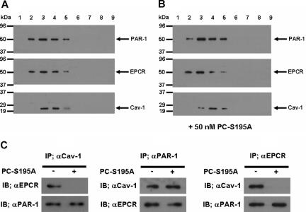
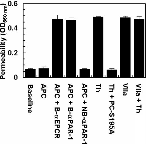
Recent studies have indicated that activated protein C (APC) may exert its cytoprotective and anti-inflammatory activities through the endothelial protein C receptor (EPCR)-dependent cleavage of protease-activated receptor 1 (PAR-1) on vascular endothelial cells. Noting that (1) the activation of protein C on endothelial cells requires thrombin, (2) relative to APC, thrombin cleaves PAR-1 with approximately 3 to 4 orders of magnitude higher catalytic efficiency, and (3) PAR-1 is a target for the proinflammatory activity of thrombin, it is not understood how APC can elicit a protective signaling response through the cleavage of PAR-1 when thrombin is present. In this study, we demonstrate that EPCR is associated with caveolin-1 in lipid rafts of endothelial cells and that its occupancy by the gamma-carboxyglutamic acid (Gla) domain of protein C/APC leads to its dissociation from caveolin-1 and recruitment of PAR-1 to a protective signaling pathway through coupling of PAR-1 to the pertussis toxin-sensitive G(i)-protein. Thus, when EPCR is bound by protein C, the PAR-1 cleavage-dependent protective signaling responses in endothelial cells can be mediated by either thrombin or APC. These results provide a new paradigm for understanding how PAR-1 and EPCR participate in protective signaling events in endothelial cells.


Figures

Figure 1
Figure 1
Binding of protein C and PC-Gla/meizothrombin to soluble EPCR and the cleavage of PAR-1 exodomain by the proteases. (A) An ELISA-based assay using the monoclonal antibody HPC4 (1 μg/mL) was used to measure the affinity of either protein C (○) or PC-Gla/meizothrombin (■) for interaction with soluble EPCR (0.5 μmol/L). (B) EA.hy926 cells were transiently transfected with ALP-PAR-1 reporter plasmid and incubated with increasing concentrations of thrombin (○), meizothrombin (□), PC-Gla/meizothrombin (■) and APC (▵) for 1 hour. The activity of soluble alkaline phosphatase (ALP) was measured as described under “PAR-1 cleavage assay.” The activity of 10 nmol/L thrombin is presented as 100%. OD, optical density. Error bars represent SD.
Figure 2
Figure 2
The permeability barrier enhancing and proapoptotic effects of thrombin are reversed by the ligand occupancy of EPCR. (A) EA.hy926 cells were incubated with increasing concentrations of thrombin (○), thrombin + anti-PAR-1 antibody (●), meizothrombin (□), PC-Gla/meizothrombin (■), thrombin + PC-S195A (50 nmol/L) (▴), and APC (▵) for 3 hours before inducing permeability with 5 nmol/L thrombin for 10 minutes. (B) The same as (A) except that after incubation of endothelial cells with increasing concentrations of thrombin (○), thrombin + PC-S195A (▴), and APC (▵), the permeability was induced with LPS (10 ng/mL) for 4 hour. (C) EA.hy926 cells were treated with increasing concentrations of thrombin (○), thrombin + PC-S195A (▴), and APC (▵) for 24 hours, followed by induction of apoptosis with staurosporine (5 μmol/L) for 4 hours. The number of apoptotic cells is expressed as the percentage of TUNEL-positive cells of the total number of nuclei. The number of TUNEL-positive cells in the absence of staurosporine was 10%-15%. (D) The same as (A) except that the concentration dependence of the protective effect of PC-S195A in the presence of 2 nmol/L thrombin (○) is measured in the permeability assay. The extent of the protective effect of 10 nmol/L APC (▵) is also presented. OD, optical density. Error bars represent SD.
Figure 3
Figure 3
The barrier-enhancing effect of TRAP and its reversal by the ligand occupancy of EPCR. (A) EA.hy926 cells were incubated with increasing concentrations of thrombin (○), thrombin + thrombomodulin fragments 456 (TM456) (●), TRAP (□), TRAP + wild-type protein C (■), TRAP + PC-S195A (▵), TRAP + wild-type protein C + blocking anti-PAR-1 (H-111) (▴), and TRAP + PC-S195A + anti-PAR-1 (H-111) (▿) for 3 hours before inducing permeability with 5 nmol/L thrombin for 10 minutes. (B) The same as (A) except that the permeability was directly monitored as a function increasing concentrations of thrombin (○), thrombin + PC-S195A (▴), TRAP (□), and TRAP + PC-S195A (■) by incubation of EA.hy926 cells with different agonists for 3 hours (the 5 nmol/L thrombin step for inducing permeability has been omitted in this assay). The first data point on the y-axis represents the baseline permeability level. OD, optical density. Error bars represent SD.
Figure 4
Figure 4
The effect of thrombin on the permeability of endothelial cells in the presence of protein C S195A and siRNA for PAR-4. (A) EA.hy926 cells were transiently transfected with siRNA for PAR-4 or control siRNA (1 μg each). After 3 days of incubation at 37°C, total cellular proteins were extracted, separated on 10% SDS-PAGE, and Western-blotted with antibodies directed to either PAR-4 or PAR-1. An antibody against actin was used as internal control. (B) EA.hy926 cells were incubated with increasing concentrations of thrombin (○), thrombin with prior treatment of cells for 3 days with control siRNA (●), thrombin with prior treatment of cells with siRNA for PAR-4 (□), thrombin + PC-S195A (■), thrombin + PC-S195A with prior treatment of cells with control siRNA (▵), and thrombin + PC-S195A with prior treatment of cells with siRNA for PAR-4 (▴) for 3 hours before inducing permeability with 5 nmol/L thrombin for 10 minutes. (C) The same as (B) except that the barrier-protective effect of APC was monitored in the absence (▵) and presence of control siRNA (●) or siRNA for PAR-4 (□). (D) The barrier-protective effect of thrombin + PC-S195A in endothelial cells is sensitive to PTX. EA.hy926 cells were incubated with increasing concentrations of thrombin (○), thrombin with prior treatment of cells with 100 ng/mL PTX for 16 hours (●), APC (□), APC with prior treatment of cells with 100 ng/mL PTX for 16 hours (■), thrombin + PC-S195A (▵), and thrombin + PC-S195A with prior treatment of cells with PTX (▴) for 3 hours before inducing permeability with 5 nmol/L thrombin. Error bars represent SD.
Figure 5
Figure 5
SDS-PAGE, immunoblotting, and coimmunoprecipitation of lipid raft/caveolae preparations derived from EA.hy926 cells with and without treatment with PC-S195A. (A) After SDS-PAGE of membrane fractions prepared by discontinuous sucrose gradient ultracentrifugation, they were immunoblotted with antibodies directed to PAR-1, EPCR, and caveolin-1. (B) The same as (A) except that the fractions were derived from EA.hy926 cells treated with 50 nmol/L PC-S195A. (C) Total cellular proteins from nontreated and PC-S195A-treated EA.hy926 cells were immunoprecipitated with anti-caveolin-1, anti-PAR-1, and anti-EPCR antibodies, separated on SDS-PAGE and immunoblotted with different pairs of the same antibodies.
Figure 6
Figure 6
The permeability barrier-enhancing effect of thrombin is reversed by the ligand occupancy of EPCR with PC-S195A, but not with factor VIIa. EA.hy926 cells were incubated with APC (10 nmol/L) and thrombin (2 nmol/L) in the absence and presence of either PC-S195A or factor VIIa (50 nmol/L) for 3 hours before inducing permeability with 5 nmol/L thrombin for 10 minutes. B-αEPCR is blocking anti-EPCR (clone RCR-252), B-αPAR-1 (H-111) is blocking anti-PAR-1, and NB-αPAR-1 (S-19) is nonblocking anti-PAR-1, which have been incubated with endothelial cells for 30 minutes before treatment with APC. Baseline represents the permeability of untreated control cells not stimulated with thrombin. Error bars represent SD.

Similar articles

Cited by

References

    1. Esmon CT. Molecular events that control the protein C anticoagulant pathway. Thromb Haemost. 1993;70:29–35. - PubMed
    1. Stenflo J. Structure and function of protein C. Semin Thromb Hemost. 1984;10:109–121. - PubMed
    1. Walker FJ, Fay PJ. Regulation of blood coagulation by the protein C system. FASEB J. 1992;6:2561–2567. - PubMed
    1. Dahlbäck B. Protein S and C4b-binding protein: Components involved in the regulation of the protein C anticoagulant system. Thromb Haemost. 1991;66:49–61. - PubMed
    1. Griffin JH, Evatt B, Zimmerman TS, Kleiss AJ, Wideman C. Deficiency of protein C in congenital thrombotic disease. J Clin Invest. 1981;68:1370–1373. - PMC - PubMed

Publication types

MeSH terms