. 2007 Dec 1;176(11):1146-53.
doi: 10.1164/rccm.200705-750OC. Epub 2007 Sep 6.

Intrauterine pulmonary hypertension impairs angiogenesis in vitro: role of vascular endothelial growth factor nitric oxide signaling

Affiliations

Intrauterine pulmonary hypertension impairs angiogenesis in vitro: role of vascular endothelial growth factor nitric oxide signaling

Jason Gien et al. Am J Respir Crit Care Med. .

Abstract

Rationale: Mechanisms that impair angiogenesis in neonatal persistent pulmonary hypertension (PPHN) are poorly understood.

Objectives: To determine if PPHN alters fetal pulmonary artery endothelial cell (PAEC) phenotype and impairs growth and angiogenesis in vitro, and if altered vascular endothelial growth factor-nitric oxide (VEGF-NO) signaling contributes to this abnormal phenotype.

Methods: Proximal PAECs were harvested from fetal sheep that had undergone partial ligation of the ductus arteriosus in utero (PPHN) and age-matched control animals. Growth and tube formation +/- VEGF and NO stimulation and inhibition were studied in normal and PPHN PAECs. Western blot analysis was performed for VEGF, VEGF receptor-2 (VEGF-R2), and endothelial NO synthase (eNOS) protein content. NO production with VEGF administration was measured in normal and PPHN PAECs.

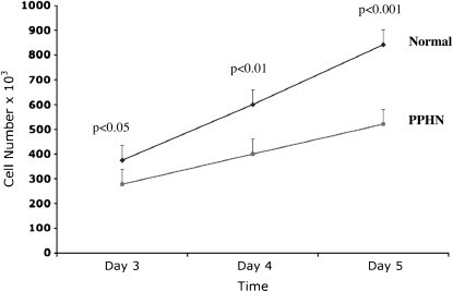
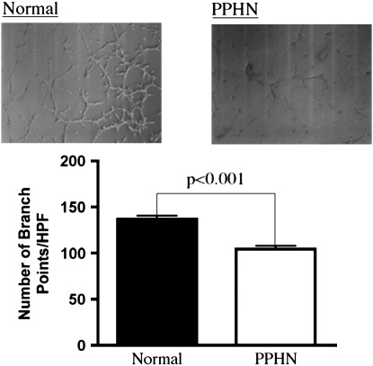
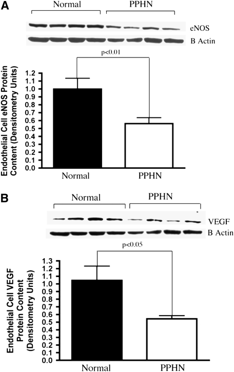
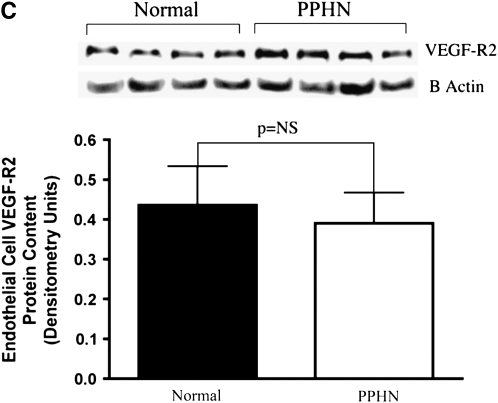
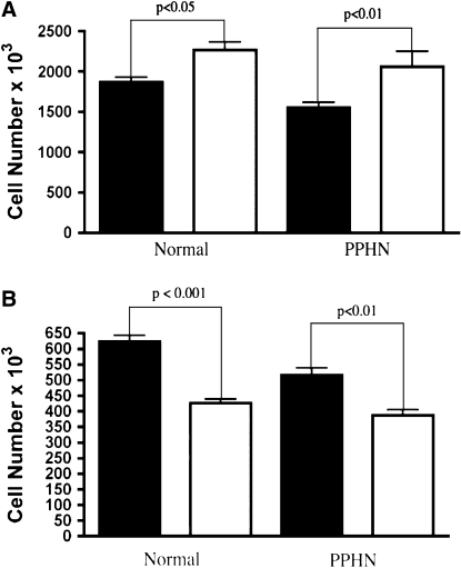
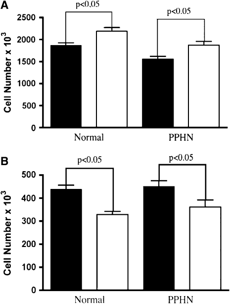
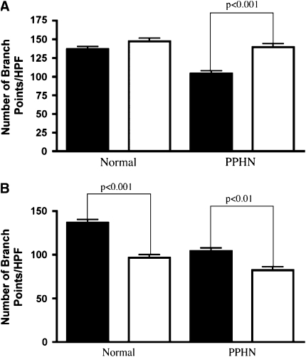
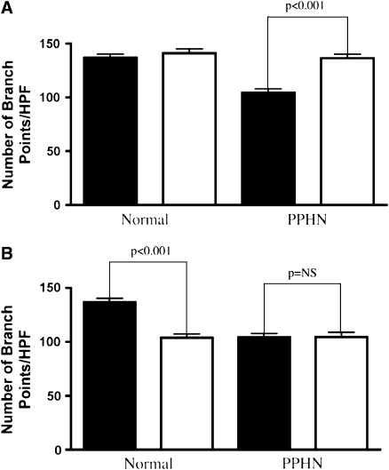
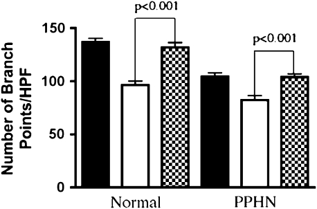
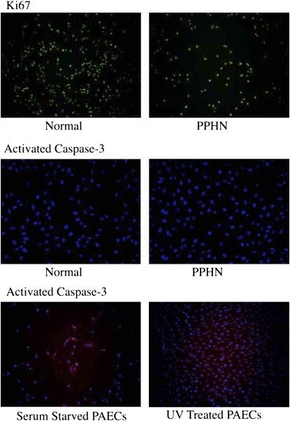
Measurements and main results: PPHN PAECs demonstrate decreased growth and tube formation in vitro. VEGF and eNOS protein expression were decreased in PPHN PAECs, whereas VEGF-R2 protein expression was not different. VEGF and NO increased PPHN PAEC growth and tube formation to values achieved in normal PAECs. VEGF inhibition decreased growth and tube formation in normal and PPHN PAECs. NOS inhibition decreased growth in normal and PPHN PAECs, but tube formation was only reduced in normal PAECs. NO reversed the inhibitory effects of VEGF-R2 inhibition on tube formation in normal and PPHN PAECs. VEGF increased NO production in normal and PPHN PAECs.

Conclusions: PPHN in utero causes sustained impairment of PAEC phenotype in vitro, with reduced PAEC growth and tube formation and down-regulation of VEGF and eNOS protein. VEGF and NO enhanced growth and tube formation of PPHN PAECs.


Figures

<b>Figure 1.</b>
Figure 1.
Decreased growth in fetal pulmonary artery endothelial cells (PAECS) from sheep with persistent pulmonary hypertension of the newborn (PPHN). Fetal ovine PAECs from normal and PPHN lambs were plated in 10% fetal bovine serum under 3% oxygen conditions and counted daily for 5 days. In comparison with normal PAECs, proliferation of endothelial cells from PPHN lambs was decreased over time in culture from Day 3 to Day 5. Error bars represent SD from mean.
<b>Figure 2.</b>
Figure 2.
Decreased tube formation in fetal pulmonary artery endothelial cells (PAECS) from sheep with persistent pulmonary hypertension of the newborn (PPHN). Fetal ovine PAECs from normal and PPHN lambs were plated on Matrigel in serum-free media under 3% oxygen conditions. The ability of PPHN cells to form vascular networks is markedly impaired when compared with normal controls. Error bars represent SD from mean. HPF = high power field.
<b>Figure 3.</b>
Figure 3.
Decreased endothelial NO synthase (eNOS) and vascular endothelial growth factor (VEGF) protein expression in fetal pulmonary artery endothelial cells (PAECs) from sheep with persistent pulmonary hypertension of the newborn (PPHN). Western blot analysis on cell lysates from PAECs from PPHN sheep demonstrate decreased eNOS (A) and VEGF (B) protein when compared with normal controls. There was no difference in kinase domain receptor (C) expression. Error bars represent SD from mean.
<b>Figure 4.</b>
Figure 4.
Effect of VEGF and SU5416 on endothelial cell growth. Ovine pulmonary artery endothelial cells (PAECs) from normal lambs and lambs with persistent pulmonary hypertension of the newborn (PPHN) were grown in 5% fetal bovine serum under 3% oxygen conditions with and without VEGF (50 ng/ml) and SU5416 (10 μM). (A) VEGF treatment stimulated growth of PAECs in both normal and PPHN cells (solid bars, control; open bars, VEGF). (B) VEGF receptor blockade with SU5416 decreased growth in both normal and PPHN PAECs (solid bars, control; open bars, SU5416). Error bars represent SD from mean.
<b>Figure 5.</b>
Figure 5.
Effect of NO gas and nitro-l-arginine (LNA) on endothelial cell growth. Fetal ovine pulmonary artery endothelial cells (PAECs) from normal lambs and lambs with persistent pulmonary hypertension of the newborn (PPHN) were grown in 5% fetal bovine serum under 3% oxygen conditions with and without NO gas (10 ppm) and LNA (4 mM). (A) As shown, NO treatment stimulated growth (solid bars, control; open bars, NO gas); and (B) NO synthase inhibition with LNA decreased growth in normal and PPHN PAECs (solid bars, control; open bars, LNA). Error bars represent SD from mean.
<b>Figure 6.</b>
Figure 6.
Effect of VEGF and SU5416 on tube formation. Fetal ovine pulmonary artery endothelial cells (PAECs) from normal sheep and sheep with persistent pulmonary hypertension of the newborn (PPHN) were plated on Matrigel in serum-free media under 3% oxygen conditions with and without VEGF treatment (50 ng/ml) and SU5416 (10 μM) treatment. (A) VEGF treatment increased the number of branch points in PPHN but not normal cells, rescuing the abnormal phenotype (solid bars, control; open bars, VEGF). (B) VEGF receptor blockade with SU5416 decreased tube formation in both normal and PPHN cells (solid bars, control; open bars, SU5416). HPF = high power field.
<b>Figure 7.</b>
Figure 7.
Effect of S-nitroso-N-acetylpenicillamine (SNAP) and nitro-l-arginine (LNA) on tube formation. Fetal ovine pulmonary artery endothelial cells (PAECs) from normal sheep and sheep with persistent pulmonary hypertension of the newborn (PPHN) were plated on Matrigel in serum-free media under 3% oxygen conditions with and without SNAP (1 μM) treatment and LNA (4 mM) treatment. (A) SNAP increased the number of branch points in PPHN but not normal cells, rescuing the abnormal phenotype (solid bars, control; open bars, SNAP). (B) NO synthase inhibition with LNA decreased tube formation in normal but not PPHN PAECs (solid bars, control; open bars, LNA).
<b>Figure 8.</b>
Figure 8.
NO rescue of SU5416-induced reduction of tube formation. Fetal ovine pulmonary artery endothelial cells (PAECs) from normal sheep and sheep with persistent pulmonary hypertension of the newborn (PPHN) were plated on Matrigel in serum-free media under 3% oxygen conditions with and without S-nitroso-N-acetylpenicillamine (SNAP) (1 μM) and SU5416 (10 μM) treatment in combination. In both normal and PPHN cells, as previously shown, blockade of VEGF activity with SU5416 treatment decreased tube formation in vitro. The addition of SNAP as an NO donor completely reversed the decrease in tube formation in both normal and PPHN cells. Solid bars, control; open bars, SU5416; cross-hatched bars, SU5416 + SNAP.
<b>Figure 9.</b>
Figure 9.
Proliferation and apoptosis were measured in normal and PPHN pulmonary arterial endothelial cells (PAECs) by immunostaining. Both normal and persistent pulmonary hypertension of the newborn (PPHN) PAECs were uniformly Ki67 positive and active caspase-3 negative, demonstrating decreased proliferation rather than increased apoptosis in PPHN PAECs. Serum-starved and ultraviolet (UV) light–treated normal PAECs were used as controls for apoptosis and were strongly positive for activated caspase-3.

References

    1. Levin DL, Heymann MA, Kitterman JA, Gregory GA, Phibbs RH, Rudolph AM. Persistent pulmonary hypertension of the newborn infant. J Pediatr 1976;89:626–630. - PubMed
    1. Geggel RL, Reid LM. The structural basis of PPHN. Clin Perinatol 1984;11:525–549. - PubMed
    1. Abman SH, Shanley PF, Accurso FJ. Failure of postnatal adaptation of the pulmonary circulation after chronic intrauterine pulmonary hypertension in fetal lambs. J Clin Invest 1989;83:1849–1858. - PMC - PubMed
    1. Villanueva ME, Zaher FM, Svinarich DM, Konduri G. Decreased gene expression of endothelial nitric oxide synthase in newborns with persistent pulmonary hypertension. Pediatr Res 1998;44:338–343. - PubMed
    1. Villamor E, Le Cras TD, Horan MP, Halbower AC, Tuder RM, Abman SH. Chronic intrauterine pulmonary hypertension impairs endothelial nitric oxide synthase in the ovine fetus. Am J Physiol Lung Cell Mol Physiol 1997;16:L1013–L1020. - PubMed

Publication types

MeSH terms