. 2007 Oct;117(10):3075-86.
doi: 10.1172/JCI32001.

The CCN family member Wisp3, mutant in progressive pseudorheumatoid dysplasia, modulates BMP and Wnt signaling

Affiliations

The CCN family member Wisp3, mutant in progressive pseudorheumatoid dysplasia, modulates BMP and Wnt signaling

Yukio Nakamura et al. J Clin Invest. 2007 Oct.

Abstract

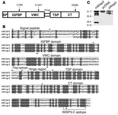
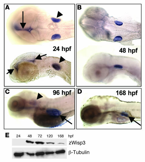
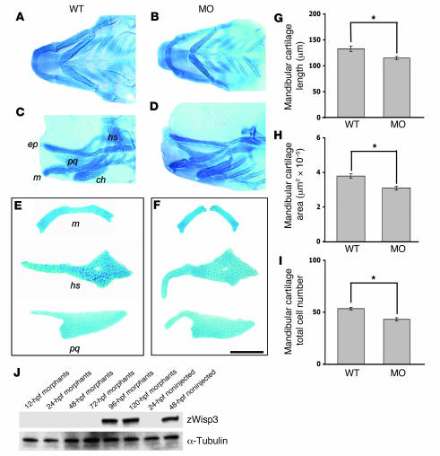
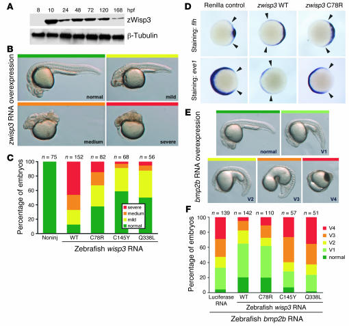
In humans, loss-of-function mutations in the gene encoding Wnt1 inducible signaling pathway protein 3 (WISP3) cause the autosomal-recessive skeletal disorder progressive pseudorheumatoid dysplasia (PPD). However, in mice there is no apparent phenotype caused by Wisp3 deficiency or overexpression. Consequently, the in vivo activities of Wisp3 have remained elusive. We cloned the zebrafish ortholog of Wisp3 and investigated its biologic activity in vivo using gain-of-function and loss-of-function approaches. Overexpression of zebrafish Wisp3 protein inhibited bone morphogenetic protein (BMP) and Wnt signaling in developing zebrafish. Conditioned medium-containing zebrafish and human Wisp3 also inhibited BMP and Wnt signaling in mammalian cells by binding to BMP ligand and to the Wnt coreceptors low-density lipoprotein receptor-related protein 6 (LRP6) and Frizzled, respectively. Wisp3 proteins containing disease-causing amino acid substitutions found in patients with PPD had reduced activity in these assays. Morpholino-mediated inhibition of zebrafish Wisp3 protein expression in developing zebrafish affected pharyngeal cartilage size and shape. These data provide a biologic assay for Wisp3, reveal a role for Wisp3 during zebrafish cartilage development, and suggest that dysregulation of BMP and/or Wnt signaling contributes to cartilage failure in humans with PPD.


Figures

Figure 1
Figure 1. Structure and sequence of Wisp3.
(A) Domain structure of Wisp3, with locations of the 3 tested disease-causing PPD missense mutations noted. Signal peptide (SP), IGFBP, VWC, thrombospondin 1 (TSP), and CT domains are shown. (B) Alignment of Wisp3 amino acid sequence for hWISP3, mWisp3, and zWisp3. Fully conserved residues across these species are shaded. Asterisks denote the residues affected by PPD-causing missense mutations. Inverted triangles denote approximate domain boundaries. Bracketed region indicates the mouse polypeptide epitope against which the WISP3-C antibody was generated. (C) Western blot of HEK293T cell conditioned media containing recombinant mWisp3, control (pcDNA), or zWisp3, separated by 12% SDS-PAGE under reducing conditions and probed with the WISP3-C antibody. WISP3-C antibody detected mWisp3 and zWisp3 (asterisk) as well as a background band (arrowhead) unique to the HEK293T culture media. A single zWisp3 band (~45 kDa) was detected, whereas 3 mWisp3 isoforms (due to N-linked glycosylation) were detected along with a carboxyterminal cleavage fragment (~28 kDa).
Figure 2
Figure 2. Expression of endogenous zWisp3.
(AD) Whole-mount in situ hybridization with an antisense zwisp3 RNA probe during early embryonic development. (A) Dorsal (top) and lateral (bottom) views of 24-hpf embryos demonstrated zwisp3 mRNA expression in the otic vesicle (arrowheads) and midline brain (arrows). (B) Ventral and lateral views of 48-hpf embryos demonstrated persistent zwisp3 expression in the otic vesicles. (C) Lateral view of 96-hpf larva showed diminished expression in the otic vesicle (arrowhead) and new expression in the developing swim bladder (arrow). (D) Lateral view of 168-hpf larva demonstrated persistent expression in the swim bladder (arrow). No zwisp3 expression was observed by in situ hybridization in embryos prior to 24 hpf (not shown). (E) Western blot of zWisp3 protein extracted from deyolked embryos and larvae. Protein (60 μg) was separated by 10% SDS-PAGE under reducing conditions and immunodetected using WISP3-C antibody or anti–β-tubulin antibody as a control. zwisp3 can be detected by RT-PCR at 6 hpf (not shown), by in situ hybridization at 24 hpf, and zWisp3 by Western blot at 48 hpf.
Figure 3
Figure 3. Pharyngeal cartilage changes in 108-hpf morphants and quantitative morphometric analysis of mandibular cartilage from wild-type and morphant larvae.
(AD) Ventral (A and B) and lateral (C and D) images of Alcian blue–stained pharyngeal cartilages in intact wild-type and morphant larva (24 ng zwisp3 translation blocking morpholino; MO). The ethmoid plate (ep), mandibular (m), palatoquadrate (pq), hyosympletic (hs), and ceratohyal (ch) cartilages are indicated. (E and F) Representative images of dissected Alcian blue–stained mandibular, palatoquadrate, and hyosympletic cartilages from wild-type and morphant larvae. Note zWisp3 morphant larvae had a smaller mandibular cartilage and abnormally shaped palatoquadrate and hyosympletic cartilages. During microdissection, these cartilages were noticeably more fragile in morphant compared with wild-type larvae. Similar findings were observed for larvae injected with the zwisp3 splice-site morpholino. Scale bar: 100 μm. (G) Mandibular length in morphants significantly decreased compared with wild-type fish. (H) Mandibular surface area in morphants significantly decreased compared with wild-type fish. (I) The number of chondrocytes per hemimandible was significantly fewer in morphants compared with wild-type fish. (J) Western blot of zWisp3 protein extracted from deyolked embryos and larvae. Protein (60 μg) was separated by 10% SDS-PAGE under reducing conditions and immunodetected using WISP3-C antibody or anti–α-tubulin antibody as a control. Note the morphants had no immunodetectable zWisp3 until 96 hpf, while zWisp3 was detected by 48 hpf in noninjected controls. *P < 0.01.
Figure 4
Figure 4. Overexpression of zWisp3 in zebrafish inhibits BMP signaling.
(A) Western blot of zWisp3 protein extracted from deyolked embryos and larvae injected with 150 pg zwisp3 RNA. Protein (40 μg) was separated by 10% SDS-PAGE under reducing conditions and detected using WISP3-C or anti–β-tubulin antibody. zWisp3 protein was detectable by 10 hpf when overexpressed. (B) Photographs of live 24-hpf embryos depicting the phenotypic classes of dorsalization. Embryos classified as mild correspond to convergence-extension classes C1 and C2, medium to C3 and C4, and severe to C5, as previously described (30). (C) Phenotype frequencies (shown in B) caused by injection of wild-type, C78R, C145Y, and Q338L zwisp3 RNA. Mutants had reduced biologic activity compared with wild-type zWisp3. (D) Photographs of 60% epiboly embryos oriented with their dorsal domains to the right. Arrowheads indicate extent of strong expression. Expression of flh was strongly expanded in wild-type zwisp3–injected embryos and less dramatically so in zwisp3 C78R–injected embryos. Expression of eve1 was strongly reduced in wild-type zwisp3–injected embryos and less dramatically reduced in C78R-injected embryos. (E) Photographs of live 24-hpf embryos depicting phenotypic classes of ventralization caused by injection of zbmp2b RNA as previously reported (30). (F) Phenotype frequencies (shown in E) caused by injection of zbmp2b RNA plus equimolar amounts of control (luciferase), zwisp3 wild-type, or zwisp3 missense mutant RNAs. C145Y and Q338L did not inhibit BMP signaling, while wild-type and C78R RNAs substantially rescued the defects caused by zbmp2b overexpression.
Figure 5
Figure 5. Overexpression of zWisp3 in zebrafish modulates canonical Wnt signaling.
(A) Photograph of a 30-hpf live embryo injected with an RNA encoding a constitutively active form of hLRP6 (hLRP6ΔN). Note the deficient formation of anterior structures compared with the uninjected control. (B) Quantification of the phenotypic classes that result from injecting wild-type hLRP6 or hLRP6ΔN with wild-type, C78R, C145Y, or Q338L forms of zwisp3. Note the severity of the hLRP6-induced phenotype was reduced when wild-type zWisp3, but not missense mutant zWisp3, was coexpressed. Also note that wild-type zWisp3 did not reduce phenotypic severity caused by expression of hLRP6ΔN. (C) Photographs of live 30-hpf embryos that had been co-injected with zwnt8 RNA and either wild-type or C78R mutant zwisp3 RNA, or hDKK1 RNA. Note wild-type zwisp3 and hDKK1 rescued the zWnt8-induced loss-of-anterior-structure phenotype. (D) Quantification of the phenotypic effects of overexpressing zWnt8 with or without wild-type or missense mutant zWisp3 or hDKK1. Note the severity of the zWnt8-induced phenotype was reduced by coexpressing wild-type zWisp3 or hDKK1, but not missense mutant zWisp3 (C78R, C145Y, and Q338L). (E) Agarose gels containing RT-PCR amplimers of zebrafish sp5l (zsp5l) and zebrafish β-actin (zβ-actin) using template RNA extracted from 80% epiboly-stage embryos injected with zWnt8 and either wild-type or C78R zwisp3 or hDKK1. Note that zWnt8-induced expression of zsp5l was reduced only when wild-type zwisp3 or hDKK1 were co-injected.
Figure 6
Figure 6. hWISP3 inhibits BMP2 signaling in mammalian cells and physically interacts with mBMP4.
(A) ALP activity in 10T1/2 cells cultured with recombinant hBMP2. hBMP2 induced ALP activity in a dose-dependent manner, and this induction was reduced when hWISP3-containing CM versus control CM was used. (B) hBMP2-mediated ALP induction in the presence of increasing amounts of hWISP3-containing CM. hWISP3 inhibited ALP induction dose dependently. (C) ALP activity in the presence of hBMP2 and 300 μl control CM, hWISP3 CM, immunodepleted hWISP3 CM, or PPD-associated mutant CM. The largest reduction in ALP activity occurred with hWISP3 CM. (D) Western blot of hWISP3 supernatants from CM mixed with protein G beads coated with increasing amounts of WISP3-C antibody. WISP3-C antibody dose-dependently immunodepleted hWISP3 protein. Positive control is supernatant after beads uncoated with antibody were used; negative control is eluate from these uncoated beads. (E) Western blots of protein precipitated with anti-myc beads probed with either anti-myc or WISP3-C antibody. Wild-type hWISP3 and the C78R missense mutant efficiently coprecipitated with mBMP4, whereas the C145Y and Q338L mutants did not. Input hWISP3 containing CM used in the co-IP experiments is shown at bottom. Asterisks indicate full-length hWISP3; arrowhead indicates a cross-reacting band observed only in CM from HEK293T cells; and double arrowhead indicates a hWISP3 cleavage product found in CM from HEK293T cells. The cleavage product did not coprecipitate with mBMP4 (not shown). (F) Western blots of protein precipitated with anti-flag beads probed with either anti-myc or WISP3-C antibody. mBMP4 coprecipitated with wild-type hWISP3.
Figure 7
Figure 7. zWisp3 physically and biologically interacts with the Wnt coreceptors LRP6 and FzD8.
(A) Ratio of firefly luciferase activity to Renilla luciferase activity in HEK293T cells transfected with TopFlash, pRL-TK, and either empty vector (pcDNA), hLRP6, mWnt1-V5, or hLRP6 plus mWnt1-V5, and then cultured in either control CM, wild-type zWisp3, zWisp3-immunodepleted CM, or PPD-associated missense mutant CM. Wild-type zWisp3 CM, but not the immunodepleted or the 3 zWisp3 mutant CMs, reduced luciferase activity. (B) Same experimental design as in A, except mWnt10b was used instead of mWnt1-V5. (C) Western blots of protein precipitated with protein G beads from mixtures of hLRP6N-Fc and zWisp3 containing CM, and probed with either WISP3-C or anti-mouse IgG antibody. Western blot of CM immunodetected with WISP3-C antibody (bottom) demonstrated comparable expression of wild-type and missense mutant zWisp3. Only wild-type zWisp3 coprecipitated with hLRP6. WISP3-C antibody detected zWisp3 (asterisk) as well as a background band (arrowhead) unique to HEK293T culture medium. (D) Western blots of individual components of the mWnt1-V5/hLRP6N-myc/mFzD8CRD-IgG complex following their IP in the presence of increasing amounts of zWisp3. hLRP6N-myc was immunoprecipitated and immunodetected using an anti-myc antibody. FzD8CRD-IgG was precipated with protein G and immunodetected using anti-mouse IgG antibody. mWnt1-V5 was immunoprecipitated and immunodetected using anti-V5 antibody, and zWisp3 was immunodetected using WISP3-C antibody. Increased zWisp3 interfered with the ability of the mWnt1-V5/hLRP6N-myc/mFzD8CRD-IgG trimeric complex to form. Asterisk in the input column of the lower panel indicates zWisp3, with the upper band representing a background band (arrowhead) unique to HEK293T culture medium.

Similar articles

Cited by

References

    1. Hurvitz J.R., et al. Mutations in the CCN gene family member WISP3 cause progressive pseudorheumatoid dysplasia. Nat. Genet. 1999;23:94–98. - PubMed
    1. Spranger J., Albert C., Schilling F., Bartsocas C. Progressive pseudorheumatoid arthropathy of childhood (PPAC): a hereditary disorder simulating juvenile rheumatoid arthritis. Am. J. Med. Genet. 1983;14:399–401. - PubMed
    1. Wynne-Davies R., Hall C., Ansell B.M. Spondylo-epiphysial dysplasia tarda with progressive arthropathy. A “new” disorder of autosomal recessive inheritance. J. Bone Joint Surg. Br. 1982;64:442–445. - PubMed
    1. el-Shanti H.E., Omari H.Z., Qubain H.I. Progressive pseudorheumatoid dysplasia: report of a family and review. J. Med. Genet. 1997;34:559–563. - PMC - PubMed
    1. Ehl S., et al. Clinical, radiographic, and genetic diagnosis of progressive pseudorheumatoid dysplasia in a patient with severe polyarthropathy. Rheumatol. Int. 2004;24:53–56. - PubMed

Publication types

MeSH terms

Substances