. 2007 Dec;13(12):2881-90.
doi: 10.1089/ten.2007.0078.

Vascularized adipose tissue grafts from human mesenchymal stem cells with bioactive cues and microchannel conduits

Affiliations

Vascularized adipose tissue grafts from human mesenchymal stem cells with bioactive cues and microchannel conduits

Michael S Stosich et al. Tissue Eng. 2007 Dec.

Abstract

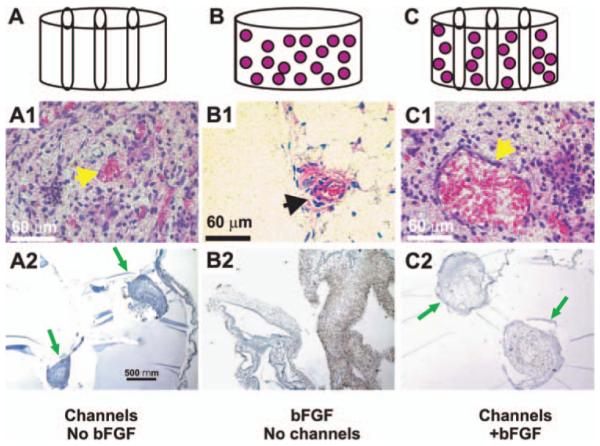
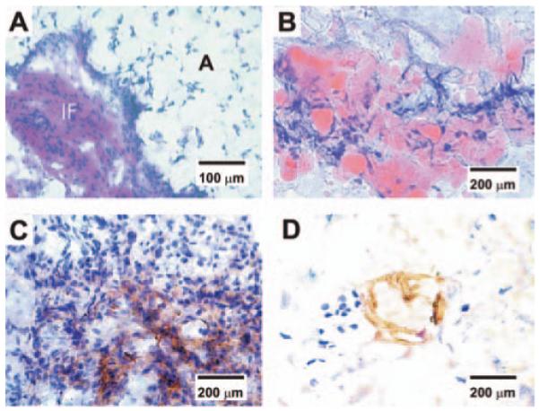
Vascularization is critical to the survival of engineered tissues. This study combined biophysical and bioactive approaches to induce neovascularization in vivo. Further, we tested the effects of engineered vascularization on adipose tissue grafts. Hydrogel cylinders were fabricated from poly(ethylene glycol) diacrylate (PEG) in four configurations: PEG alone, PEG with basic fibroblast growth factor (bFGF), microchanneled PEG, or both bFGF-adsorbed and microchanneled PEG. In vivo implantation revealed no neovascularization in PEG, but substantial angiogenesis in bFGF-adsorbed and/or microchanneled PEG. The infiltrating host tissue consisted of erythrocyte-filled blood vessels lined by endothelial cells, and immunolocalized to vascular endothelial growth factor (VEGF). Human mesenchymal stem cells were differentiated into adipogenic cells, and encapsulated in PEG with both microchanneled and adsorbed bFGF. Upon in vivo implantation subcutaneously in immunodeficient mice, oil red O positive adipose tissue was present and interspersed with interstitial fibrous (IF) capsules. VEGF was immunolocalized in the IF capsules surrounding the engineered adipose tissue. These findings suggest that bioactive cues and/or microchannels promote the genesis of vascularized tissue phenotypes such as the tested adipose tissue grafts. Especially, engineered microchannels may provide a generic approach for modifying existing biomaterials by providing conduits for vascularization and/or diffusion.


Figures

FIG. 1
FIG. 1
In vivo implantation of PEG hydrogel. PEG hydrogel was fabricated in four configurations: PEG alone, PEG with bFGF, microchanneled PEG, or both bFGF-adsorbed and microchanneled PEG. (A) PEG hydrogel molded into 6×4 mm (diameter×height) cylinder (without either bFGF or microchannels). (B) PEG hydrogel with three microchannels. (C) PEG hydrogel cylinder with microchannels and adsorbed with both 0.5 μg/μL bFGF and three microchannels. Following in vivo implantation subcutaneously in the dorsum of immunodeficient mice, the harvested PEG hydrogel samples showed distinct histological features. (A1) PEG hydrogel with microchannels but without bFGF showed host tissue infiltration primarily in the lumen of microchannels, and scarcely in the rest of PEG hydrogel. The infiltrating host tissue includes erythrocyte-filled blood vessels that are lined by endothelial cells (arrow). (A2) VEGF was immunolocalized only to host-derived tissue within the lumen of microchannels, indicating the vascular nature of the infiltrating host tissue. Arrows point to microchannels and the infiltrating host tissue. (B1) PEG hydrogel with bFGF but without microchannels showed apparently random and isolated islands of infiltrating host tissue (arrow). There is random infiltration of host tissue into bFGF-adsorbed PEG hydrogel. The infiltrating host tissue includes vascular structures with erythrocyte-filled blood vessels that are lined by endothelial cells (arrow). (B2) VEGF was immunolocalized to host-derived tissue within PEG hydrogel (without microchannels). (C1) PEG hydrogel with both microchannels and bFGF showed host tissue infiltration only in the lumen of microchannels, but scarcely in the rest of the PEG hydrogel. The infiltrating host tissue includes vascular structures with erythrocyte-filled blood vessels that are lined by endothelial cells (arrow). (C2) VEGF was immunolocalized only to host-derived tissue within the lumen of microchannels. Since no cells were delivered in any of the PEG hydrogel samples in this experiment, tissue infiltration following in vivo implantation must be derived from the host. Arrows point to microchannels. Color images available online at www.liebertpub.com/ten.
FIG. 2
FIG. 2
Adipogenic differentiation of human mesenchymal stem cells (hMSCs) in oil red O staining for lipid vacuoles. Top row (A–E): hMSCs without adipogenic differentiation over 35 days. Bottom row (F–J): hMSCs in adipogenic induction medium over 35 days. Oil red O staining is negative for hMSCs during 35 days of culture. In contrast, hMSCs treated in adipogenic medium showed gradually intense oil red O staining over the observed 35 days. Color images available online at www.liebertpub.com/ten.
FIG. 3
FIG. 3
Glycerol content of hMSCs and hMSC-derived adipogenic cells. Glycerol content was quantified by ELISA per our previous methods. Glycerol content of hMSC-derived adipogenic cells was significantly higher than hMSCs at days 28 and 35, suggesting that hMSC-derived adipogenic cells continue to mature into adipocytes with the capacity to synthesize glycerol.
FIG. 4
FIG. 4
In vivo implantation of bFGF and microchanneled PEG hydrogel loaded with adipogenic cells derived from hMSCs. Diagrams (top row) and corresponding representative photographs at the time of harvest of in vivo samples. (A) PEG hydrogel molded into 6×4 mm (width×height) cylinder (without either bFGF or microchannels). (B) PEG hydrogel cylinder loaded with both 0.5 μg/μL bFGF and three microchannels, but without the delivery of cells. (C) PEG hydrogel cylinder loaded with both 0.5 μg/μL bFGF and three microchannels, in addition to the encapsulation of adipogenic cells that have been derived from human mesenchymal stem cells at a cell seeding density of 3×10⁶ cells/mL. Following in vivo implantation subcutaneously in the dorsum of immunodeficient mice, the harvested PEG hydrogel samples showed distinct histological features. (A’) PEG hydrogel cylinder without either microchannels or bFGF showed somewhat transparent appearance. (B’) PEG hydrogel cylinder with both bFGF and three microchannels, but without delivered cells, showed darker color and a total of three openings of microchannels (arrows) that are confirmed to be areas of host cell infiltration histologically (c.f., Fig. 1A1, 1A2). (C’) PEG hydrogel cylinder with both microchannels and bFGF in addition to encapsulated hMSC-derived adipogenic cells showed the opening of microchannels (red color and pointed with arrows) that are confirmed to be areas of host cell infiltration histologically in Figure 5. Color images available online at www.liebertpub.com/ten.
FIG. 5
FIG. 5
Histological and immunohistochemical characterization of vascularized adipose tissue from human mesenchymal stem cells. (A) Hematoxylin and eosin staining revealed IF tissue interposing between foam-like space labeled with A for adipose tissue. The presence of adipose tissue is confirmed in (B), showing substantial oil red O positive staining in PEG hydrogel encapsulating hMSC-derived adipogenic cells, in addition to bFGF and built-in microchannels. In contrast, there is no evidence of adipogenesis in PEG hydrogel with bFGF and built-in microchannels, but with hMSCs (without adipogenic differentiation). (C) Positive immunolocalization of VEGF antibody in the IF tissue interposing areas of adipogenesis, indicating the presence of vascular supply. (D) Positive immunolocalization of lectin WGA in the interstitial fibrous tissue interposing areas of adipogenesis, serving as further indication of the presence of vascular endothelial cells. Color images available online at www.liebertpub.com/ten.

References

    1. Alhadlaq A, Mao JJ. Mesenchymal stem cells: isolation and therapeutics. Stem Cells Dev. 2004;13:436–448. - PubMed
    1. Caplan AI. Mesenchymal stem cells. J Orthop Res. 1991;9:641–650. - PubMed
    1. Marion NW, Mao JJ. Mesenchymal stem cells and tissue engineering. Methods Enzymol. 2006;420:339–361. - PMC - PubMed
    1. Pittenger MF, Mackay AM, Beck SC, Jaiswal RK, Douglas R, Mosca JD, Moorman MA, Simonetti DW, Craig S, Marshak DR. Multilineage potential of adult human mesenchymal stem cells. Science. 1999;284:143–147. - PubMed
    1. Sekiya I, Larson BL, Vuoristo JT, Cui JG, Prockop DJ. Adipogenic differentiation of human adult stem cells from bone marrow stroma (MSCs) J Bone Miner Res. 2004;19:256–264. - PubMed

Publication types

Substances

LinkOut - more resources