. 2007 Sep;65(6):1534-46.
doi: 10.1111/j.1365-2958.2007.05887.x.

Dual localization pathways for the engulfment proteins during Bacillus subtilis sporulation

Affiliations

Dual localization pathways for the engulfment proteins during Bacillus subtilis sporulation

Stefan Aung et al. Mol Microbiol. 2007 Sep.

Abstract

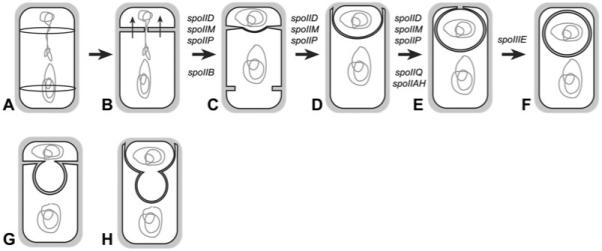
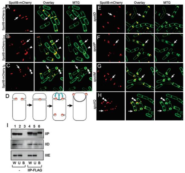
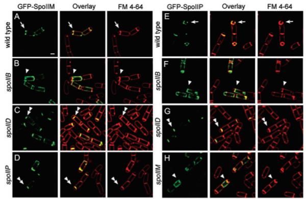
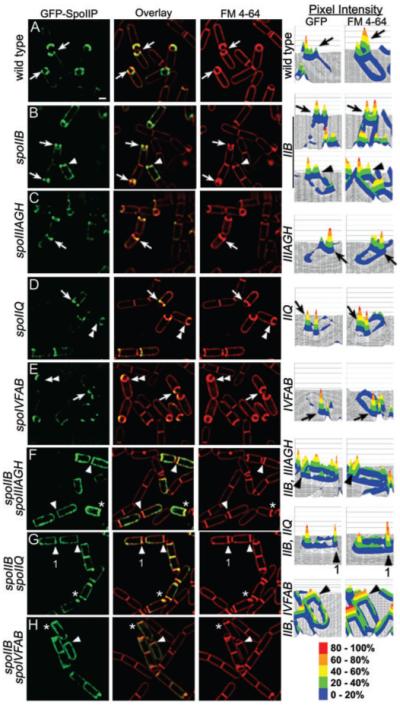
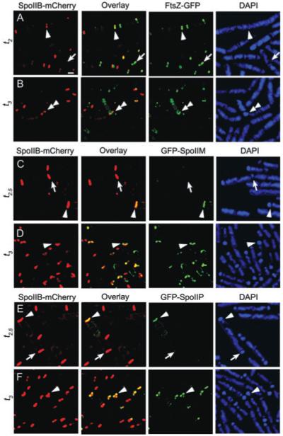
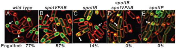
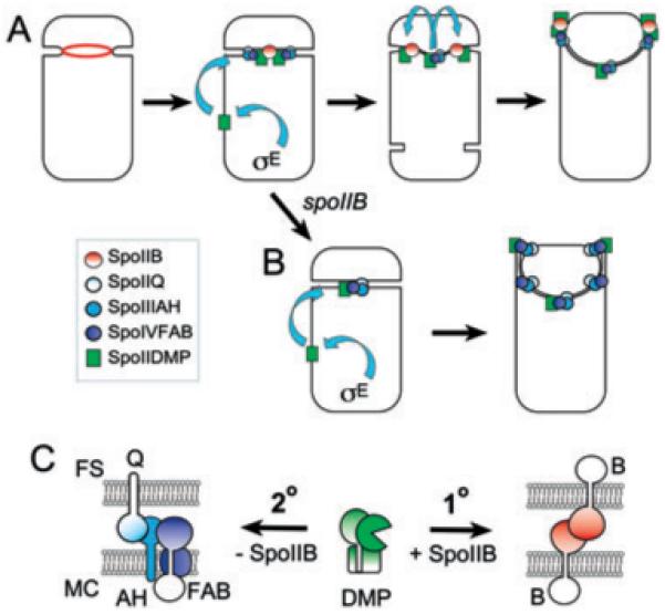
Engulfment in Bacillus subtilis is mediated by two complementary systems, SpoIID, SpoIIM and SpoIIP (DMP), which are essential for engulfment, and the SpoIIQ-SpoIIIAGH (Q-AH) zipper, which provides a secondary engulfment mechanism and recruits other proteins to the septum. We here identify two mechanisms by which DMP localizes to the septum. The first depends on SpoIIB, which is recruited to the septum during division and provides a septal landmark for efficient DMP localization. However, sporangia lacking SpoIIB ultimately localize DMP and complete engulfment, suggesting a second mechanism for DMP localization. This secondary targeting pathway depends on SpoIVFA and SpoIVFB, which are recruited to the septum by the Q-AH zipper. The absence of a detectable localization phenotype in mutants lacking only SpoIVFAB (or Q-AH) suggests that SpoIIB provides the primary DMP localization pathway while SpoIVFAB provides a secondary pathway. In keeping with this hypothesis, the spoIIB spoIVFAB mutant strain has a synergistic engulfment defect at septal thinning (which requires DMP) and is completely defective in DMP localization. Thus, the Q-AH zipper both provides a compensatory mechanism for engulfment when DMP activity is reduced, and indirectly provides a compensatory mechanism for septal localization of DMP when its primary targeting pathway is disrupted.


Figures

Fig. 1
Fig. 1
The sporulation pathway of B. subtilis. A. Early in sporulation, two FtsZ rings assemble at each pole of the cell. B. The sporulation septum separates the smaller forespore and larger mother cell, and traps the forespore chromosome, which is pumped into the forespore by SpoIIIE (arrows). C. The first step of engulfment, septal thinning, commences as the peptidoglycan within the septum (grey) is thinned, a step requiring the mother cell-expressed proteins SpoIID, SpoIIM, SpoIIP, and facilitated by SpoIIB. D. Next, the mother cell membrane migrates up and around the forespore, which also requires DMP and, under certain conditions, the Q-AH zipper. E and F. (E) Ultimately, the migrating membrane meets and (F) fuses at the forespore pole, a step requiring the SpoIIIE DNA translocase (Sharp and Pogliano, 1999). Proteins shown above the arrow are always essential for the step, while those shown below slow but do not completely block engulfment under some conditions. G. Mutants completely defective in septal thinning (i.e. spoIID, spoIIM or spoIIP null mutations) produce a bulge phenotype in which the growing forespore pushes through the unthinned septum. H. Mutants that slow septal thinning (i.e. spoIIB null mutations) also produce a bulge, but the engulfing membranes are ultimately able to migrate around septal peptidoglycan.
Fig. 2
Fig. 2
Localization of SpoIIB–mCherry. Live cells expressing SpoIIB–mCherry (red) and stained with the membrane stain Mitotracker Green (green). Sporulation was induced by re-suspension at 30°C and samples were harvested at the indicated time. A–C. SpoIIB–mCherry in wild type (KP1026) (A) at t2 (2 h after the initiation of sporulation), (B) at t2.5 and (C) at t3. SpoIIB localizes to the septum (arrow), and to the potential second division site (arrowhead) at the opposite pole. In engulfing sporangia, SpoIIB localizes to the leading edge of the engulfing membrane (C, arrow), where it remains until the completion of engulfment (C, double arrowhead). D. Cartoon representation of SpoIIB–mCherry during engulfment. E–H. SpoIIB–mCherry in (E) spoIID (KP1027), (F) spoIIP (KP1028), (G) spoIIM (KP1029) and (H) spoIIQ (KP1030) strains. SpoIIB–mCherry is restricted to the septal midpoint in spoIID and spoIIP mutants (arrows, E and F), but moves to the edge of the septum in the spoIIM mutant (arrow, G), and shows wild-type localization in the spoIIQ mutant (H). I. Co-immunoprecipitation of SpoIID by SpoIIP-FLAG. KP4156 (spoIIP-FLAG; lanes 4–6) and the negative control strain PY79 (wild type; lanes 1–3) were solubilized by 0.5% digitonin after 2.5 h of sporulation at 37°C and then subjected to immunoprecipitation using anti-FLAG antibodies. Proteins were visualized by Western blotting using anti-SpoIIP (top), anti-SpoIID (middle) and anti-SpoIIIE (negative control; bottom). W, U and B represent whole cell, unbound and bound fractions respectively. Scale bar in (A), 1 μm.
Fig. 3
Fig. 3
Localization of GFP–SpoIIM and GFP–SpoIIP in mutant backgrounds. Localization of GFP–SpoIIM (A–D, green) and GFP–SpoIIP (E–H, green) in sporangia sampled 1 h and 45 min after the initiation of sporulation by re-suspension (t1.75). Membranes are stained with FM 4-64 (red). A–D. GFP–SpoIIM localization in (A) wild type (KP721), (B) spoIIB (KP762), (C) spoIID (KP763) and (D) spoIIP (KP722) strains. GFP–SpoIIM localizes at the leading edge of the engulfing mother cell membrane in wild type (arrow), but is randomly localized in the spoIIB strain (arrowheads) and restricted to the septal midpoint in spoIID and spoIIP strains (double arrowheads). E–H. Localization of GFP–SpoIIP in (E) wild type (KP722), (F) spoIIB (KP774), (G) spoIID (KP775) and (H) spoIIM (KP776) strains. GFP–SpoIIP localizes to the leading edge and assembles distinct foci at the leading edge and within the septum of wild type (arrow), but shows mostly random distribution in spoIIB and spoIIM strains (arrowheads) and is trapped at the middle of the septum in the spoIID strain (double arrowhead). Scale bar in (A), 1 μm.
Fig. 4
Fig. 4
Localization of GFP–SpoIIP in the presence of untagged SpoIIP to rescue synergistic engulfment defects. Localization of GFP–SpoIIP (green) in sporangia also expressing untagged SpoIIP sampled at t1.75. Membranes are stained with FM 4-64 (red). Pixel intensities (fourth column) of GFP–SpoIIP signals are represented as surface plots from the highest intensity (red) to the lowest (blue), as described in Experimental procedures. A. In wild type (KP1035), GFP–SpoIIP localized to the leading edge of the engulfing mother cell membrane with some foci forming at the septal midpoint and around the forespore (arrows). B. In the spoIIB mutant (KP1036), GFP–SpoIIP localizes to the leading edge in engulfing sporangia and to the edges of septal bulges (arrows). Non-engulfing sporangia show additional delocalized GFP–SpoIIP fluorescence (arrowhead). C–E. GFP–SpoIIP in (C) spoIIIAGH (KP1037), (D) spoIIQ (KP1038) and (E) spoIVFAB (KP1039) strains. Nearly wild-type localization is seen in these strains with a slight increase in mother cell fluorescence (arrows) and fewer clear foci at the septal midpoint (double arrowheads). F–H. GFP–SpoIIP in (F) spoIIB spoIIIAGH (KP1040), (G) spoIIB spoIIQ (KP1041) and (H) spoIIB spoIVFAB (KP1042) strains. GFP–SpoIIP shows a more random distribution (arrowheads) with some accumulation at regions with increased membrane fluorescence (*) and septa. Scale bar in (A), 1 μm.
Fig. 5
Fig. 5
Colocalization of SpoIIB–mCherry with FtsZ–GFP, GFP–SpoIIM and GFP–SpoIIP. Sporulation of SpoIIB–mCherry strains was conducted at 30°C, and the sporangia were counterstained with the DNA stain DAPI (blue). A and B. Colocalization of SpoIIB–mCherry (red) and FtsZ–GFP (green) in strain KP1043 induced to sporulate by re-suspension at 30°C. Samples were collected at t2 (A) or t3 (B). FtsZ-GFP localizes to polar division sites first (arrow) followed by SpoIIB–mCherry (arrowhead). SpoIIB–mCherry remains localized after FtsZ–GFP is lost from the completed septum (double arrowhead). C and D. Colocalization of SpoIIB–mCherry (red) and GFP–SpoIIM (green) in strain KP1032 at t2.5 (C) or t3 (D). SpoIIB–mCherry (KP1032) localizes to the septum first (arrow), followed by GFP–SpoIIM (arrowheads). E and F. Colocalization of SpoIIB–mCherry and GFP–SpoIIP in strain KP1031 at t2.5 (E) or t3 (F). SpoIIB–mCherry localizes to the septum first (arrow), followed by GFP–SpoIIP (arrowhead). Scale bar in (A), 1 μm.
Fig. 6
Fig. 6
Synergistic engulfment defect in the absence of SpoIIB and SpoIVFAB. The completion of engulfment was followed using a membrane fusion assay using the membrane impermeable stain FM 4-64 (red) and the membrane permeable stain Mitotracker Green (MTG, green). During engulfment FM 4-64 stains all the membranes (arrowhead), while after engulfment, MTG but not FM 4-64 stains the forespore membranes (arrow). While wild type engulfs smoothly, mutants with septal thinning defects show the bulging of the forespore into the mother cell resulting in a bubble of membrane (double arrowheads). Strains were sporulated by resuspension at 37°C and scored for the percent of sporangia completing membrane fusion at t3.5. (A) wild type (PY79), (B) spoIVFAB (KP1013), (C) spoIIB (KP343) shows both bulges and complete engulfment, (D) spoIIB spoIVFAB (KP1014) shows no engulfment and mostly sporangia with bulges, (E) spoIIP (KP513) also shows a complete defect in septal thinning. Scale bar in (A), 1 μm.
Fig. 7
Fig. 7
Model for DMP localization. A. SpoIIB (red ring and circles) is targeted to the septum during cytokinesis (ring) and remains at the septum. After septation, DMP (green square) are synthesized by the mother cell transcription factor σE, and recruited to the septum by SpoIIB. The Q-AH zipper forms by the interaction of the forespore protein SpoIIQ (light blue) with the mother cell protein SpoIIIAH (royal blue), which recruits the SpoIVFA and SpoIVFB proteins (purple) that are required for late mother cell gene expression. SpoIIB is primarily responsible for localization of DMP to the leading edge of the engulfing membrane, while SpoIVFAB allow the DMP proteins to assemble foci behind the leading edge. B. In the absence of SpoIIB, the Q-AH zipper can indirectly mediate DMP localization via SpoIVFAB. C. Our results suggest that there are two localization pathways for DMP, a primary SpoIIB-dependent pathway and a compensatory SpoIVFAB-dependent pathway. SpoIIB is synthesized before polar septation (Margolis et al., 1993; Perez et al., 2000) and is therefore drawn here as though the protein is present in both daughter cells, although this has not been experimentally tested.

Similar articles

Cited by

References

    1. Abanes-De Mello A, Sun YL, Aung S, Pogliano K. A cytoskeleton-like role for the bacterial cell wall during engulfment of the Bacillus subtilis forespore. Genes Dev. 2002;16:3253–3264. - PMC - PubMed
    1. Blaylock B, Jiang X, Rubio A, Moran CP, Jr, Pogliano K. Zipper-like interaction between proteins in adjacent daughter cells mediates protein localization. Genes Dev. 2004;18:2916–2928. - PMC - PubMed
    1. Broder DH, Pogliano K. Forespore engulfment mediated by a ratchet-like mechanism. Cell. 2006;126:917–928. - PMC - PubMed
    1. Chastanet A, Losick R. Engulfment during sporulation in Bacillus subtilis is governed by a multi-protein complex containing tandemly acting autolysins. Mol Microbiol. 2007;64:139–152. - PubMed
    1. Chen JC, Viollier PH, Shapiro L. A membrane metalloprotease participates in the sequential degradation of a Caulobacter polarity determinant. Mol Microbiol. 2005;55:1085–1103. - PubMed

Publication types

MeSH terms

Substances

LinkOut - more resources